Autoimmune disease model animal
a model animal and autoimmune disease technology, applied in the field of autoimmune disease model animals, can solve the problems of no mice producing antibodies capable of reacting to mouse dsg3 protein, and achieve the effect of showing the phenotype of autoimmune disease, reducing muscle power, and facilitating and objectively evaluating the effectiveness
- Summary
- Abstract
- Description
- Claims
- Application Information
AI Technical Summary
Benefits of technology
Problems solved by technology
Method used
Image

Examples
example 1
Production of Recombinant Mouse Dsg3 Protein
[0054]A cDNA encoding the entire extracellular domain of mouse Dsg3 (Genbank U86016) was amplified by PCR using appropriate primers (5′-CCGAGATCTCCTATAAATATGACCTGCCTCTTCCCTAGA-3′ / SEQ ID NO: 1, 5′-CGGGTCGACCCTCCAGGATGACTCCCCATA-3′ / SEQ ID NO: 2) and using a phage clone containing mouse Dsg3 cDNA (a gift from Dr. Jouni Uitto) as a template; amplified fragment was subcloned (pEVmod-mDsg3-His) by replacing it with human Dsg3 cDNA in pEVmod-Dsg3-His vector (Ishii, K., et al., J. Immunol. 159:2010–2017 (1997)). A recombinant baculo-protein, mouse rDsg3, was prepared as described previously (Amagai, M. et al., J. Clin. Invest. 94:59–67 (1994); Amagai, M. et al., J. Invest. Dermatol. 104:895–901 (1995)).
example 2
Immunization of Wild-Type DSG3+ / + Mouse with Mouse Dsg3 Protein
[0055]First, attempts were made to produce antibodies against Dsg3 protein in a variety of wild-type mouse strains after immunizing with human or mouse rDsg3 (Table 1).
[0056]Mice were sensitized with 5 μg of purified mouse or human rDsg3 by intraperitoneal injection with complete Freund's adjuvant (CFA). Then booster immunization was carried out every week, 3 or 7 times, with mouse or human rDsg3 using incomplete Freund's adjuvant (IFA). An ELISA test for antibody production was conducted 3 days after each booster immunization.
[0057]In ELISA assay for blood IgG against mouse Dsg3 protein (mDsg3) or human Dsg3 protein (hDsg3) in mice, mouse or human rDsg3 was used as a coating antigen. More specifically, a 96-well microtiter plate was coated with 100 μl of 5 μg / ml purified mouse or human rDsg3 at 4° C. overnight. All serum samples were diluted 50 to 5,000 times and then incubated on a 96-well ELISA plate at room temperatu...
example 3
Immunization of DSG3− / − Mouse and DSG3+ / − Mouse with Mouse Dsg3 Protein
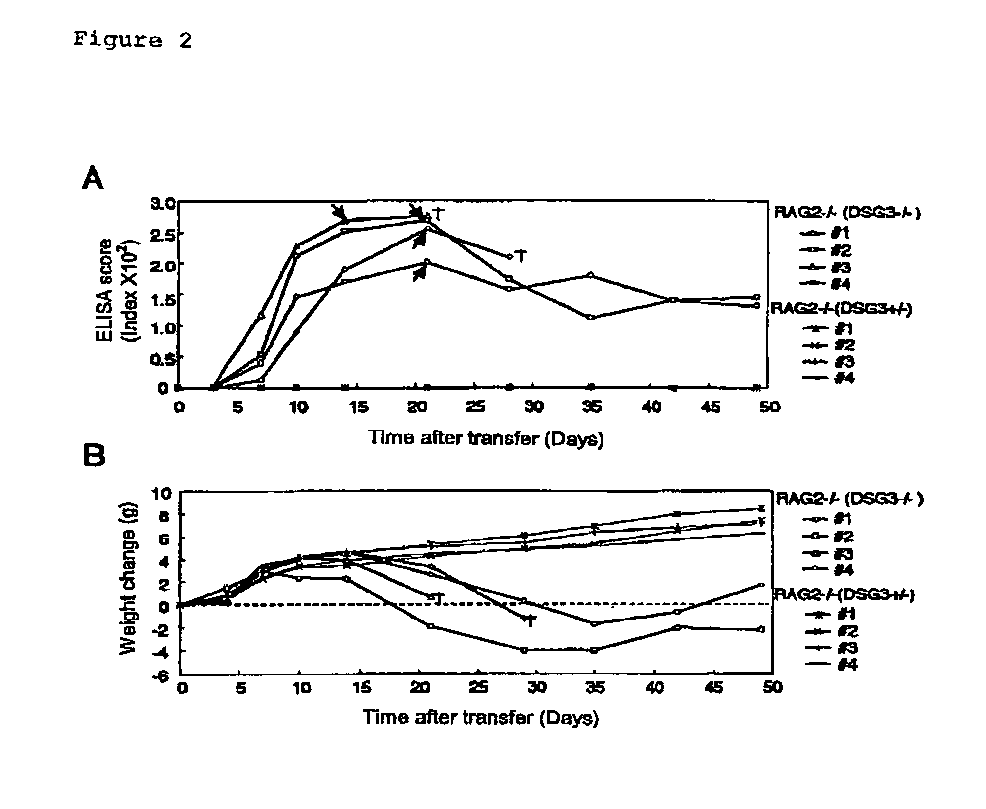
[0062]DSG3− / − mice were prepared by mating male DSG3− / − mice with female DSG3+ / − mice (Koch, P. J., et al., J. Cell Biol. 137:1091–102 (1997)). RAG2− / − mice, which had been obtained by back-crossing with B6.SJL-ptprc over 10 generations, were provided from Taconic (German Town, N.Y.) (Schulz, R.-J. et al., J. Immunol. 157:4379–4389 (1996)).
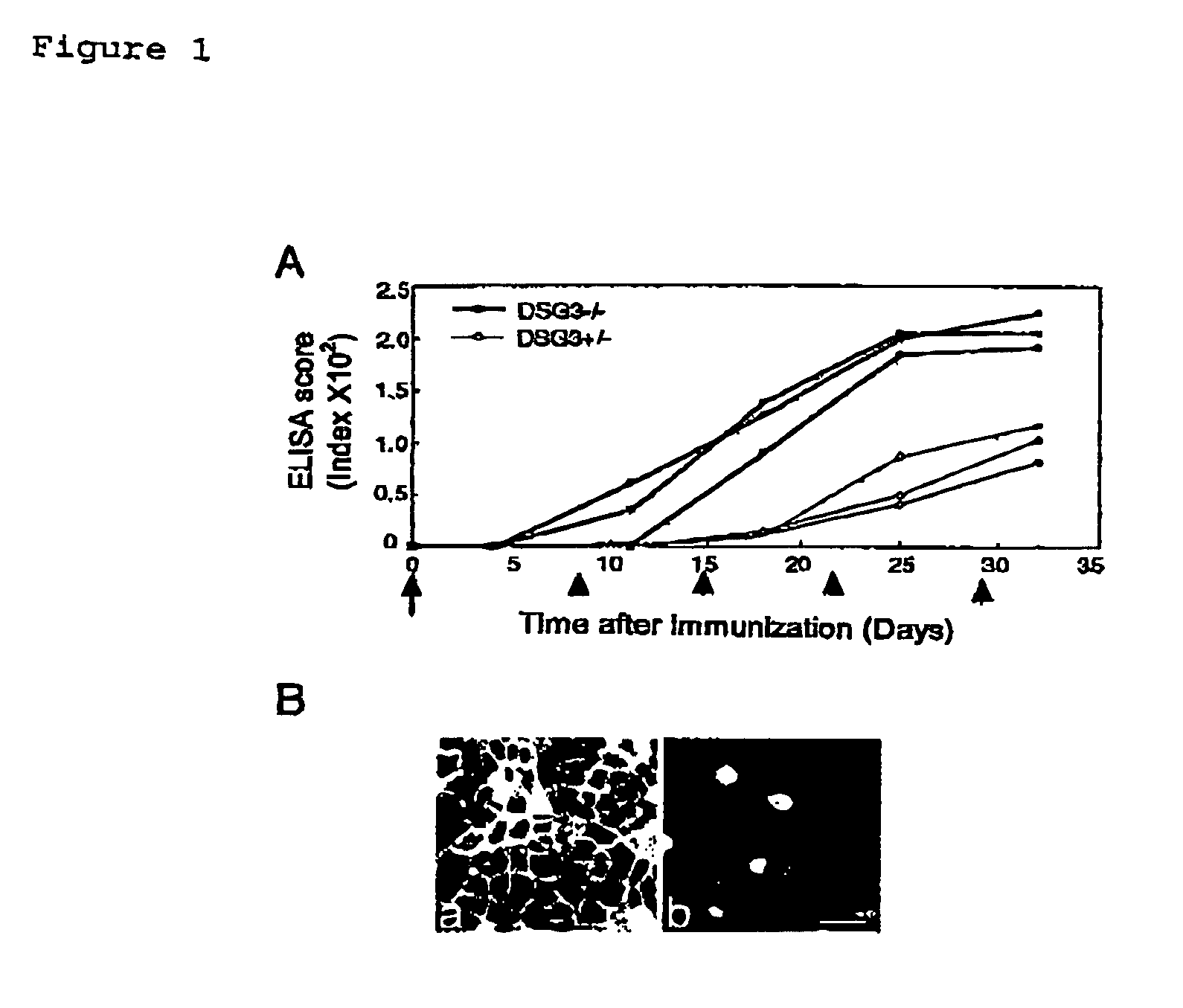
[0063]ELISA scores for mouse rDsg3 were determined after immunizing DSG3− / − mouse with mouse rDsg3 in order to verify the absence of immuno-tolerance to Dsg3 protein in DSG3− / − mouse.
[0064]Both DSG3− / − mice and DSG3+ / − mice were sensitized with 5 μg of purified mouse rDsg3 by using complete Freund's adjuvant (0 day), and then booster was carried out with mouse rDsg3 by using incomplete Freund's adjuvant after 8, 15, 22, and 28 days. The antibody production was tested by ELISA using mouse rDsg3 as a coating antigen in the same manner as in Example 2.
[0065]The production of anti-...
PUM
| Property | Measurement | Unit |
|---|---|---|
| Time | aaaaa | aaaaa |
| Angle | aaaaa | aaaaa |
| Angle | aaaaa | aaaaa |
Abstract
Description
Claims
Application Information
 - R&D Engineer
- R&D Manager
- IP Professional
- Industry Leading Data Capabilities
- Powerful AI technology
- Patent DNA Extraction
Browse by: Latest US Patents, China's latest patents, Technical Efficacy Thesaurus, Application Domain, Technology Topic.
© 2024 PatSnap. All rights reserved.Legal|Privacy policy|Modern Slavery Act Transparency Statement|Sitemap