
Autoimmune disease model animal

a model animal and autoimmune disease technology, applied in the field of autoimmune disease model animals, can solve the problems that no mice produced antibodies capable of reacting, and achieve the effects of reducing muscle power, showing the phenotype of autoimmune disease, and facilitating and objectively evaluating the effectiveness

Inactive Publication Date: 2006-07-27
KEIO UNIV
View PDF0 Cites 0 Cited by
  • Summary
  • Abstract
  • Description
  • Claims
  • Application Information

AI Technical Summary

Benefits of technology

[0039] The model animal of the present invention can show phenotype of autoimmune disease through the stable production of antibody reacting to the antigen protein for the autoimmune disease or sustained activation of T cell.
[0044] Further, immune cells can be obtained from the thymus, lymph node, spleen, liver, intestinal epithelium, peripheral blood, etc. but are not limited to those from the tissues. The spleen abundantly contains mature immune cells and thus is a preferable organ for the immune cells. It is preferable that the animal (donor) from which immune cells are prepared and the animal (recipient) to which lymphocytes derived from the immune cells are transferred belong to a same species and have a same genetic background thereby preventing the onset of GVHD which may cause tissue destruction in the recipient.
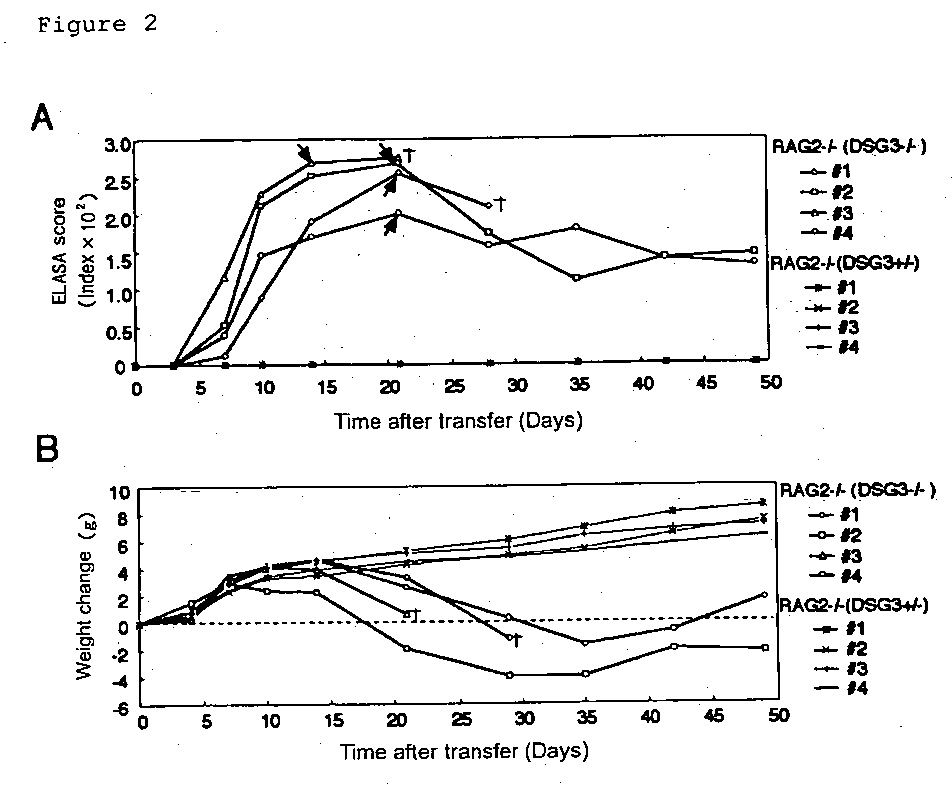
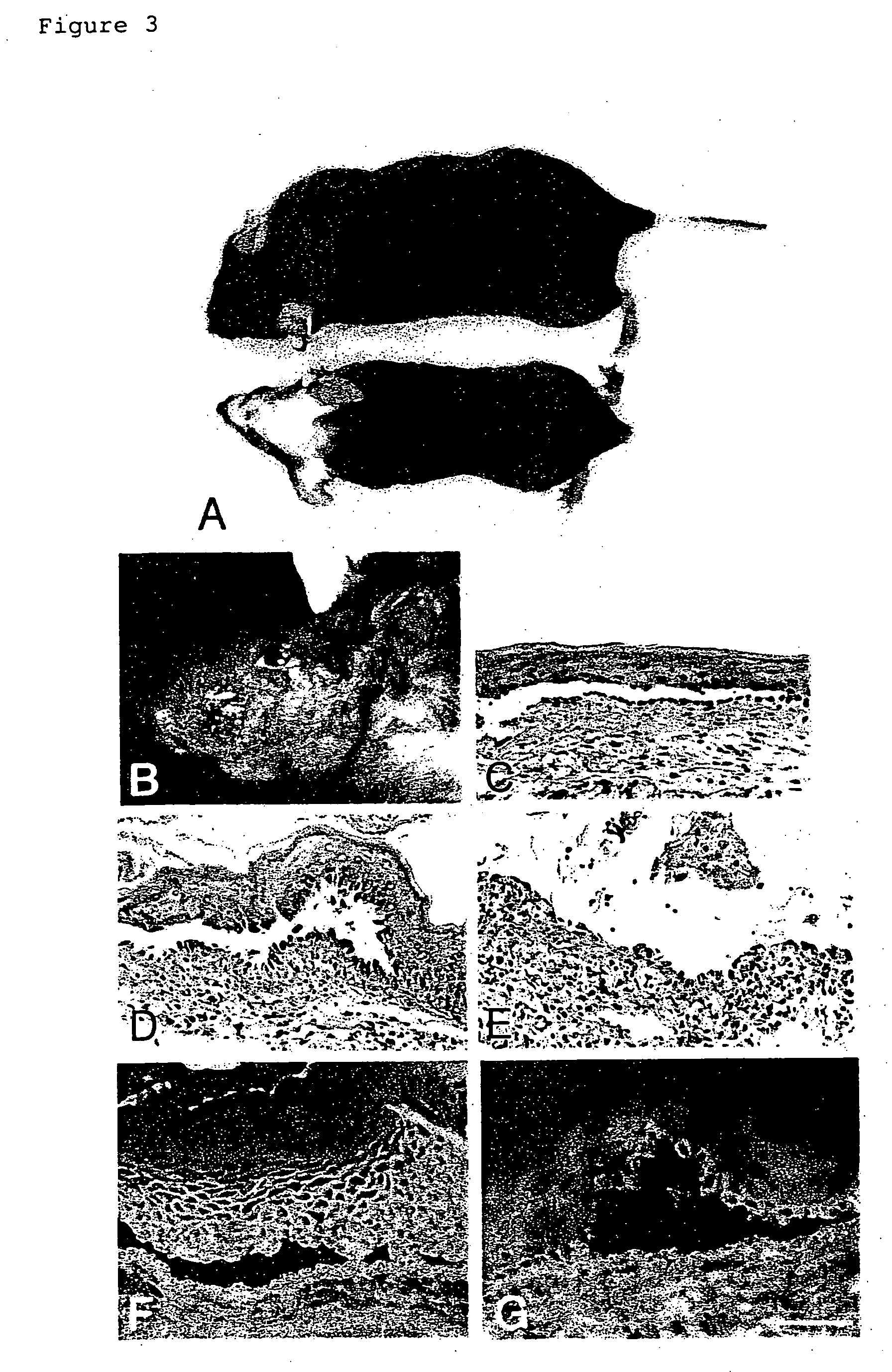
[0047] The model animal created in accordance with the present invention can show phenotype of autoimmune disease through the stable production of antibody reacting to the antigen protein for the autoimmune disease or sustained activation of T cell. In the model animal for pemphigus vulgaris, major phenotype includes weight loss and reversible telogen hair loss. Further, among autoimmune diseases other than pemphigus vulgaris, phenotype may include reduced muscle power in myasthenia gravis; anemia in autoimmune hemolytic anemia; hyperthyroidism in Basedow's disease; hypothyroidism in Hashimoto's disease; nephropathy and pulmonary disorders in Goodpasture's syndrome; glucosuria in autoimmune diabetes mellitus; and neuroparalysis in multiple sclerosis.
[0048] One can use these model animals for developing therapeutic agents or methods for the diseases, administering to them test compounds of interest for therapeutic effects on autoimmune diseases and observing phenotypes thereof. Particularly the major phenotype includes weight loss and reversible telogen hair loss in pemphigus vulgaris model mouse prepared in accordance with the present Example and the phenotypes last over 6 months, and therefore it is possible to readily and objectively evaluate the effectiveness of each therapeutic agent or method based on the observation without sacrificing the mouse. In addition, these model mice are very useful for clarifying cellular mechanism underlying the production of antibody against the antigen protein.

Problems solved by technology

However, with this method, no mice produced antibody capable of reacting to mouse Dsg3 protein (Table 1) and showed phenotype of pemphigus vulgaris at all.

Method used


Image

  • Autoimmune disease model animal
  • Autoimmune disease model animal
  • Autoimmune disease model animal

Examples


example 1

Production of Recombinant Mouse Dsg3 Protein

[0054] A cDNA encoding the entire extracellular domain of mouse Dsg3 (Genbank U86016) was amplified by PCR using appropriate primers (5′-CCGAGATCTCCTATAAATATGACCTGCCTCTTCCCTAGA-3′ / SEQ ID NO: 1, 5′-CGGGTCGACCCTCCAGGATGACTCCCCATA-3′ / SEQ ID NO: 2) and using a phage clone containing mouse Dsg3 cDNA (a gift from Dr. Jouni Uitto) as a template; amplified fragment was subcloned (pEVmod-mDsg3-His) by replacing it with human Dsg3 cDNA in pEVmod-Dsg3-His vector (Ishii, K., et al., J. Immunol. 159:2010-2017 (1997)). A recombinant baculo-protein, mouse rDsg3, was prepared as described previously (Amagai, M. et al., J. Clin. Invest. 94:59-67 (1994); Amagai, M. et al., J. Invest. Dermatol. 104:895-901 (1995)).

example 2

Immunization of Wild-Type DSG3+ / + Mouse with Mouse Dsg3 Protein

[0055] First, attempts were made to produce antibodies against Dsg3 protein in a variety of wild-type mouse strains after immunizing with human or mouse rDsg3 (Table 1).

[0056] Mice were sensitized with 5 μg of purified mouse or human rDsg3 by intraperitoneal injection with complete Freund's adjuvant (CFA). Then booster immunization was carried out every week, 3 or 7 times, with mouse or human rDsg3 using incomplete Freund's adjuvant (IFA). An ELISA test for antibody production was conducted 3 days after each booster immunization.

[0057] In ELISA assay for blood IgG against mouse Dsg3 protein (mDsg3) or human Dsg3 protein (hDsg3) in mice, mouse or human rDsg3 was used as a coating antigen. More specifically, a 96-well microtiter plate was coated with 100 μl of 5 μg / ml purified mouse or human rDsg3 at 4° C. overnight. All serum samples were diluted 50 to 5,000 times and then incubated on a 96-well ELISA plate at room tem...

example 3

Immunization of DSG3− / − Mouse and DSG3+ / − Mouse with Mouse Dsg3 Protein

[0061] DSG3− / − mice were prepared by mating male DSG3− / − mice with female DSG3+ / − mice (Koch, P. J., et al., J. Cell Biol. 137:1091-1102 (1997)). RAG2− / − mice, which had been obtained by back-crossing with B6.SJL-ptprc over 10 generations, were provided from Taconic (German Town, N.Y.) (Schulz, R.-J. et al., J. Immunol. 157:4379-4389 (1996)).

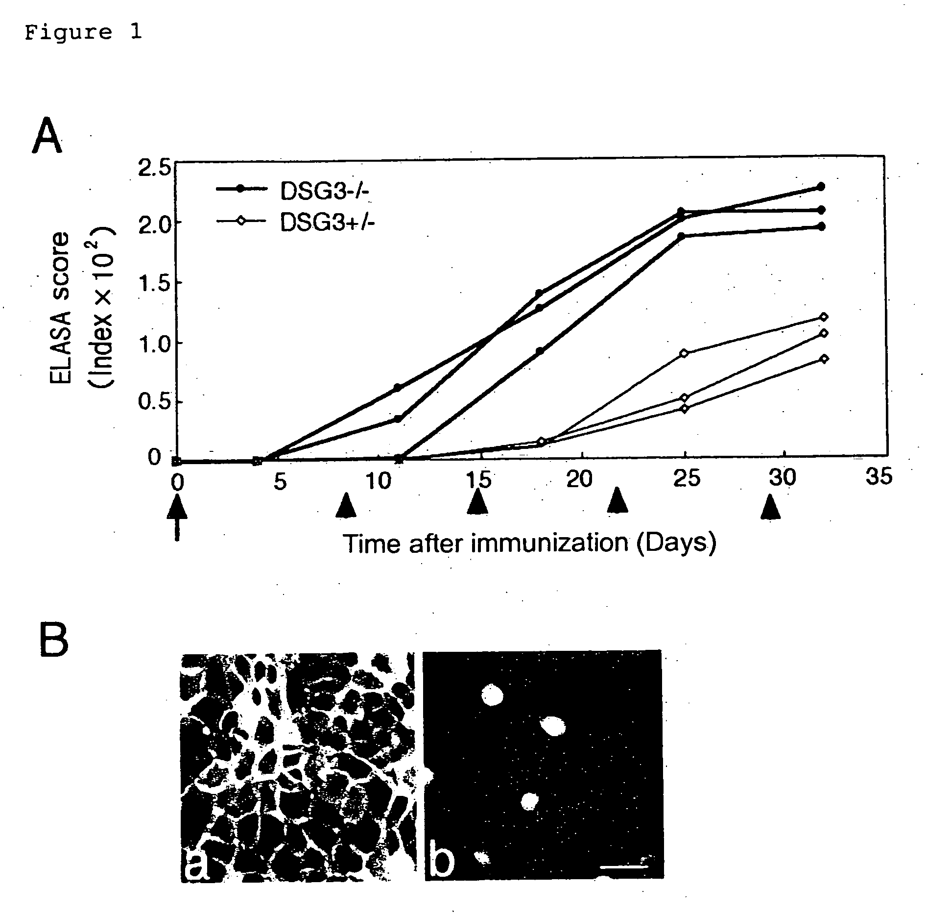
[0062] ELISA scores for mouse rDsg3 were determined after immunizing DSG3− / − mouse with mouse rDsg3 in order to verify the absence of immuno-tolerance to Dsg3 protein in DSG3− / − mouse.

[0063] Both DSG3− / − mice and DSG3+ / − mice were sensitized with 5 μg of purified mouse rDsg3 by using complete Freund's adjuvant (0 day), and then booster was carried out with mouse rDsg3 by using incomplete Freund's adjuvant after 8, 15, 22, and 28 days. The antibody production was tested by ELISA using mouse rDsg3 as a coating antigen in the same manner as in Example 2.

[0064] The production...


PUM

PropertyMeasurementUnit
adhesionaaaaaaaaaa
indirect fluorescent antibody techniqueaaaaaaaaaa
weight lossaaaaaaaaaa

Abstract

Pemphigus vulgaris (PV) is an autoimmune disease with a possible fatality of the skin and mucosae which is induced by an antibody against desmoglein 3 (Dsg3). Persistent production of anti-Dsg3 IgG can be induced by adoptively transferring spleen cells of a DSG3− / − mouse immunized with rDsg3 into an RAG2− / − immunodeficient mouse expressing Dsg3 protein. This IgG in the blood binds to the Dsg3 protein in vivo, induces the breakage of intercellular adhesion of keratinocytes and thus brings about the phenotype of pemphigus vulgaris involving the formation of blisters in the oral mucosa and the disappearance of resting hair. These effects are sustained over 6 months. By using this method, active disease model animals relating to various autoimmune diseases can be constructed.

Description

[0001] The present application is a continuation of U.S. patent application Ser. No. 09 / 937,739, which claims the benefit of PCT International Application No. PCT / JP00 / 02023, filed Mar. 30, 2000, which claims benefit of Japanese Patent Application No. 11 / 91408, filed Mar. 31, 1999, which are hereby incorporated in their entirety.TECHNICAL FIELD [0002] The present invention relates to autoimmune disease model animals and methods for producing them. BACKGROUND ART [0003] Pemphigus vulgaris (PV) is an autoimmune disease with involvement of skin and mucous membrane blistering, which is sometimes fatal, and is histologically characterized by blistering in the epidermis as well as immunopathologically characterized by the presence of autoantibody IgG to the cell surface of keratinocyte (Stanley, J. R. Pemphigus. In Dermatology in General Medicine. I. M. Freedberg, A. Z. Eisen, K. Wolff, K. F. Austen, L. A. Goldsmith, S. I. Katz, and T. B. Fitzpatrick, eds. McGraw-Hill, New York, 654-666 (...

Claims


Application Information

Patent Timeline
Patent Type & Authority Applications(United States)
IPC IPC(8): A01K67/027C07K14/705C07K16/28C12N15/09
CPCA01K67/0271A61K2039/505C07K14/705C07K16/28
Inventor AMAGAI, MASAYUKINISHIKAWA, TAKEJISUZUKI, HARUMIKOYASU, SHIGEO
Owner KEIO UNIV
Learn More