Method for quantitative video-microscopy and associated system and computer software program product

Active Publication Date: 2006-06-20
TRIPATH IMAGING INC
View PDF25 Cites 34 Cited by
  • Summary
  • Abstract
  • Description
  • Claims
  • Application Information

AI Technical Summary

Benefits of technology

[0021]Such imaging techniques as described herein, otherwise known as multi-spectral imaging techniques, when particularly adapted to color imaging, allow a substantially real time, or video rate, processing and viewing of the sample. The use of, for example, a RGB color CCD camera allows acquisition and processing time for sample images to be performed at a video rate, typically 40 millisecond per frame, which provides a considerable advantage as compared to prior art imaging techniques which generally exhibit field of view acquisition and processing times of over 1 second. Where an RGB camera is used by the system, image acquisition through the different channels is performed in parallel and look-up tables (LUT) can be generated so as to map the possible RGB color input values to predetermined concentrations and/or transmittance of each of various dyes. Thus, such capabilities may, for example, enhance processing speed and facilitate real time processing for display purposes.
[0022]Accordingly, an a posteriori evaluation of the image can be performed to evaluate the efficiency of an a priori known dye combination used to solve the linear equations for each pixel. That is, an evaluation as detailed herein also provides a confidence evaluation for each pixel in that the color and intensity measured at the given pixel may be justified by a combination of the a priori known dyes. Such a confidence evaluation can be expressed as the inverse to the closest match in the theoretical model. In a situation in which there are fewer dyes to evaluate than input channels (less unknown parameters than equations, e.g. one counter stain+one marker (=2 d

Problems solved by technology

However, the maximum number of markers that may be used in a study is restricted by several factors.
For example, the spectral overlapping of the colors used to reveal the respective markers may be a limiting factor because dyes may absorb throughout a large portion of the visible spectrum.
Accordingly, the higher the number of dyes involved in a study, the higher the risk of spectral overlapping.
Further, the spectral resolution of the acquisition device may be a limiting factor and the minimal color shift that the device is able to detect must be considered.
In addition, immunochemistry, as well as chemistry in ISH, are generally considered to exhibit poor sensitivity when quantification of a marker must be achieved.
However, the calibration standard is usually only applicable to a particular specimen, such as a specific cell or a structure of a specific type which is known to exhibit constant features with respect to the standard, and may be of limited utility when applied to a sample of a different nature.

Method used


Image

  • Method for quantitative video-microscopy and associated system and computer software program product
  • Method for quantitative video-microscopy and associated system and computer software program product
  • Method for quantitative video-microscopy and associated system and computer software program product

Examples


Embodiment Construction

[0027]The present invention now will be described more fully hereinafter with reference to the accompanying drawings, in which preferred embodiments of the invention are shown. This invention may, however, be embodied in many different forms and should not be construed as limited to the embodiments set forth herein; rather, these embodiments are provided so that this disclosure will be thorough and complete, and will fully convey the scope of the invention to those skilled in the art. Like numbers refer to like elements throughout.

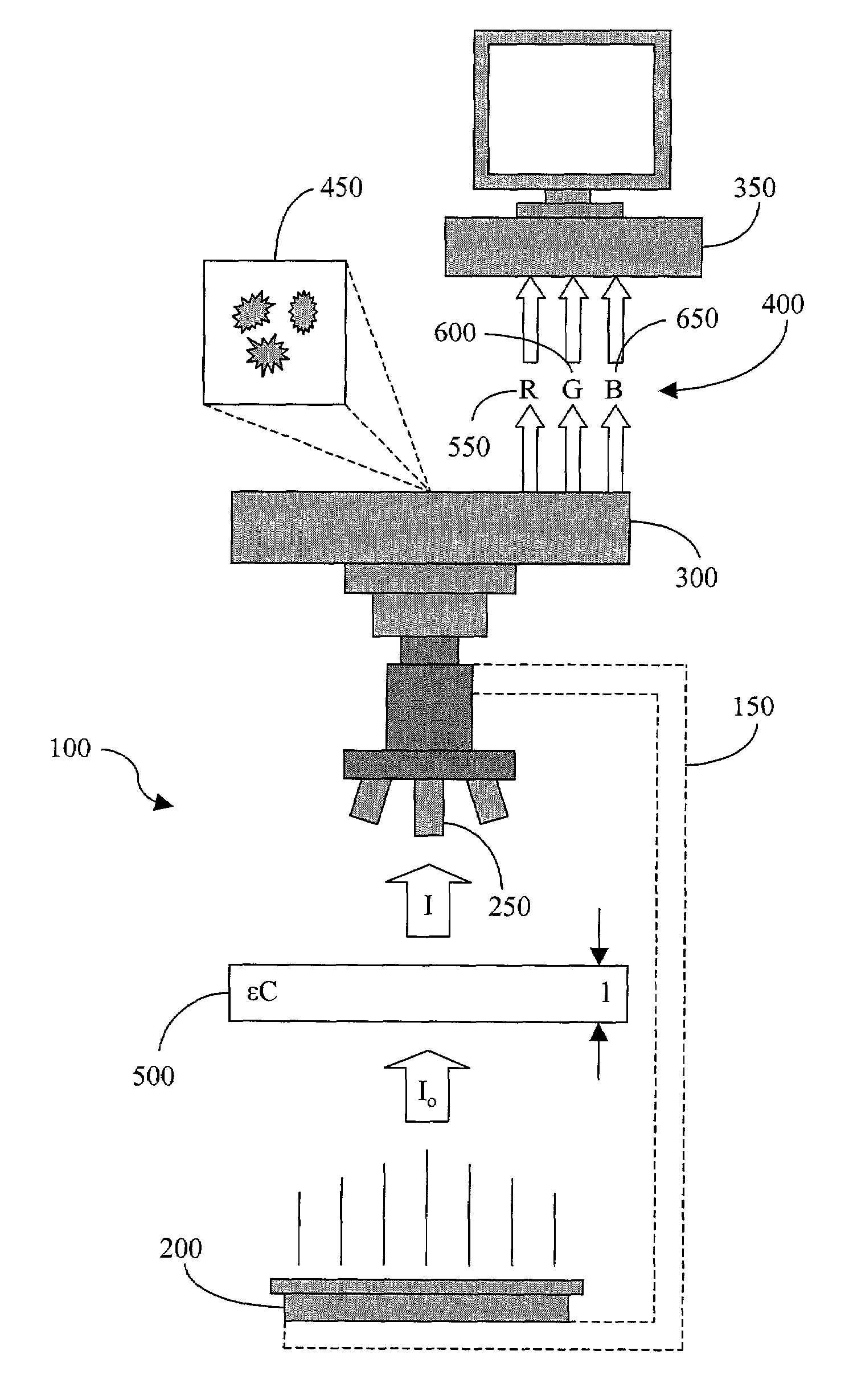
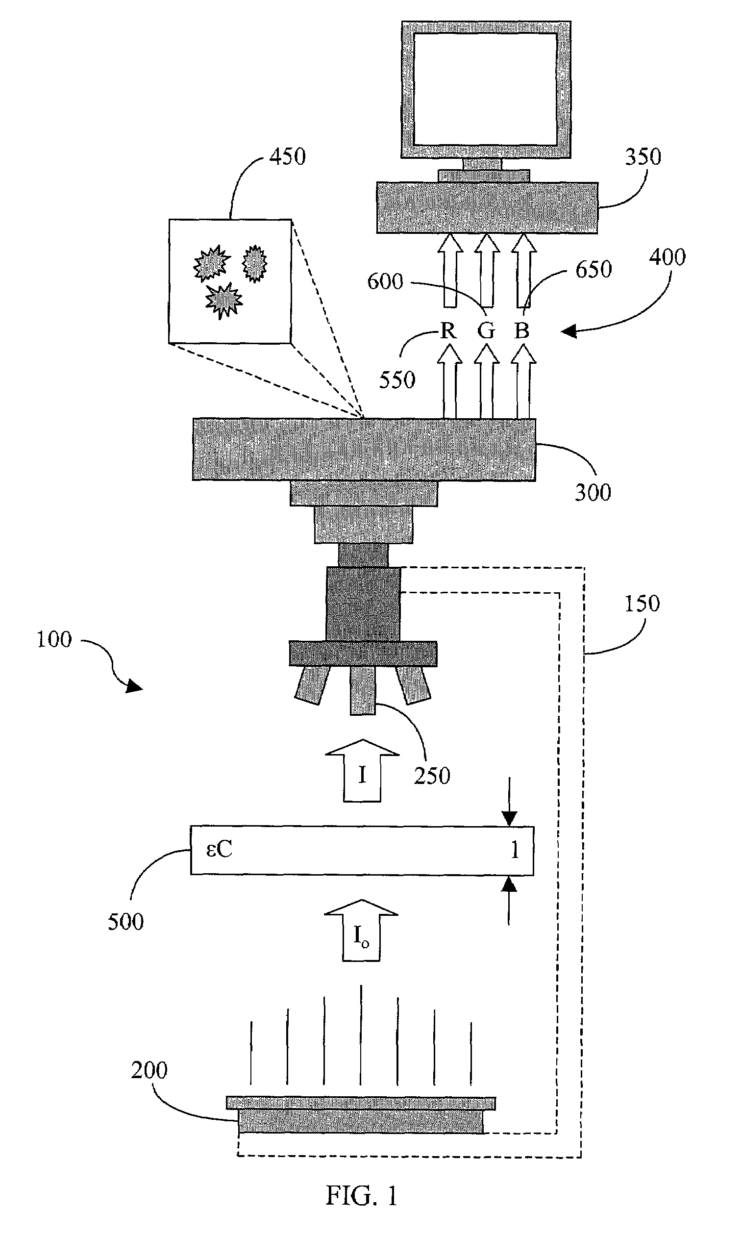
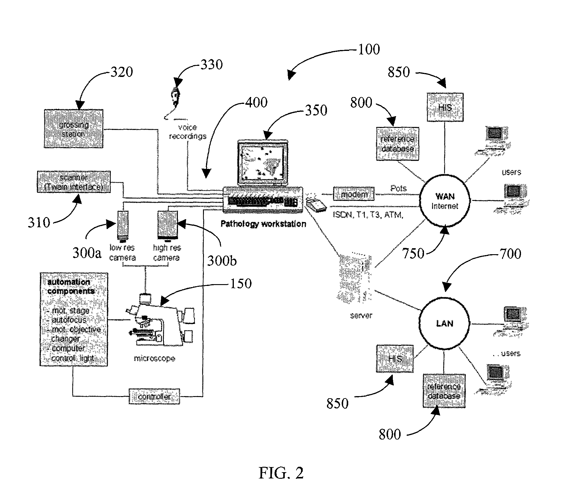
[0028]The platform for the evaluation of biological samples via image analysis is increasingly shifting from a general-purpose image analyzer to a more, and often highly, specialized dedicated “pathology workstation.” Such workstations are typically designed to facilitate routine work, often combining many of the tools needed to provide a pathologist with the necessary information to determine the best possible results. One example of such a workstation is...


PUM


Abstract

A method of determining an amount of at least one molecular specie comprising a sample is provided, each molecular specie being indicated by a dye. The amount of the molecular specie is determined from an image of the sample captured as image data by a color image acquisition device in a video-microscopy system. An optical density of the sample is first determined in each of a red, green, and blue channel at a particular pixel in the image. A corresponding optical density matrix is thereafter formed for that pixel. The optical density matrix is then multiplied by the inverse of a relative absorption coefficient matrix so as to form a resultant matrix for the pixel. The relative absorption coefficient matrix comprises a relative absorption coefficient for each dye, independently of the sample, in each of the red, green, and blue channels. The resultant matrix thus comprises the amount of each molecular specie, as indicated by the respective dye, for that pixel. Associated systems and computer software program products are also provided.

Description

FIELD OF THE INVENTION[0001]The present invention relates to image analysis and, more particularly, to a method for quantitative video-microscopy in cellular biology and pathology applications and an associated system and computer software program product therefor.BACKGROUND OF THE INVENTION[0002]Effective analysis of microscopic images is essential in cellular biology and pathology, particularly for detection and quantification of genetic materials such as, for example, genes or messenger RNA, or the expression of this genetic information in the form of proteins such as through, for example, gene amplification, gene deletion, gene mutation, messenger RNA molecule quantification, or protein expression analyses. Gene amplification is the presence of too many copies of the same gene in one cell, wherein a cell usually contains two copies, otherwise known as alleles, of the same gene. Gene deletion indicates that less than two copies of a gene can be found in a cell. Gene mutation indi...

Claims


Application Information

Patent Timeline
IPC IPC(8): G06K9/00G01J3/51G01N21/27G01N21/64G01N33/48G01N33/483G02B21/36G06F19/00G06T7/00G06T7/60
CPCG01N21/27G06T7/60G06K9/00127G06T2207/10056G06V20/69
Inventor MARCELPOIL, RAPHAELGAHM, THOMAS
Owner TRIPATH IMAGING INC
Why Eureka
Try Eureka