Ultrasound diagnosis apparatus that adjusts a time phase between a plurality of image series

a technology of ultrasonic diagnosis and image series, applied in ultrasonic/sonic/infrasonic image/data processing, instruments, applications, etc., can solve the problems of outstandingly short interval specimens to undergo cardiac examinations are likely to suffer from arrhythmias, and may not always be constant between r-waves of electrocardiographic complexes

Active Publication Date: 2007-02-13
TOSHIBA MEDICAL SYST CORP
View PDF10 Cites 21 Cited by
  • Summary
  • Abstract
  • Description
  • Claims
  • Application Information

AI Technical Summary

Problems solved by technology

When, however, a time phase of the ultrasound images is determined (or set) based on the electrocardiographic complex in a conventional manner, intervals between R-waves of an electrocardiographic complex may not always be constant.
Particularly, specimens to undergo cardiac examinations are likely to suffer from an arrhythmia.
Further, even if it is a normal healthy person, it is obvious that intervals between R-waves of the electrocardiographic complex are outstandingly short after the exercise stress has been given to the person.
As described in the above example, when a diastolic period and/or a systolic period temporally varies, it is d

Method used


Image

  • Ultrasound diagnosis apparatus that adjusts a time phase between a plurality of image series
  • Ultrasound diagnosis apparatus that adjusts a time phase between a plurality of image series
  • Ultrasound diagnosis apparatus that adjusts a time phase between a plurality of image series

Examples


first embodiment

(First Embodiment)

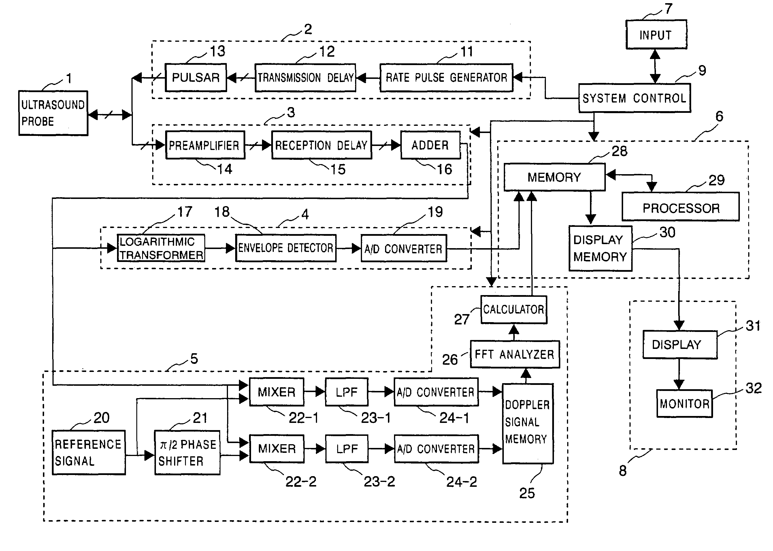
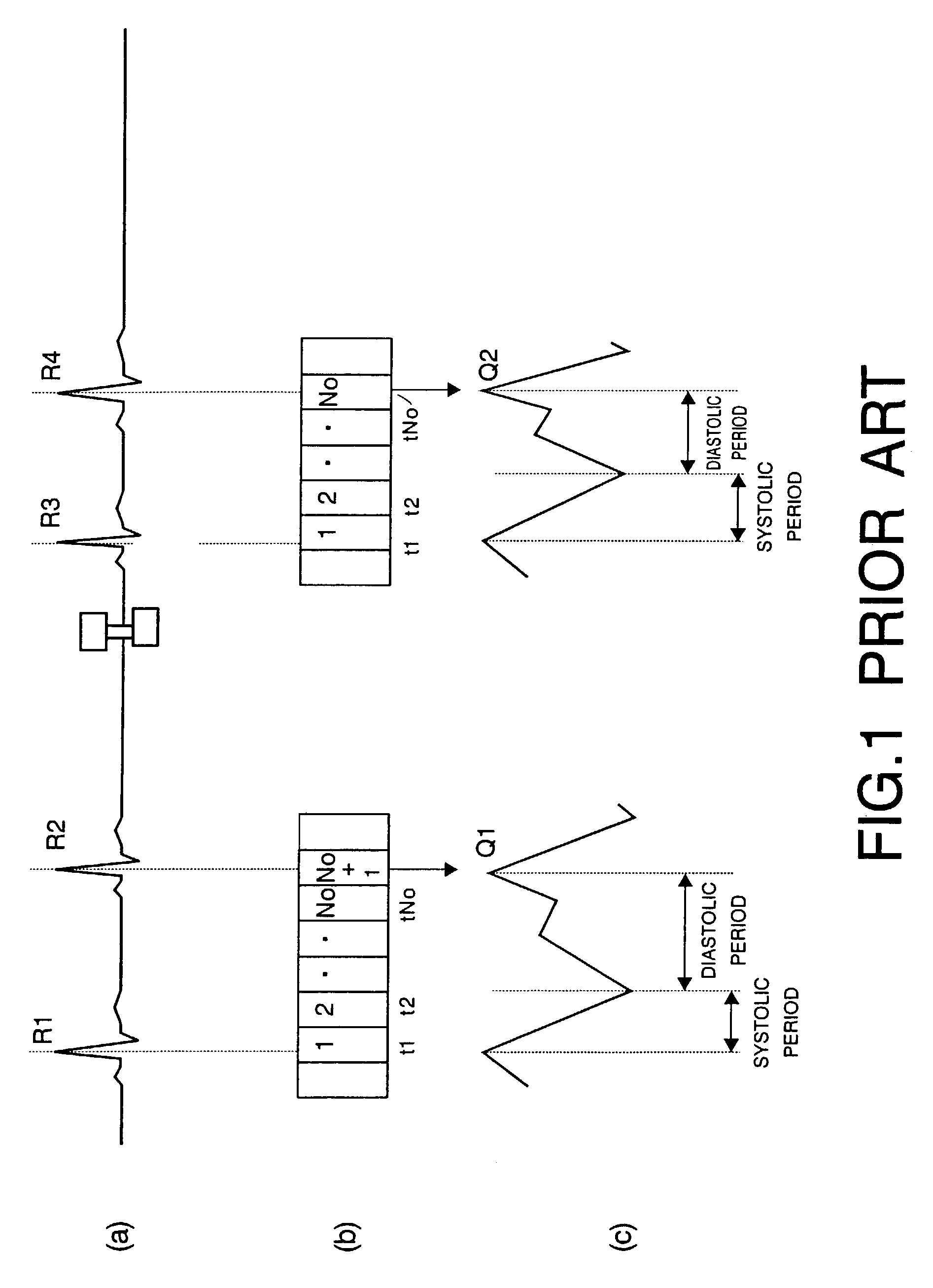
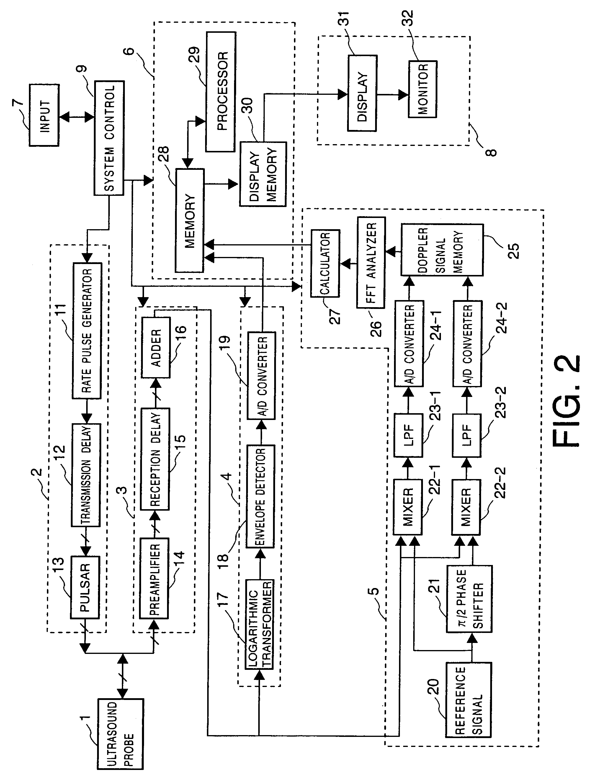
[0042]An ultrasound diagnosis apparatus according to a first embodiment of the present invention will be described with reference to FIGS. 2 to 10. In the first embodiment of the present invention, a volume of an inner space of a heart chamber is measured for each image included in two (kinds of) sequential image data (or two different series of images). The two sequential image data are obtained for a same specimen under two different conditions, respectively. The two sequential image data may comprise first sequential image data and second sequential image data. The first sequential image data may represent various time-series aspects of a four-chamber view of the heart. The second sequential image data may represent various time-series aspects of a two-chamber view of the heart. The four-chamber view is a view of the heart orthogonal with the two-chamber view. The view results from ultrasound scanning. After the volume measurements, a first systolic period and a...

second embodiment

(Second Embodiment)

[0109]Next, a second embodiment of the present invention will be described with reference to FIGS. 2, and 10 to 14. In the first embodiment of the present invention, it has been described to improve measurement accuracies, adjusting a time phase between two kinds of sequential B-mode image data, when various measurements are performed on the two kinds of sequential B-mode image data obtained under two different conditions, respectively. According to the second embodiment of the present invention, however, it will be described to adjust a time phase between the two kinds of sequential B-mode image data obtained under two different conditions and to display two kinds of sequential images in time phase side by side (or in parallel). The two kinds of sequential images to be displayed may be two kinds of sequential images resulting from the synthesis between the two kinds of sequential B-mode image data and two kinds of sequential Doppler-mode image data corresponding ...


PUM


Abstract

An ultrasound diagnosis apparatus comprises an insonifier, a receiver, and a processor. The insonifier is configured to insonify an ultrasound to a specimen. The receiver is configured to receive an echo signal from the specimen resulting from the ultrasound. The processor is in communication with the receiver. Further, the processor is configured to process the echo signal so as to obtain a first series of images under a first condition and a second series of images under a second condition. The processor is also configured to measure a first physical value on the first series of images and a second physical value on the second series of images. The processor is further configured to adjust a time phase of the second series of images relative to a time phase of the first series of images based on the first physical value and the second physical value.

Description

CROSS-REFERENCE TO RELATED APPLICATION[0001]This application is based upon and claims the benefit of priority from prior Japanese Patent Application No. P2002-176893, filed on Jun. 18, 2002, the entire contents of which are incorporated herein by reference.FIELD OF THE INVENTION[0002]The present invention relates to an ultrasound diagnosis apparatus which acquires a plurality of image series under respective different conditions. The ultrasound diagnosis apparatus is operative to adjust a time phase between the plurality of image series. The present invention further relates to a method of adjusting the time phase between the plurality of image series acquired in a medical equipment.BACKGROUND OF THE INVENTION[0003]An ultrasound diagnosis apparatus insonifies ultrasound pulses to a patient or an object (hereinafter referred to as a specimen). The ultrasound pulses are generated from transducers built in an ultrasound probe. The ultrasound diagnosis apparatus then receives echo signa...

Claims


Application Information

Patent Timeline
IPC IPC(8): A61B8/00A61B8/08A61B8/14
CPCA61B8/14A61B8/463G01S7/52087G01S7/52085A61B8/488A61B8/543
Inventor YONEYAMA, NAOKI
Owner TOSHIBA MEDICAL SYST CORP
Why Eureka
Try Eureka