Systems and methods for automated analysis of cells and tissues

a technology of cells and tissues and automated analysis, applied in the field of systems and methods for automated analysis of cells and tissues, to achieve the effects of rapid assessment of the prognostic benefit of biomarkers, easy optical analysis of array of biological samples, and increased applicability

Inactive Publication Date: 2007-05-15
YALE UNIV
View PDF13 Cites 170 Cited by
  • Summary
  • Abstract
  • Description
  • Claims
  • Application Information

AI Technical Summary

Benefits of technology

[0009]In another aspect, the invention features an algorithm that facilitates the optical analysis of an array of biological samples, despite image irregularities, distortions, varying topologies, and the absence of one or more elements.
[0010]Analysis of patient samples according to the systems and processes described herein can be useful diagnostically (e.g. to identify patients who have a particular disease, have been exposed to a particular toxin or are responding well to a particular therapeutic or organ transplant) and prognostically (e.g. to identify patients who are likely to develop a particular disease, respond well to a particular therapeutic or be accepting of a particular organ transplant). As new and better markers of disease become identified in the post-genomic era, the instant described processes, which not only quantitate the markers, but also determine their relative location within a cell, will increase in applicability.
[0011]Automated analysis of cell containing preparations, as described herein, can provide a rapid assessment of the prognostic benefit of biomarkers. In addition, these automated techniques can identify associations that are typically not revealed using manual techniques. Also, automated analysis can better discern subtle differences in staining intensity, particularly at the upper and lower extremes. The ability to detect low level expression and distinguish it from no expression can provide important prognostic information. Furthermore, analysis of the sub-cellular distribution of certain biomarkers may elucidate previously unrecognized associations with patient survival.

Problems solved by technology
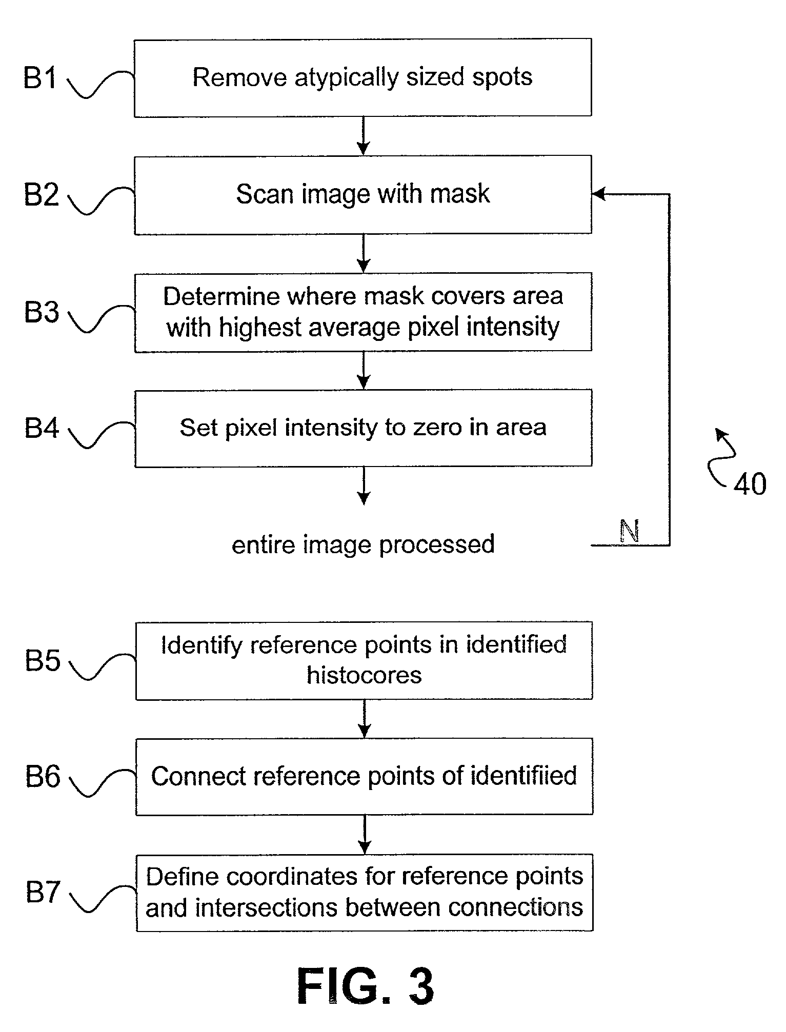
Furthermore, analysis of the sub-cellular distribution of certain biomarkers may elucidate previously unrecognized associations with patient survival.

Method used


Image

  • Systems and methods for automated analysis of cells and tissues
  • Systems and methods for automated analysis of cells and tissues
  • Systems and methods for automated analysis of cells and tissues

Examples


example 1

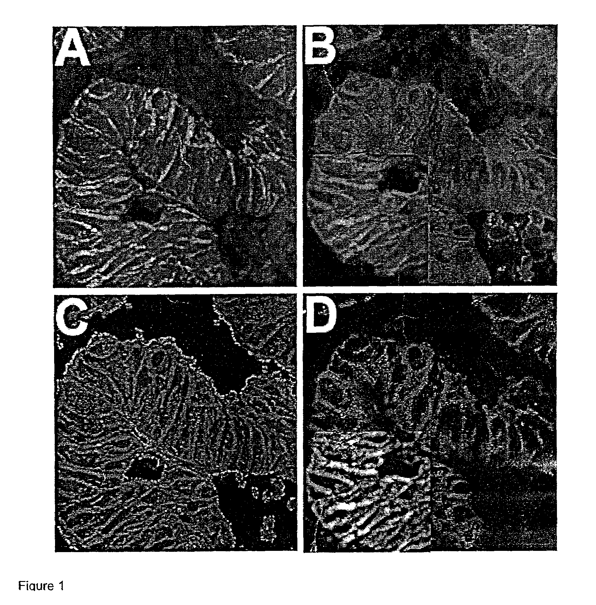
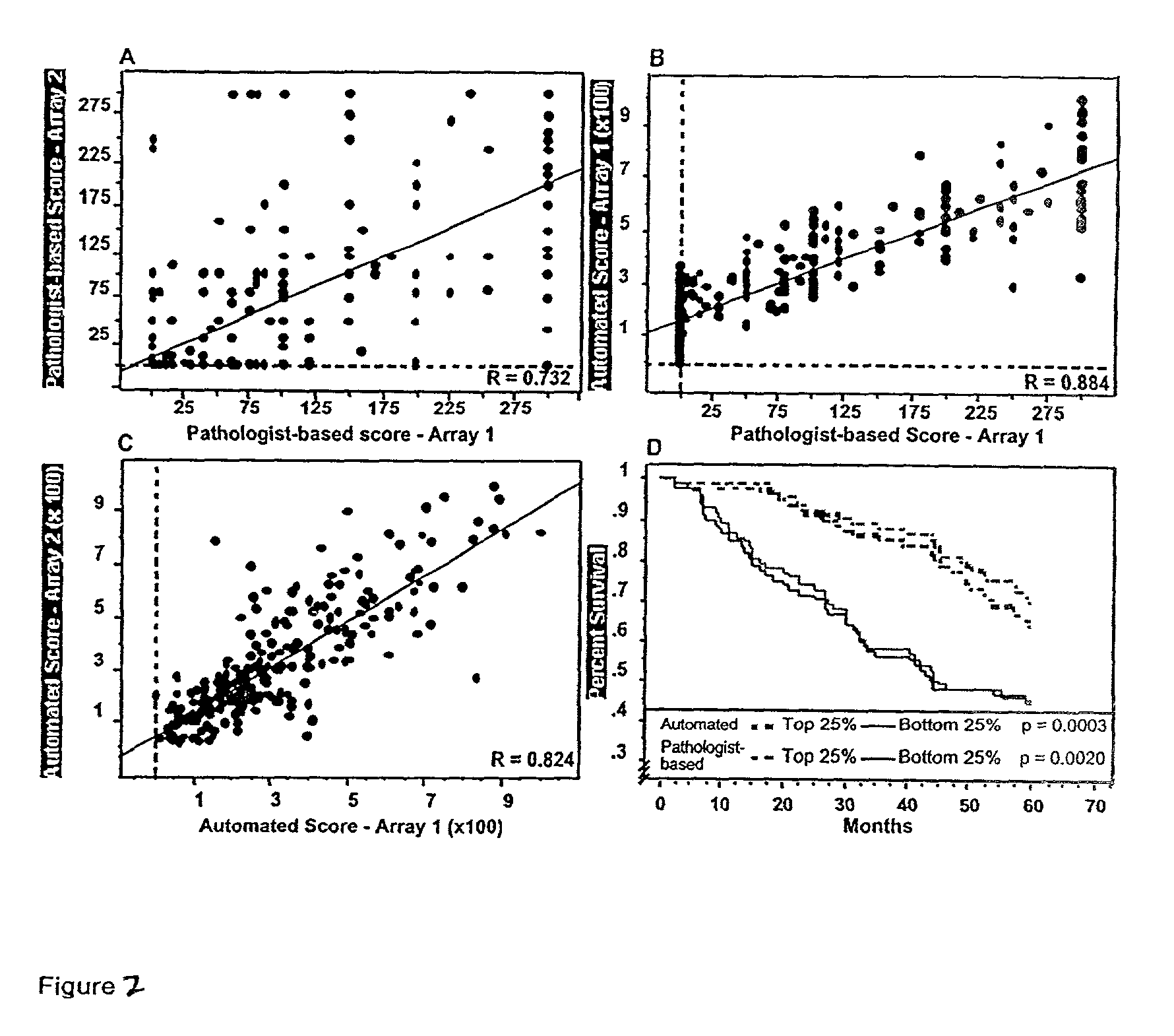
Construction of Tissue Microarrays for a Survival Analysis of the Estrogen Receptor (ER) and HER2 / neu and for Analysis of Nuclear Associated Beta-catenin

[0108]Tissue microarray design: Paraffin-embedded formalin-fixed specimens from 345 cases of node-positive invasive breast carcinoma were identified. Areas of invasive carcinoma, away from in situ lesions and normal epithelium, were identified and three 0.6 cm punch “biopsy” cores were taken from separate areas. Each core was arrayed into a separate recipient block, and five-micron thick sections were cut and processed as previously described (Konenen, J. et al., Tissue microarrays for high-throughput molecular profiling of tumor specimens, (1987) Nat. Med. 4:844–7). Similarly, 310 cases of colon carcinoma were obtained and arrayed, as previously described (Chung, G. G. et al., Clin. Cancer Res. (In Press)).

[0109]Immunohistochemistry: Pre-cut paraffin-coated tissue microarray slides were deparaffinized and antigen-retrieved by press...


PUM

PropertyMeasurementUnit
thicknessaaaaaaaaaa
diameteraaaaaaaaaa
diameteraaaaaaaaaa

Abstract

Systems and methods for rapidly analyzing cell containing samples, for example to identify morphology or to localize and quantitate biomarkers are disclosed.

Description

[0001]Under 35 USC § 119(e)(1), this application claims the benefit of prior U.S. provisional application 60 / 285,155, filed Apr. 20, 2001, and U.S. provisional application 60 / 334,723, filed Oct. 31, 2001, the contents of which are incorporated herein by reference.[0002]Work described herein was supported in part by grant No. 5K08ES011571 awarded by the National Institute of Health. The U.S. Government may therefore have certain rights in the invention.1. BACKGROUND OF THE INVENTION [0003]Tissue microarray technology offers the opportunity for high throughput analysis of tissue samples (Konen, J. et al., Nat. Med. 4:844–7 (1998); Kallioniemi, O. P. et al., Hum. Mol. Genet. 10:657–62 (2001); Rimm, D. L. et al., Cancer J. 7:24–31 (2001)). For example, the ability to rapidly perform large scale studies using tissue microarrays can provide critical information for identifying and validating drug targets / prognostic markers (e.g. estrogen receptor (ER) and HER2 / neu) and candidate therapeut...

Claims


Application Information

Patent Timeline
Patent Type & Authority Patents(United States)
IPC IPC(8): G01N33/48G01N21/64G01N21/27
CPCG01N21/6458G02B21/367G06T7/0012G06T7/401G06K9/00127G06T7/0014G02B21/16G06T2207/20036G06T2207/30096G06T2207/30024G06T2207/30072G06T2207/20221G06T2207/20101G06T7/41G06V20/69
Inventor RIMM, DAVID L.CAMP, ROBERT L.
Owner YALE UNIV
Why Eureka
Try Eureka