Treatment of glial tumors with glutamate antagonists
a glutamate antagonist and tumor technology, applied in the field of glial tumors, can solve the problems of not teaching or enabling the use of glutamate antagonists in brain tumors, ikonomidou does not treat glial cell tumors, etc., to prolong the life of patients with glial tumors, slow the expansion of glial tumors, and inhibit the excitatory activity of glutamate
- Summary
- Abstract
- Description
- Claims
- Application Information
AI Technical Summary
Benefits of technology
Problems solved by technology
Method used
Image
Examples
example 1
Primary Cultures and Cell Lines
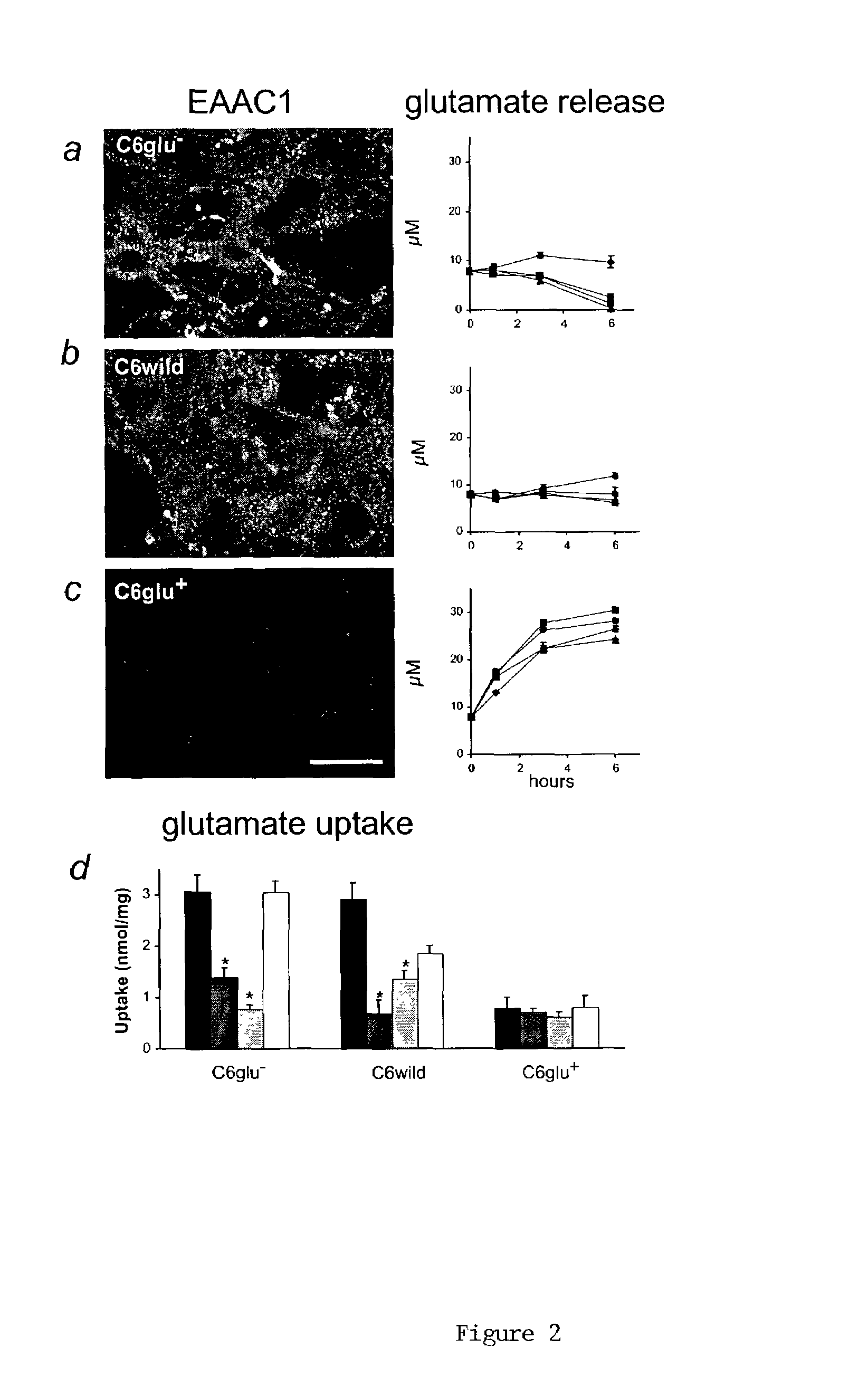
[0050]Cortical neurons were prepared from Wistar rats (16 days gestation; Taconic, Germantown, N.Y.) and plated in 24-well plates (Nedergaard, M., “Direct Signaling From Astrocytes to Neurons in Cultures of Mammalian Brain Cells,”Science, 263:1768–1771 (1994), which is hereby incorporated in its entirety). Cortical astrocytes were prepared from 1-day postnatal rate pups (Cotrina et al., “Astrocytic Gap Junctions Remain Open During Ischemic Conditions,”J. Neurosci., 18:2520–2537 (1998), which is hereby incorporated in its entirety), whereas C6 and RG2 glioma cells were obtained from American Type Culture Collection (Lin et al., “Ga-Junction-Mediated Propagation and Amplification of Cell Injury,”Nature Neurosci., 1:494–500 (1998), which is hereby incorporated in its entirety) (Manassas, Va.). All cultures were grown in DMEM / F12 supplemented with 10% FBS, 20 mM glucose and antibiotics.
example 2
Cocultures of Neurons and Glioma Aggregates
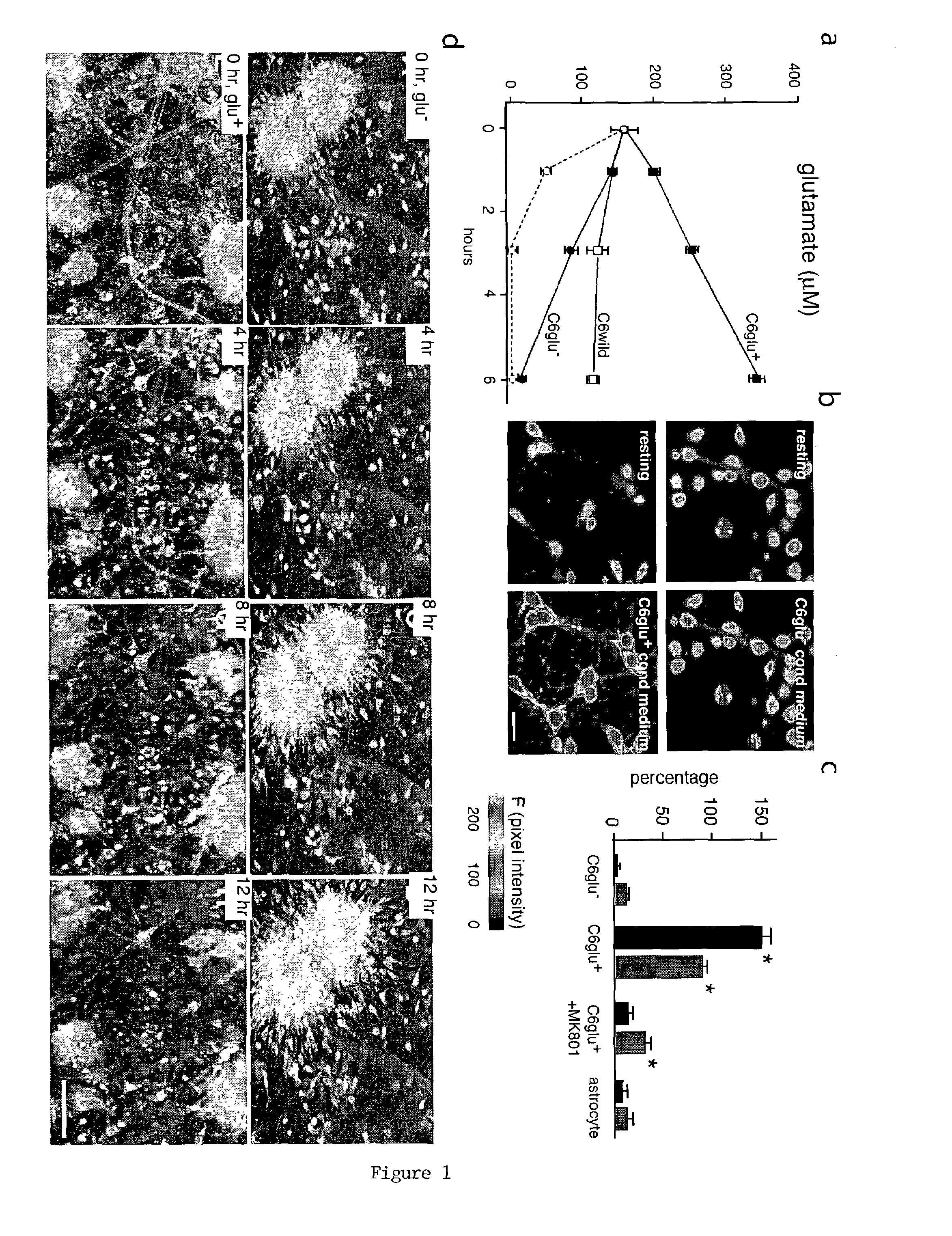
[0051]Glioma cell aggregates were produced by plating 100,000 C6Glu+ or C6Glu−cells in plates with non-adhesive substrate (ultra low cluster 3473; Costar, Cambridge, Mass.). Twenty-four hours later, the glioma aggregates were loaded with calcein AM (green, 2 μM, excitation 488 nm) and transferred to neuronal cultures. Cortical neuronal cultures were preloaded with the cell tracker Sytox 62 (white, 0.1 μM, excitation 647 nm). Loss of neuronal viability was quantified as the percentage of neurons that displayed nuclear staining with propidium iodide (red, 1 μM, excitation 567 nm) at 12 or 24 hours after coculturing (Cotrina et al., “Astrocytic Gap Junctions Remain Open During Ischemic Conditions,”J. Neurosci., 18:2520–2537 (1998); Nedergaard, M., “Direct Signaling From Astrocytes to Neurons in Cultures of Mammalian Brain Cells,”Science, 263:1768–1771 (1994), which are hereby incorporated in its entirety).
example 3
Calcium Measurements, Cell Proliferation, and Migration Assays
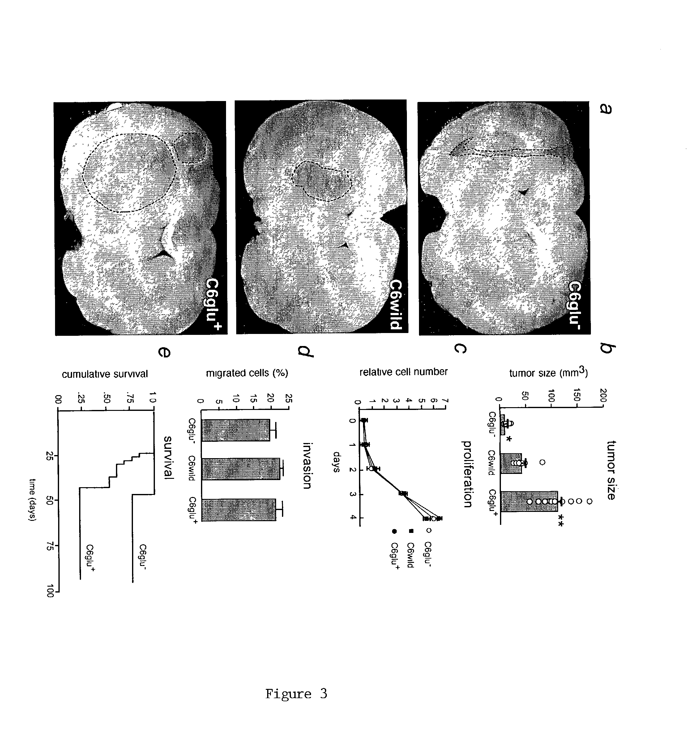
[0052]Ten-day-old cortical neurons were loaded with the calcium indicator, fluo-3 AM (5 μM, 1 h), and the fluorescence signal was detected by confocal microscopy (MRC1000, BioRad, Richmond, Calif.) as described (Lin et al., “Ga-Junction-Mediated Propagation and Amplification of Cell Injury,”Nature Neurosci., 1:494–500 (1998); Zhang et al., “Tamoxifen-Induced Enhancement of Calcium Signaling in Glioma and MCF-7 Breast Cancer Cells,”Cancer Res., 60:5395–5400 (2000), which are hereby incorporated in their entirety). 100 μl conditioned medium from the C6 clones was added to the culture (initial media volume was 300 μl). Proliferation of the C6 subclones was determined by cell counting or by the Alamar blue assay (Biosource, Camarillo, Calif.) and cellular invasiveness evaluated using Matrigel-coated transwell inserts as described (Zhang et al., “Direct Gap Junction Communication Between Malignant Glioma Cells and Astrocytes,”...
PUM
| Property | Measurement | Unit |
|---|---|---|
| volume | aaaaa | aaaaa |
| volume | aaaaa | aaaaa |
| pH | aaaaa | aaaaa |
Abstract
Description
Claims
Application Information
 - R&D
- Intellectual Property
- Life Sciences
- Materials
- Tech Scout
- Unparalleled Data Quality
- Higher Quality Content
- 60% Fewer Hallucinations
Browse by: Latest US Patents, China's latest patents, Technical Efficacy Thesaurus, Application Domain, Technology Topic, Popular Technical Reports.
© 2025 PatSnap. All rights reserved.Legal|Privacy policy|Modern Slavery Act Transparency Statement|Sitemap|About US| Contact US: help@patsnap.com


