
Fluorescein based sensors for tracking nitric oxide in live cells

a technology of fluorescein and live cells, applied in the direction of instruments, organic chemistry, material analysis through optical means, etc., can solve the problems of complicated deciphering of these roles and implications, limited use of fluorescence technology for a particular application, and complicating the delineation of these various processes. , to achieve the effect of increasing brightness

Active Publication Date: 2009-02-24
MASSACHUSETTS INST OF TECH
View PDF4 Cites 4 Cited by
  • Summary
  • Abstract
  • Description
  • Claims
  • Application Information

AI Technical Summary

Benefits of technology

[0005]The feasibility of using fluorescence technology for a particular application is often limited by the availability of an appropriate fluorescent sensor. There are a number of features that are desirable in fluorescent sensors, some of which may or may not be present in any particular sensor. First, fluorescent sensors should produce a perceptible change in fluorescence upon binding a desired analyte. Second, fluorescent sensors should selectively bind a particular analyte. Third, to allow concentration changes to be monitored, fluorescent sensors should have a Kd near the median concentration of the species under investigation. Fourth, fluorescent sensors, especially when used intracellularly, should produce a signal with a high brightness, the product of the quantum yield and extinction coefficient. Fifth, the wavelengths of both the light used to excite the fluorescent molecule (excitation wavelengths) and of the emitted light (emission wavelengths) are often important. If possible, for intracellular use, a fluorescent sensor should have excitation wavelengths exceeding 340 nm to permit use with glass microscope objectives and prevent UV-induced cell damage, and have emission wavelengths approaching 500 nm to avoid autofluorescence from native substances in the cell and allow use with typical fluorescence microscopy optical filter sets. Excitation and emission at even longer wavelengths, approaching the near-IR, are also of value. Sixth, ideal sensors should allow for passive and irreversible loading into cells. Finally, ideal sensors should exhibit increased fluorescence with increasing levels of analyte.
[0007]Since the discovery in the 1980s that nitric oxide (NO) is the endothelium-derived relaxing factor (EDRF), postulated biological roles for NO have continued to proliferate. For example, in addition to cardiovascular signaling, NO also seems to function as a neurotransmitter that may be important in memory and as a weapon to fight infection when released by immune system macrophages. Uncovering these roles and deciphering their implications is complicated by the array of reactions that this gaseous molecule undergoes. In a biological environment, NO can react with a host of targets, including dioxygen, oxygen radicals, thiols, amines and transition metal ions. Some of the products formed, such as NO2 and NO+, are pathophysiological agents, whereas others, such as S-nitrosothiols, may in fact themselves be NO-transfer agents. Transition metal centers, especially iron in oxyhemoglobin, can rapidly scavenge free NO, thereby altering the amount available for signaling purposes.

Problems solved by technology

The feasibility of using fluorescence technology for a particular application is often limited by the availability of an appropriate fluorescent sensor.
Uncovering these roles and deciphering their implications is complicated by the array of reactions that this gaseous molecule undergoes.
The concentration-dependent lifetime of NO as well as its ability to diffuse freely through cellular membranes further complicate the delineation of these various processes.
The Griess assay, for instance, is useful for estimating total NO production, but it is not very sensitive, cannot give real-time information and only measures the stable oxidation product nitrite.
It too is therefore incapable of monitoring intracellular NO.
However, existing imaging agents are not capable of direct, immediate, and selective NO detection, three highly desirable properties.

Method used


Image

  • Fluorescein based sensors for tracking nitric oxide in live cells
  • Fluorescein based sensors for tracking nitric oxide in live cells
  • Fluorescein based sensors for tracking nitric oxide in live cells

Examples


example 1

Synthesis of Copper-Fluorescein Sensors

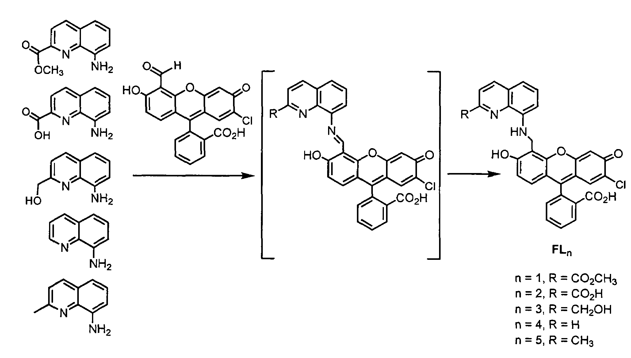
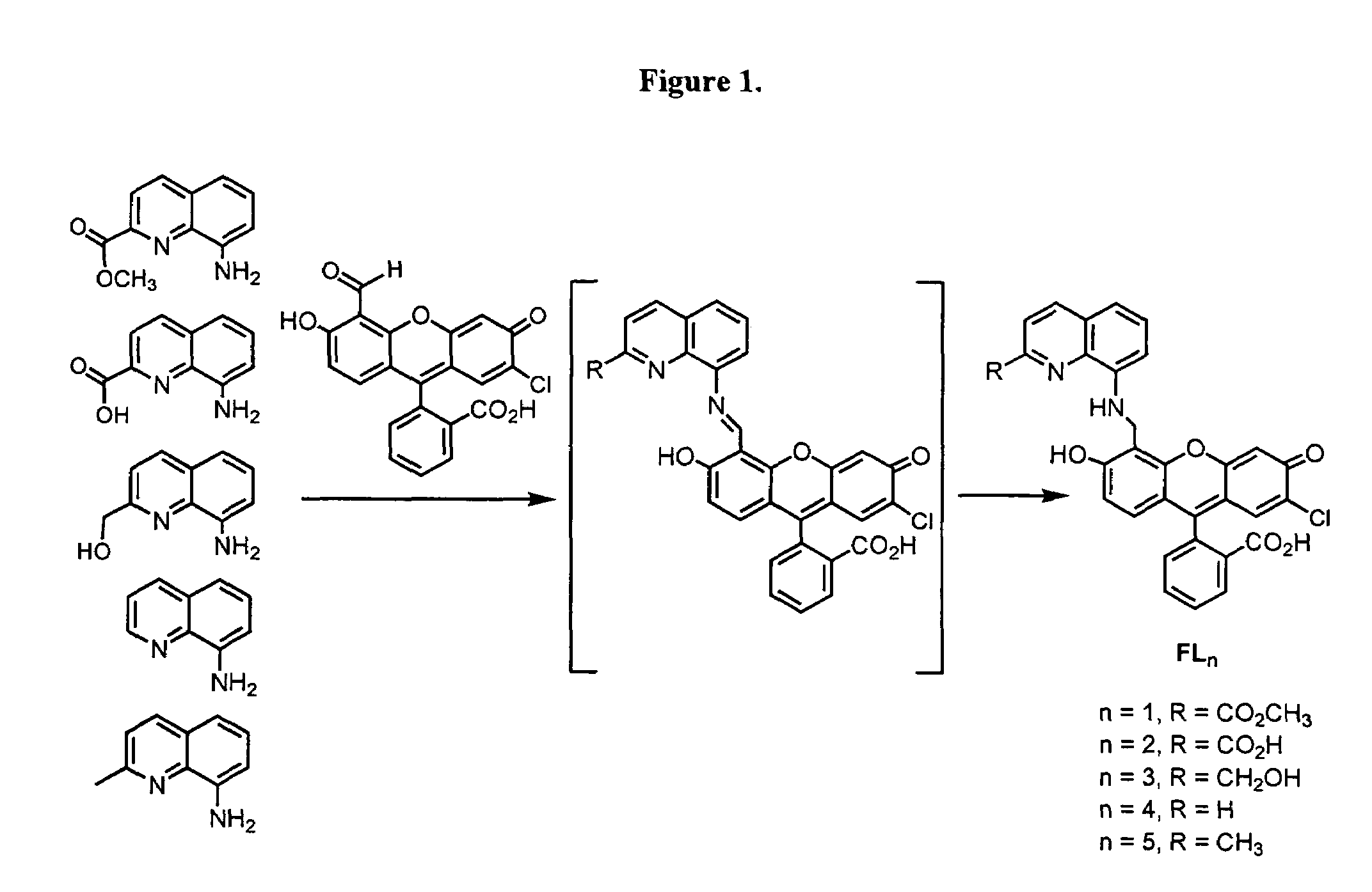
[0290]FIG. 1 depicts the synthetic strategies for the five fluorescein ligands FLn (n=1-5).

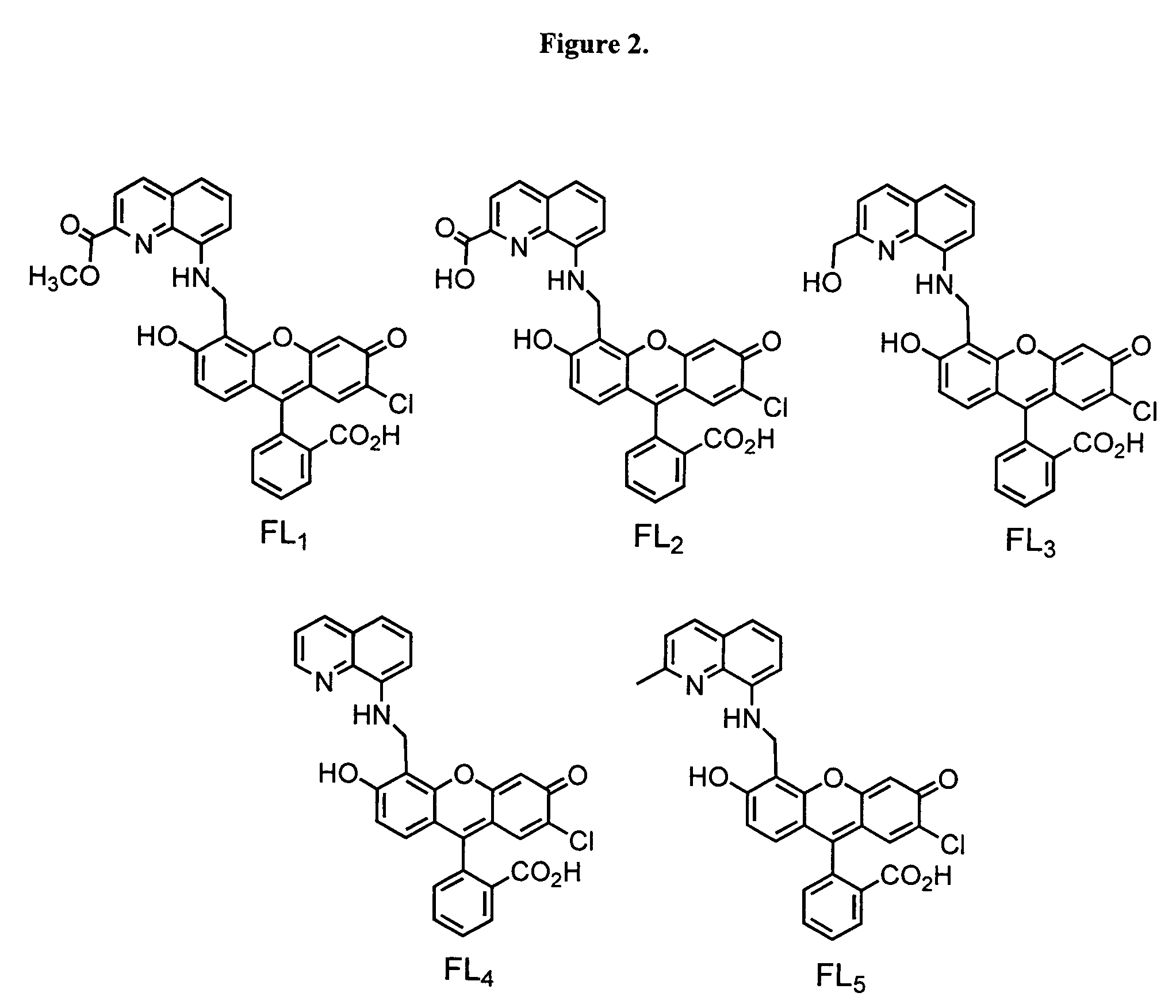
[0291]FIG. 2 depicts the structures of the five fluorescein ligands.

[0292]Fluorescence studies of the five sensors are depicted in FIG. 3.

example 2

Detailed Synthetic Method for Copper-Fluorescein Sensor Cu(FL5)

[0293]Derivatized fluorescein molecules are excellent biosensors because they excite and emit in a region of the visible spectrum that is relatively free of interference. We prepared the fluorescein-based ligand FL5 (FIG. 4) by reacting 7′-chloro-4′-fluorescein-carboxaldehyde with 8-aminoquinaldine. We generated the Cu(II) fluorescein-based NO probe Cu(FL5) (FIG. 4) in situ reacting FL5 (prepared as described below) awith CuCl2 in a 1:1 ratio in buffered aqueous solution (50 mM PIPES, pH 7.0, 100 mM KCl). The Cu(II) species exhibited a blue-shifted λmax at 499 nm (ε=4.0×10⁴ M−1cm−1), compared to that of FL5 (504 nm, ε=4.2×10⁴ M−1cm−1). A Job's plot was constructed to evaluate the nature of the FL5:Cu(II) complex by following the UV-vis absorption spectral change at 512 nm in pH 7.0 buffered solution. A break at 0.5 indicated the formation of a 1:1 complex. The negative ion electrospray mass spectrum of this species displ...

example 3

Fluorescence and Mechanistic Studies of Cu(FL5) with NO

[0296]The fluorescence of a 1 μM FL5 solution was diminished by 18±3% upon introduction of an equimolar quantity of CuCl2 at 37° C. Addition of excess NO to a buffered aqueous Cu(FL5) solution led to an immediate 11.2±1.5-fold increase in fluorescence (FIG. 5). Fluorescence was also enhanced when Cu(FL5) was allowed to react with the NO-releasing chemical agent S-nitroso-N-acetyl-D,L-penicillamine (SNAP) at pH 7.0 (50 mM PIPES, 100 mM KCl) over a 30 min time interval. This result demonstrates rapid NO detection with significant turn-on emission at a physiologically relevant pH. The lower detection limit for NO is 5 nM. The fluorescence response of the Cu(FL5) probe detects NO specifically over other reactive species present in biological systems, including O2−, ClO−, H2O2, HNO, NO2−, NO3−, and ONOO− (FIG. 6).

[0297]A commercially available NO probe, DAF-2 (o-diaminofluorescein), was used for comparison with Cu(FL5). The fluoresce...


PUM

PropertyMeasurementUnit
emission wavelengthsaaaaaaaaaa
excitation wavelengthsaaaaaaaaaa
pHaaaaaaaaaa

Abstract

The present invention is directed, in part, to coordination complexes for detecting analytes, and methods of making and using the same.

Description

CROSS-REFERENCE TO RELATED APPLICATIONS[0001]This application claims the benefit of U.S. Provisional Application No. 60 / 704,511, filed Aug. 1, 2005, the contents of which are hereby incorporated by this reference in their entirety.GOVERNMENT SUPPORT[0002]The invention was made with government support awarded by the National Science Foundation under NSF Grant No. CHE-0234951. Accordingly, the U.S. Government has certain rights in this invention.INTRODUCTION[0003]I. Fluorescent Sensors[0004]Fluorescence technology has revolutionized cell biology and many areas of biochemistry. In certain instances, fluorescent molecules may be used to trace molecular and physiological events in living cells. Certain sensitive and quantitative fluorescence detection devices have made fluorescence measurements an ideal readout for in vitro biochemical assays. In addition some fluorescence measurement systems may be useful for determining the presence of analytes in environmental samples. Finally, becaus...

Claims


Application Information

Patent Timeline
Patent Type & Authority Patents(United States)
IPC IPC(8): G01N21/76C07D311/82G01N33/20C12Q1/68
CPCC07D405/12
Inventor LIPPARD, STEPHEN J.LIM, MI HEE
Owner MASSACHUSETTS INST OF TECH
Learn More