Bi-Specific Therapeutic Proteins for Tissue Repair

Active Publication Date: 2017-04-06
SILVER CREEK PHARMA INC
View PDF0 Cites 2 Cited by
  • Summary
  • Abstract
  • Description
  • Claims
  • Application Information

AI Technical Summary

Benefits of technology

[0011]Within still further aspects, methods are provided for treating pathological tissue damage in a patient, comprising administering a pharm

Problems solved by technology

The resulting lack of oxygen causes irreversible tissue damage (necrosis and apoptosis), due to the inability of the heart to sufficiently activate endogenous regeneration programs and self-repair.
Such tissue damage is a leading cause of congestive heart failure, a condition in which the heart is no longer capable of effectiv

Method used


Image

  • Bi-Specific Therapeutic Proteins for Tissue Repair
  • Bi-Specific Therapeutic Proteins for Tissue Repair
  • Bi-Specific Therapeutic Proteins for Tissue Repair

Examples


Example

Example 1. Bi-Specific Fusion Proteins can be Engineered to have Reduced Potency on Healthy Cells

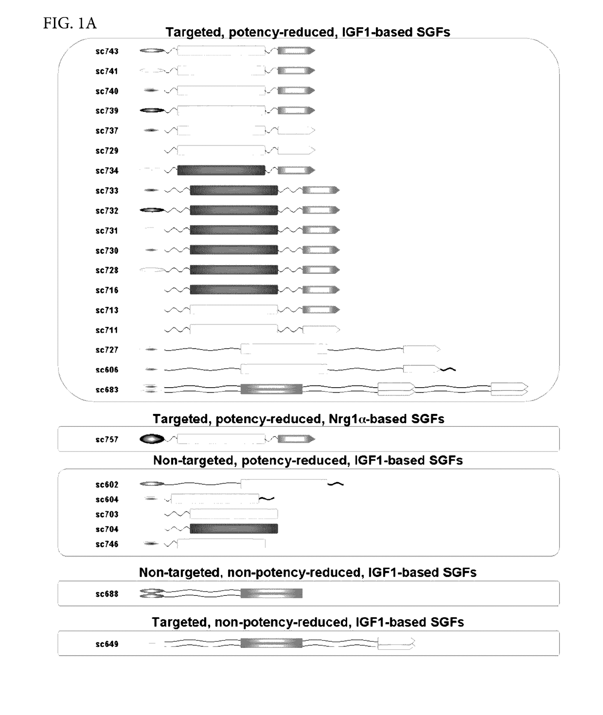
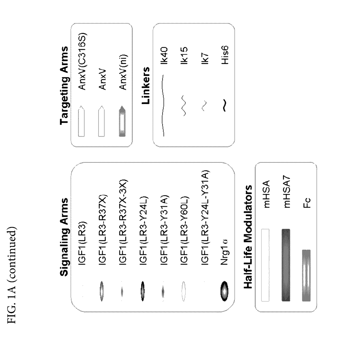
[0485]In order to enable targeting / selectivity for damaged cells, IGF1 was engineered to have reduced potency on healthy cells as compared to wt IGF-1 (FIGS. 2A-2B). Potency is defined as the concentration needed to achieve the half maximal level of pAKT signaling (pAKT EC50). In some embodiments, the IGF1 engineered variants were engineered using the IGF-1 (LR3) variant which contains a 13 amino acid N-terminal extension and a substitution of Arginine for Glutamate at position 3. The Arginine for Glutamate substitution was added to prevent binding of the fusion protein comprising the IGF-1 variant to IGF binding proteins (IGFBPs) and does not significantly affect potency (see FIGS. 2A-2B, EC50 of wt IGF1 is 1.22±0.74 vs. EC50 of IGF-1 (LR3) is 0.73±0.35).

[0486]A potency reduction of 6 fold or more was achieved by:[0487]1. Substituting amino acids. In some embodiments, the tyrosine resid...

Example

Example 2: Targeted, Potency Reduced Bi-Specific Fusion Proteins Selectively Signal (i.e, Exhibit a Potency Shift) in Cells Containing Target Molecule Compared to Cells without Target Molecule

[0498]The ability of phosphatidylserine (PS)-targeted, potency-reduced bi-specific proteins to selectively signal on cells containing the target molecule PS was measured in healthy (which does not display PS at the cell surface) vs. damaged (which displays PS at the cell surface) pluripotent stem cell derived cardiomyocytes (Cellular Dynamics International) and signaling was quantified by accumulation of phosphorylated AKT (FIGS. 3A, 3B and 3C).

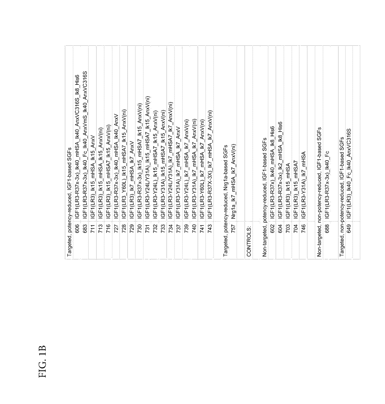
[0499]Phosphatidylserine (PS)-targeted, potency-reduced bi-specific proteins (having a 6-fold or greater potency reduction compared to wt IGF-1, (e.g. SGFs 743, 741, 740, 739, 733, 732, 731, 730, 729, 728, 727, 716, 713, 711, 606, SEQ ID NOs: 86, 85, 84, 83, 82, 81, 80, 79, 78, 77, 76, 75, 74, 73, and 70, respectively) were compared against non-targeted ...

Example

Example 3: Reduction of Hypoxia-Induced Apoptosis in Human Cardiomyocytes In Vitro Using Bi-Specific Proteins

[0511]FIG. 4 shows the reduction of apoptosis using fusions protein SGF 740 in an in vitro human cardiomyocyte hypoxia-induced apoptosis assay.

[0512]The bi-specific protein SGF 740 (SEQ ID NO: 84) comprising from the N terminus to the C-terminus: a variant of IGF-1 (LR3, Y31A), a 7 amino acid linker lk7, a variant of HSA (mHSA: C58S, K420E, and N527Q), a 7 amino acid linker lk7, and a non-internalizing variant of annexin A5 (ni-AnxV: R63A, K40A, K101A, E138A, D139G, N160A) was used in vitro to assess its efficacy. Apoptosis was induced by culturing cells at 1% oxygen for 48 hours. Fusion protein SGF 740 was added at the start of the hypoxia period. Caspase activity was measured. Caspases are a family of aspartate-specific, cysteine proteases that serve as the primary mediators of apoptosis. Apoptotic caspases are activated upon the receipt of either an extrinsic or an intrins...


PUM

PropertyMeasurementUnit
Angleaaaaaaaaaa
Ratioaaaaaaaaaa

Abstract

Bi-specific fusion proteins with therapeutic uses are provided, as well as pharmaceutical compositions comprising such fusion proteins, and methods for using such fusion proteins to repair or regenerate damaged or diseased tissue.

Description

CROSS REFERENCE TO RELATED APPLICATIONS[0001]This application claims the benefit of and priority to U.S. Provisional Application Ser. No. 62 / 236,169, filed Oct. 2, 2015, U.S. Provisional Application Ser. No. 62 / 237,889, filed Oct. 6, 2015, U.S. Provisional Application Ser. No. 62 / 322,910, filed Apr. 15, 2016, each of which is incorporated herein by reference in its entirety.STATEMENT REGARDING FEDERALLY SPONSORED RESEARCH[0002]This invention was made with government support under contract number 1R43HL124678-01A1 awarded by National Institute of Health (NIH) SBIR program. The government has certain rights in the invention.REFERENCE TO SEQUENCE LISTING[0003]This specification includes a sequence listing submitted herewith, which includes the file entitled 132463-010303 / US_ST25.txt having the following size: 413,847 bytes which was created Sep. 30, 2016, the contents of which are incorporated by reference herein.TECHNICAL FIELD[0004]The present invention relates generally to proteins ...

Claims


Application Information

Patent Timeline
IPC IPC(8): C07K14/65C07K14/765C07K14/47
CPCC07K14/65C07K14/47A61K38/00C07K2319/33C07K14/765C07K2319/035C07K2319/75A61P1/16A61P1/18A61P11/00A61P13/12A61P17/00A61P19/00A61P19/02A61P19/08A61P25/00A61P43/00A61P7/00A61P9/00
Inventor ANTIPOV, LAURA D.J.ESHGHI, SHAWDEEKUCHENBECKER, KRISTOPHER M.MILLARD, BJORN L.ONSUM, MATTHEW D.NICKERSON, ANDREA D.STOWE, TIMOTHY R.ZHANG, YAN
Owner SILVER CREEK PHARMA INC
Why Eureka
Try Eureka