Method and system for identifying optimal image within a series of images that depict a moving organ

a technology of optimal image and series, applied in the field of medical image processing devices, can solve the problems of inaccurate measurement, inability to correlate ecg signal to desired coronary state, impairing image results and consequently clinical assessments, etc., and achieve the effect of reducing or eliminating motion artifacts

Inactive Publication Date: 2009-09-08
MEDTRONIC INC
View PDF75 Cites 260 Cited by
  • Summary
  • Abstract
  • Description
  • Claims
  • Application Information

AI Technical Summary

Benefits of technology

[0017]The invention provides a novel method of evaluating the cyclic motion of an organ from a series of images of any source and provides implementations of such a method that decrease or eliminate motion artifacts. Specifically, we present a novel method and system for selecting optimal images for the process of 3D reconstruction of the coronaries. We further provide a method and system for replacing the need for ECG Gating by an analysis of the heart movement.

Problems solved by technology

These methods use ECG triggering that synchronize image acquisition to ECG events (for example end diastole) in order to decrease motion artifacts that decrease image resolution and image quality, thus impairing the image result and consequently clinical assessments.
While it is well known and widely covered in the literature that 2D angiography has some inherent drawbacks, mainly presenting and measuring projected objects, which result in inaccurate measurements, methods are available for performing three-dimensional reconstruction of the arteries from the series of the two-dimensional images obtained.
For example, the ECG signal, in many cases, is hard to correlate to a desired state of the coronaries.
Furthermore, when reviewing recorded angiographic films, often the E.C.G signal is unavailable.
It is also evident that imaging a moving organ poses great difficulties for all modalities, impairing quantitative results and clinical assessment.

Method used


Image

  • Method and system for identifying optimal image within a series of images that depict a moving organ
  • Method and system for identifying optimal image within a series of images that depict a moving organ
  • Method and system for identifying optimal image within a series of images that depict a moving organ

Examples


embodiment

Preferred Embodiment

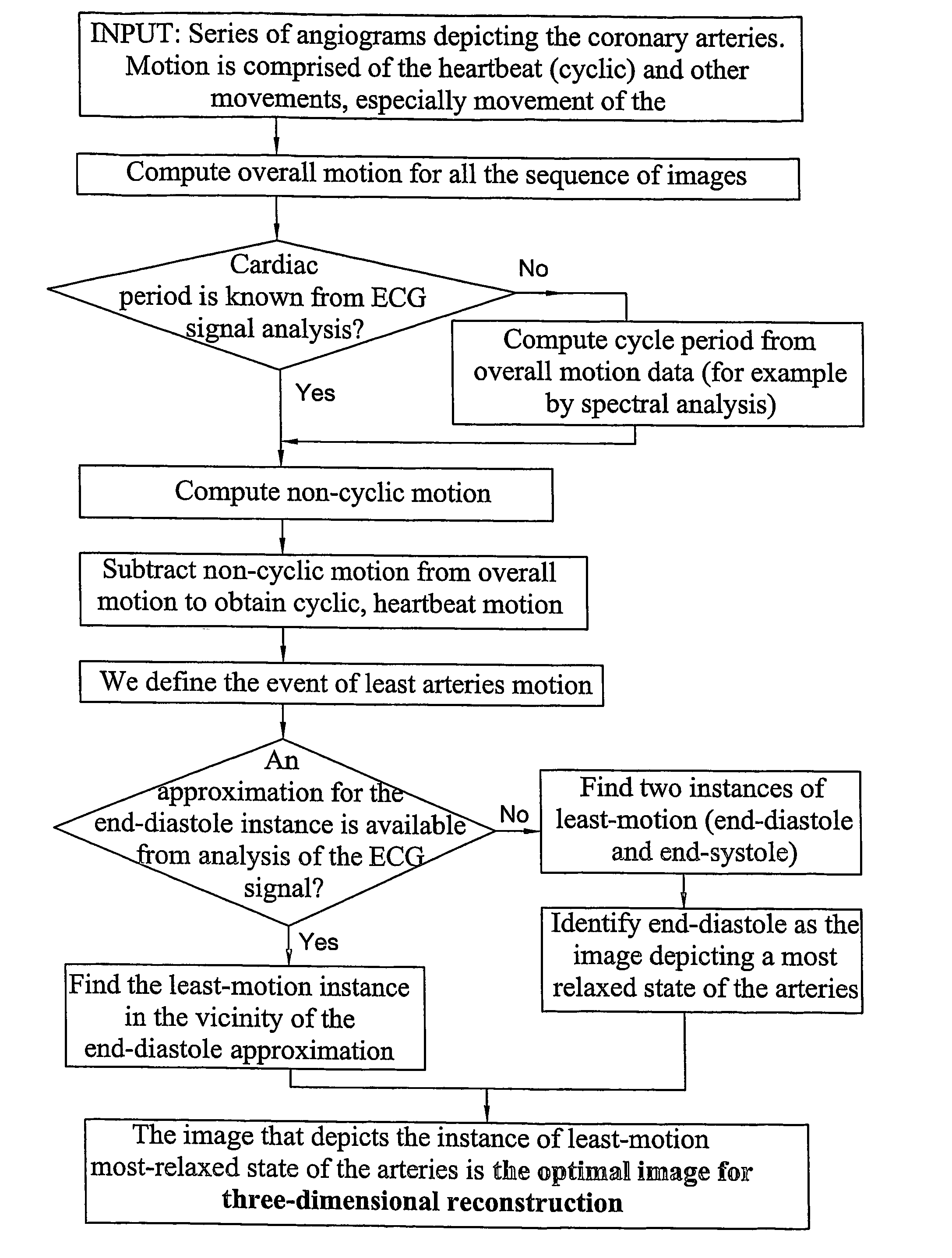
[0059]We suggest a preferred embodiment for an application of three-dimensional reconstruction of coronary vessels from a procedure of conventional angiography. In order to reconstruct a three-dimensional image of the arteries, it is necessary to obtain at least two two-dimensional images of the arteries in the same phase of the heartbeat, for example at end-diastole. Therefore, image acquisition is usually synchronized to an ECG signal. This procedure involves simultaneous recordings of the video signal from the X-ray camera and the patient's ECG signal. We present here a novel method for identifying the end-diastole instance, equivalent to ECG-gating, without relying solely, if at all, on the ECG signal.

[0060]Let IM1, IM2 . . . IMn be n images of a catheterization-acquired run.

[0061]Let m be the number of frames per cardiac cycle, either known in advance or computed as detailed in the above-described method for estimating the organ's motion.

[0062]Let IMk be the...


PUM


Abstract

A method and system for quantifying a cyclic motion within a series of images depicting a moving object subject to composite motion containing a cyclic component and a non-cyclic component of lower frequency than the cyclic component. Composite motion is computed as well as the non-cyclic component as the integral of motion over a motion cycle. The non-cyclic component is subtracted from the composite motion so as to obtain the cyclic component

Description

FIELD OF THE INVENTION[0001]This invention relates to medical image processing devices.BACKGROUND OF THE INVENTION[0002]Medical imaging devices are often used to image moving organs. Cardiac image processing devices, in particular, are always used to image moving organs, either the heart (via ultrasound imaging for example), or the coronaries (via angiography for example). Many of these imaging processing devices are used to quantify the motion either as an indication by itself or as part of an image-processing algorithm.[0003]An image processing device for Left Ventricle Analysis is used to evaluate Ejection Fraction, which is the percentage of the blood pumped out during each heartbeat. Left Ventricle Analysis involves computing the Left Ventricle volume from an angiogram (taken from a cine-angio sequence of images). The Left Ventricle volume is computed once for the heart in its systolic phase and once for the heart in its diastolic phase. Ejection Fraction is estimated from the ...

Claims


Application Information

Patent Timeline
Patent Type & Authority Patents(United States)
IPC IPC(8): G06K9/00G06T7/20G06T11/00
CPCG06T7/20G06T11/005A61B5/7285A61B6/503A61B6/504G06T2211/412G06T2211/404A61B6/541
Inventor ZARKH, MICHAELKLAIMAN, MOSHE
Owner MEDTRONIC INC
Why Eureka
Try Eureka