Pulmonary nodule detection in a chest radiograph

a chest radiograph and pulmonary nodule technology, applied in the field of computer assisted detection, can solve the problems of reducing the ability to detect nodules near the clear lung field boundary, and affecting the detection accuracy of chest radiographs

Inactive Publication Date: 2009-11-24
CARESTREAM HEALTH INC
View PDF21 Cites 6 Cited by
  • Summary
  • Abstract
  • Description
  • Claims
  • Application Information

AI Technical Summary

Benefits of technology

[0013]An objective of the present invention to provide a method of detecting pulmonary nodules in a chest radiograph that provides for the detection of nodules that overlap with ribs and other bones in the image.
[0014]Another objective of the present invention is to provide a method of detecting pulmonary nodules in a chest radiograph that provides for the detection of nodules that occur at the boundary of the clear lung field.

Problems solved by technology

However, for a variety of reasons including viewer fatigue and nodule occlusion by ribs, nodules can go undetected in chest radiographs.
The method employs a soft tissue image in addition to a standard radiograph, which is not always available.
However, it has been viewed that applying one or more thresholds to an image may not be sufficient means of nodule segmentation.
This techniques involves discarding pixels outside the clear lung field which may reduce the ability to detect nodules near the clear lung field boundary.
The effectiveness of this approach has been considered to be limited because the characteristics of abnormal anatomy can not be anticipated when the templates are created.
Although automatic detection of pulmonary nodules in a chest radiograph has been a topic of research for several years, it remains a challenging problem for several reasons.
In a projection chest radiograph normal anatomy and pulmonary nodules are superimposed making them difficult to distinguish.

Method used


Image

  • Pulmonary nodule detection in a chest radiograph
  • Pulmonary nodule detection in a chest radiograph
  • Pulmonary nodule detection in a chest radiograph

Examples


Embodiment Construction

[0028]The following is a detailed description of the preferred embodiments of the invention, reference being made to the drawings in which the same reference numerals identify the same elements of structure in each of the several figures.

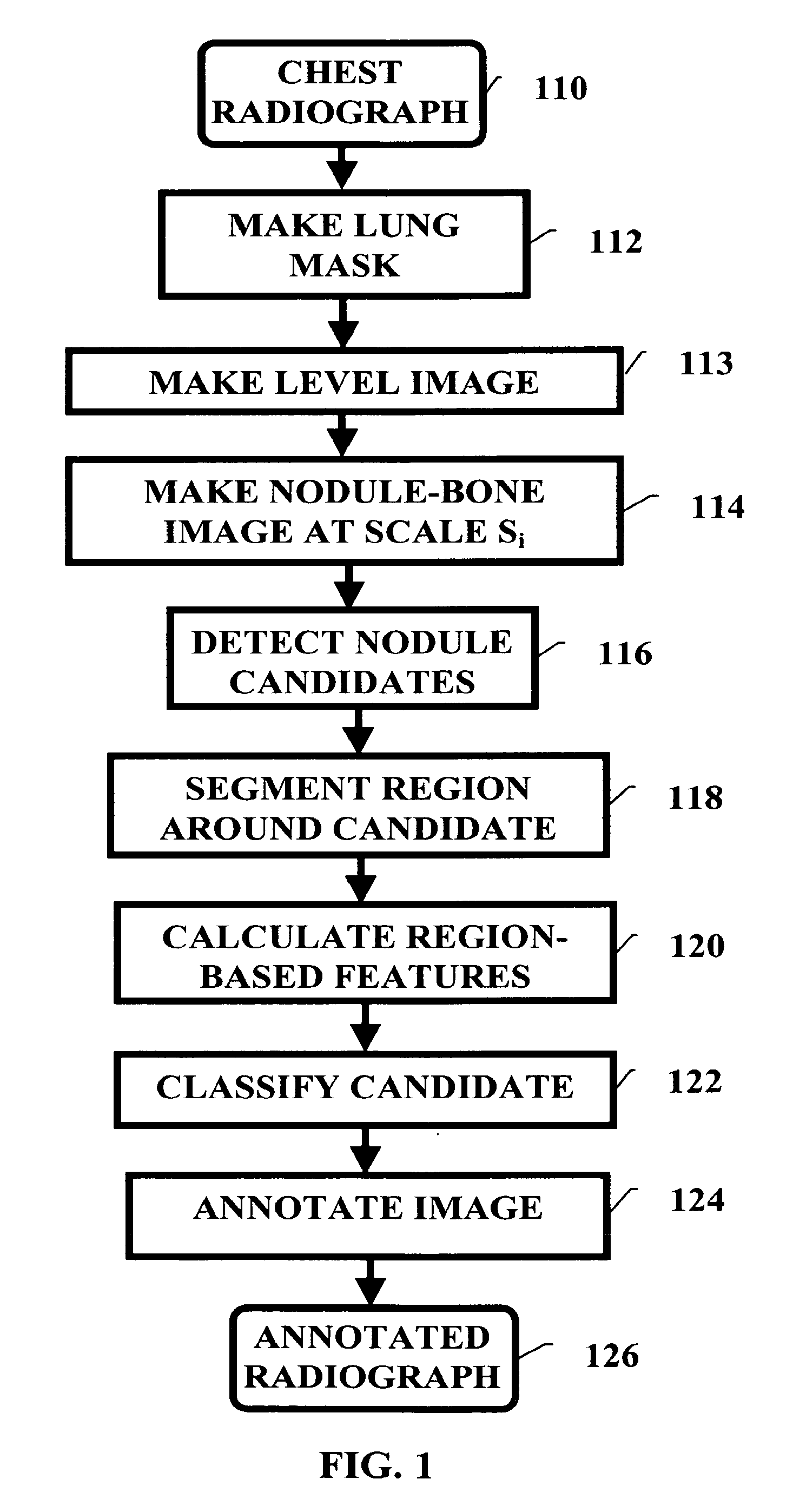
[0029]FIG. 1 shows a flowchart generally illustrating a method of pulmonary nodule detection in accordance with the present invention.

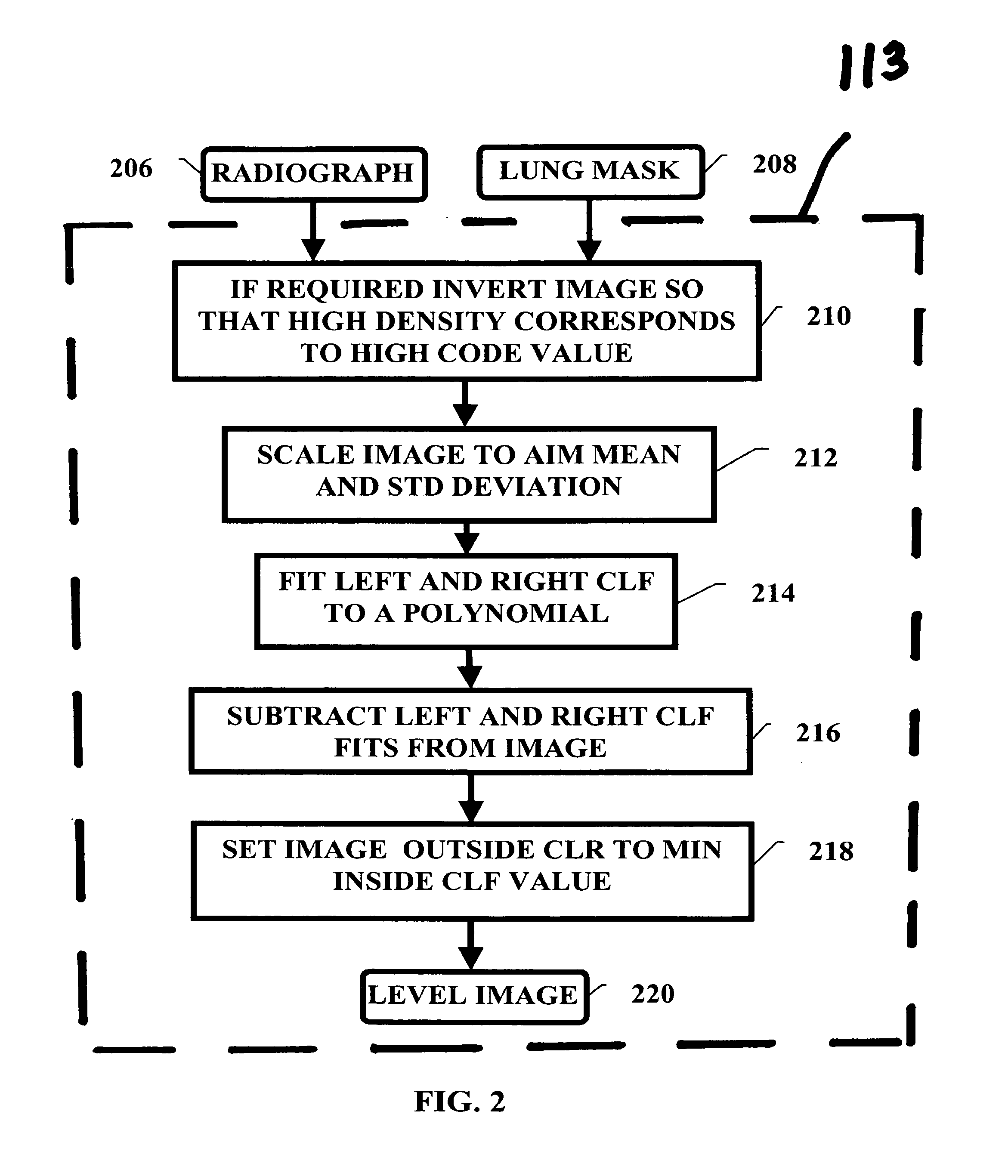
[0030]A chest radiograph (i.e., a chest image) is an input to a nodule detection system, as shown at step 110. The code value metric of the radiograph may be log exposure at the image detector; P-values as described in Part 14 of the DICOM standard; or any other image representation. If desired, the image can be scaled so that the spacing between pixels corresponds to a distance in the image plane of 0.171 mm.

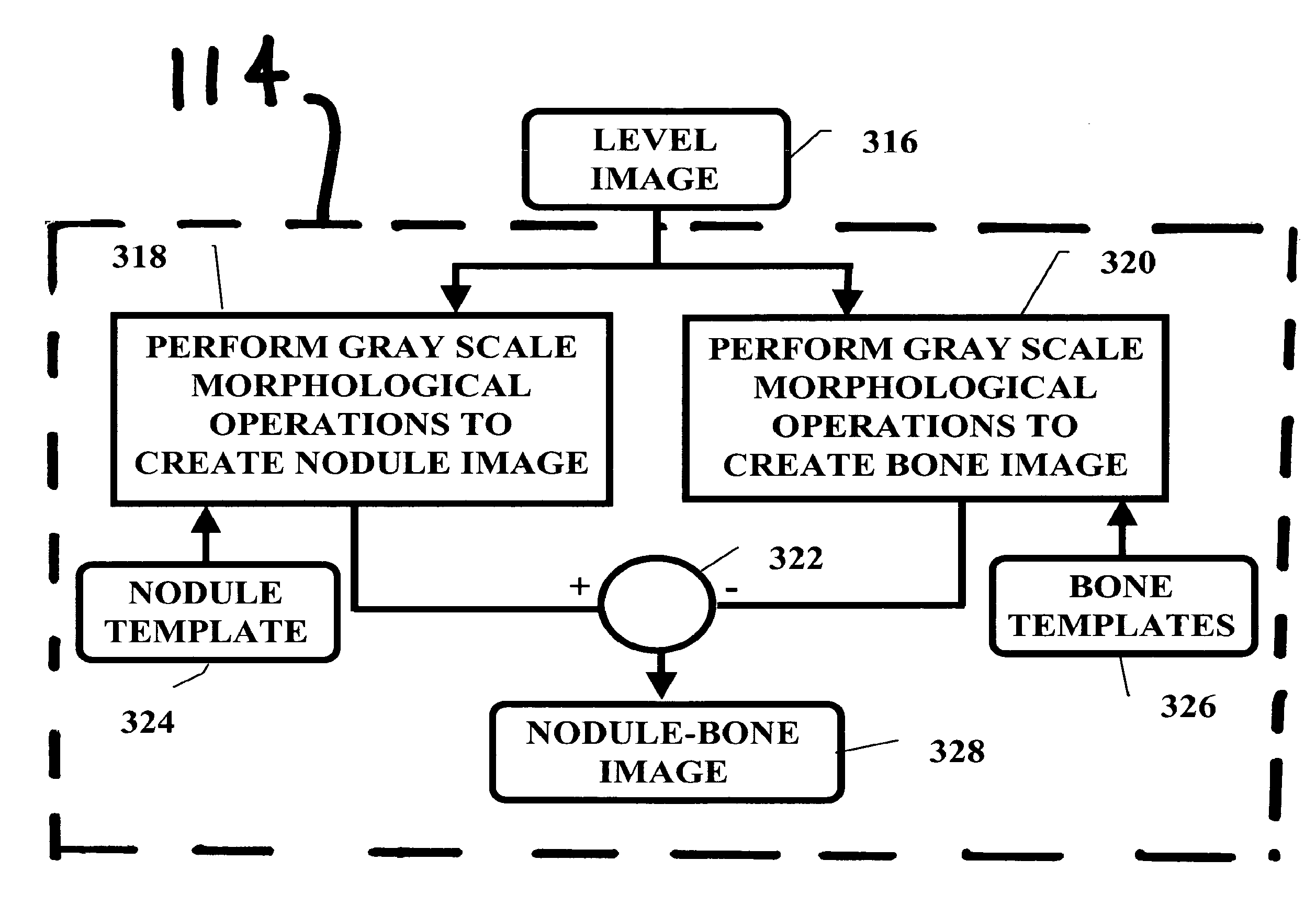
[0031]In step 112 a lung mask is calculated from the chest radiograph which indicates the clear lung field (CLF) in the image. Generally, the clear lung field is divided into a left and right clear lung field tha...


PUM


Abstract

A method of generating a pulmonary nodule image from a chest radiograph. The method includes the steps of: producing a map of a clear lung field; removing low frequency variation from the clear lung field to generate a level image; and performing at least one grayscale morphological operation on the level image to generate a nodule-bone image. Pulmonary nodules can be detected using the nodule-bone image by the further steps of: pulmonary nodules from a chest radiograph. The method includes the steps of: identifying candidate nodule locations in the nodule-bone image; segmenting a region around each candidate nodule location in the nodule-bone image; and using the features of the segmented region to determine if a candidate is a nodule.

Description

FIELD OF THE INVENTION[0001]The invention relates generally to the field of computer aided detection, and more particularly to the detection of pulmonary nodules in a chest radiograph.BACKGROUND OF THE INVENTION[0002]Lung cancer affects both men and women. At least one set of statistics has claimed that 90,363 men and 65,606 women died from lung cancer in the United States in 2001. It is believed that the early detection of lung cancer can increase the five-year survival rate from 12% to 70%. Screening for lung cancer can help with early detection.[0003]Projection radiographs of the chest can be used for screening for lung cancer. In a chest radiograph, lung cancer appears as opaque, lumpy, nodules within the lung. When nodules in chest radiographs are detected, further steps can taken to diagnose the pulmonary nodule as benign or malignant and treat the patient accordingly. However, for a variety of reasons including viewer fatigue and nodule occlusion by ribs, nodules can go undet...

Claims


Application Information

Patent Timeline
Patent Type & Authority Patents(United States)
IPC IPC(8): G06K9/00
CPCG06T7/0012G06T2207/30061
Inventor SCHILDKRAUT, JAY S.HEATH, MICHAEL D.SENN, ROBERT A.REVELLI, JR., JOSEPH F.LUCK, BRETTE
Owner CARESTREAM HEALTH INC
Why Eureka
Try Eureka