Method for improved image segmentation

a segmentation method and image technology, applied in image analysis, image enhancement, instruments, etc., can solve the problems of complex and time-consuming currently known image segmentation techniques, and the difficulty of finding the ideal threshold for each object in an image,

Active Publication Date: 2010-03-30
CYTYC CORP
View PDF7 Cites 44 Cited by
  • Summary
  • Abstract
  • Description
  • Claims
  • Application Information

AI Technical Summary

Benefits of technology

[0016]The present invention provides for a method to identify each object in an image under a plurality of threshold values, the method comprising steps of: (a) sorting pixels in the image based on a range of attribute values of the pixels, wherein the range of attribute values correspond to the plurality of threshold values; (b) adding pixels, one by one, to a labeling image to create new objects or update old objects, starting with an extreme point in the range of attribute values; (c) calculating features of the created new objects and the updated old objects; (d) matching the calculated features of the created new objects and the updated old objects with a pre-defined criteria; (e) outputting the created new objects and the updated old objects on an output image if an acceptance criteria is satisfied for the features; and (f) performing step b-e repeatedly until a stopping point is reached, wherein the stopping point chosen from any of the following: another extreme point in the range of values, a point representing background pixels values in the range of attribute values or a point representing pixel values not related to the new objects and the updated old objects.

Problems solved by technology

Finding an ideal threshold for each object in an image is a difficult task.
Often a single threshold value is not optimal for multiple objects with varying darkness values within an entire image.
Currently known techniques for image segmentation are often complex and time consuming.
These techniques do not always yield high accuracy in the segmentation process, particularly if there is little contrast between the object to be located and the background surrounding it.
Consequently, current segmentation algorithms often fail to locate objects properly.
In cell image analysis, for example, a cell nucleus might be incorrectly segmented because the located boundary is too large or too small.
This can result in false positive events (the instrument incorrectly calls a normal object suspicious) or false negative events (the instrument misses a true suspicious object).

Method used


Image

  • Method for improved image segmentation
  • Method for improved image segmentation
  • Method for improved image segmentation

Examples


Embodiment Construction

[0023]While this invention is illustrated and described in a preferred embodiment, the invention may be applied and produced in many different configurations. There is depicted in the drawings, and will herein be described in detail, a preferred embodiment of the invention, with the understanding that the present disclosure is to be considered as an exemplification of the principles of the invention and the associated functional specifications for its construction and is not intended to limit the invention to the embodiment illustrated. Those skilled in the art will envision many other possible variations within the scope of the present invention.

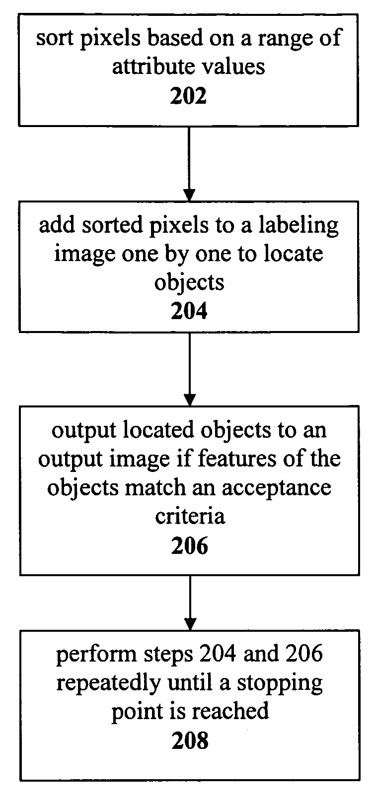
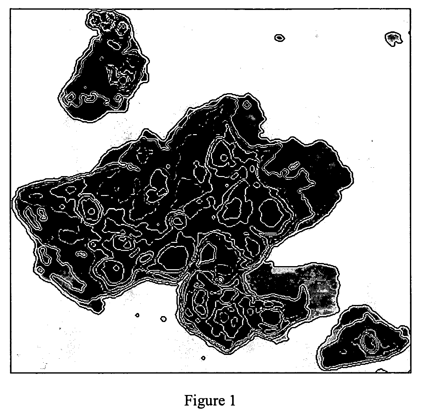
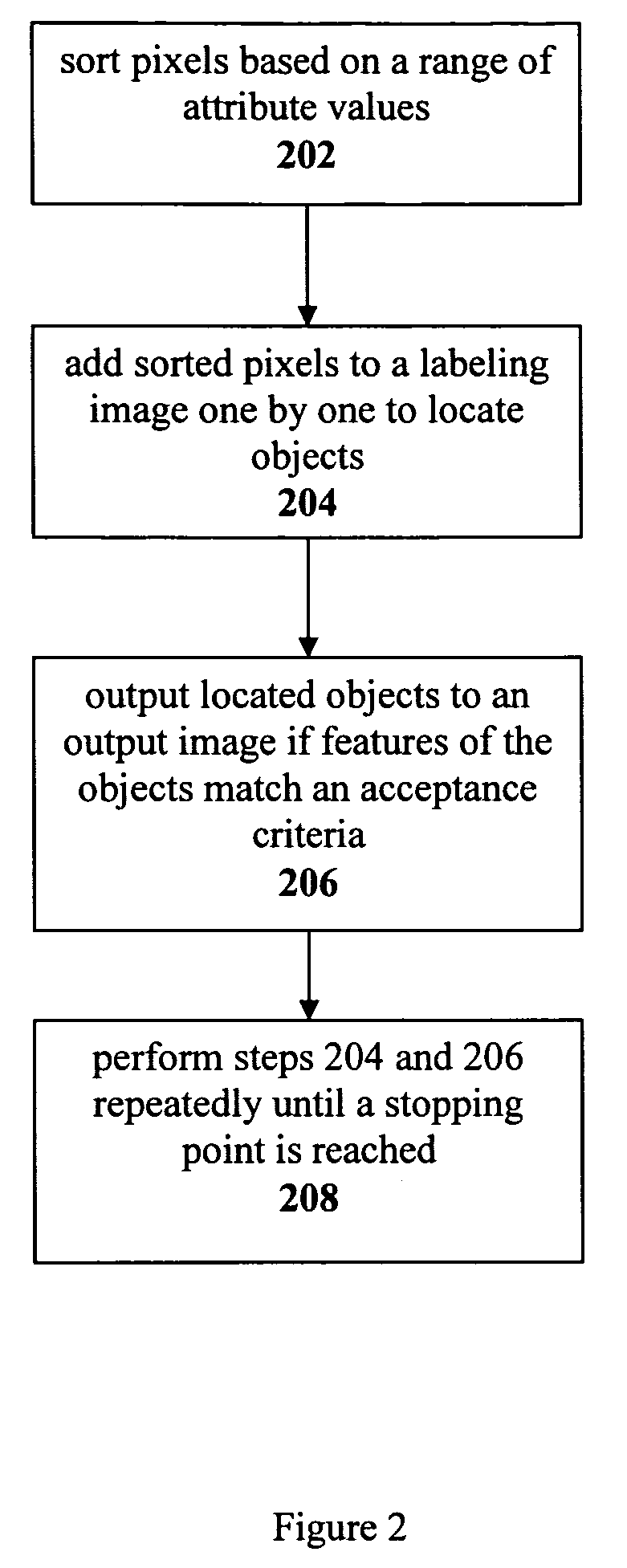
[0024]An improved automated image segmentation technique is described herein for identification of object boundaries in a digital two dimensional image. While the image segmentation technique described herein identifies nuclei, the technique itself can be applied to identifying any object in a digital image, such as cytoplasms or tissue str...


PUM


Abstract

An improved image segmentation algorithm is provided that identifies each object in an image. Pixels of the image are sorted based on a range of attribute values of the image. These pixels are then added to a labeling image one by one starting with an extreme point in the range of attribute values. Features are calculated for each object located and these features are matched with pre-defined acceptance criteria. If there is a match, the object is output to an output image. The steps of adding pixels to the image, evaluating the features of the resulting objects and outputting the objects are repeated until a stopping point is reached.

Description

RELATED APPLICATION[0001]This application claims priority benefit and is a non-provisional of U.S. Provisional Application No. 60 / 642,110 filed Jan. 10, 2005, which is incorporated herein by reference in its entirety.Government Rights[0002]This invention was made with U.S. Government support under NIH Grant Number RR018046 awarded by the National Institutes of Health. The U.S. Government may have certain rights in this invention.BACKGROUND OF THE INVENTION[0003]1. Field of Invention[0004]The present invention relates generally to the field of image analysis. More specifically, the present invention is related to a novel image segmentation method.[0005]2. Discussion of Prior Art[0006]In pathology or cytology, it is often desirable to locate and measure cells or nuclei using an automated or semi-automated instrument. Such instruments may be used for research, or for screening. An example of the latter is the screening for cervical cancer using the Papanicolou test (or Pap test). These...

Claims


Application Information

Patent Timeline
Patent Type & Authority Patents(United States)
IPC IPC(8): G06K9/34G06K9/46
CPCG06K9/342G06T7/0083G06T2207/30024G06T2207/10072G06T7/12G06V20/695G06V10/267G06T5/00G06V10/40G06F18/00
Inventor ZAHNISER, MICHAEL
Owner CYTYC CORP
Why Eureka
Try Eureka