
Algorithm for the automatic determination of optimal pacing intervals

a technology of automatic determination and optimal pacing interval, which is applied in the direction of electrotherapy, heart stimulators, therapy, etc., can solve the problems of atrial tachycardia, insufficient filling of left ventricle, and inability to achieve optimal pacing of both left and right ventricles at the same time, so as to achieve optimal cardiac output

Inactive Publication Date: 2010-05-04
MEDTRONIC INC
View PDF15 Cites 19 Cited by
  • Summary
  • Abstract
  • Description
  • Claims
  • Application Information

AI Technical Summary

Benefits of technology

[0014]A second method according to the present invention measures impedance change taken between times near the A-event and near the V-pace. The impedance change from A-event to V-pace is an impedance decrease as the ventricle fills with conductive blood. This method attempts to maximize the atrial contribution to ventricular filling by producing the largest absolute change in volume (delta Z) between the A-event and V-pace, through manipulation of the AV interval. The impedance data can be obtained from measurements taken shortly after the A-pace and near the V-pace. The AV interval can be varied in a binary search to converge on maximum impedance change indicative of maximum ventricular output. In some methods, only one or a small number of impedance measurements are taken after the A-pace, and shortly before, during, or shortly after the V-pace. The impedance change can be determined by subtracting the impedance taken near the V-pace from the impedance taken near the A-pace. The small number of carefully timed measurements can substantially reduce the power required to determine the impedance change.
[0015]In one method, a single impedance measurement is taken shortly after A-event and a single measurement taken near the V-pace. In some methods, the impedance change is measured for several heart beats at the same AV interval, and the average maximum impedance change over several heartbeats used. In yet another method, the impedance waveforms from several beats are averaged together, after being aligned on the atrial or ventricular pacing pulse or sense, and the maximum impedance change determined from the averaged waveform. The impedance change from ventricular expansion after atrial contraction can be maximized as a function of AV interval to maximize the atrial contribution to ventricular filling. The AV interval can be varied to bracket to maximum impedance change over several beats. In one method, a search algorithm is used to rapidly converge on the maximum impedance change from both time directions. In a preferred method, a binary search is used to converge on the maximum impedance change. The binary search can provide rapid convergence in few heart beats requiring little power consumption.
[0017]In some methods, the impedance change is measured for several heart beats at the same AV interval, and the average maximum impedance change over several heartbeats used. In yet another method, the impedance waveforms from several beats are averaged together, after being aligned on the atrial or ventricular pacing pulse or sense, and the maximum impedance change determined from the averaged waveform. The impedance change from expansion to contraction can be maximized as a function of AV interval to maximize the cardiac output. The AV interval can be varied to bracket the maximum impedance change over several beats. In one method, a search algorithm is used to rapidly converge on the maximum impedance change from both time directions. In a preferred method, a binary search is used to converge on the maximum impedance change. The binary search can provide rapid convergence in few heart beats requiring little power consumption.
[0018]A fourth method varies the right ventricle to left ventricle (VV) interval to converge on an impedance maximum indicative of minimum cardiac volume at end systole. This method attempts to maximize the change in ventricular volume and largest change in impedance by producing the greatest ventricular filling and maximal ventricular emptying over the ventricular ejection period through manipulation of the VV interval. The maximum impedance in a cycle is obtained as an indication of the minimum cardiac volume associated with end systole. The impedance measurements can be taken at multiple times after V-pace until next A-pace. The time difference between pacing the right and left ventricle, the ventricle to ventricle (VV) interval, can be varied to find the VV interval having the maximum impedance. The paced VV interval can be varied to bracket the maximum impedance over several heart beats. In one method, a search algorithm is used to rapidly converge on the paced VV interval having the maximum impedance. In a preferred method, a binary search is used to converge on the paced VV interval having the maximum impedance change. The binary search can provide rapid convergence in few heart beats. In some methods, the maximum impedance is measured for several heart beats at the same VV interval, and the average maximum impedance over several heartbeats used. In yet another method, the impedance waveforms from several beats is averaged together after being aligned on the atrial or ventricular pacing pulse or sense, and the maximum impedance determined from the averaged waveform.
[0025]In still another method, preferably during a period of little movement, most preferably during sleep, the pacing device tracks the breathing cycle, through any means appropriate, including an impedance signal or accelerometer signal. Logic executing in the implanted device can time or “gate” the taking of impedance data to occur only near the inspiration peak or expiration nadir, preferably during the expiration nadir. Taking the impedance during these time regions, windows, or ranges of relatively little impedance change can effectively eliminate most of the impedance change due to breathing. In some embodiments, only one or two cardiac cycles per breath are measured for impedance.

Problems solved by technology

It can be seen that if contractions in the left ventricle and the right ventricle are about the same time, then left AV synchrony is way off, with the left ventricle not having adequate time to fill up.
Further, it is known that patients with IAB are susceptible to retrograde activation of the left atrium, with resulting atrial tachycardia.
Thus, while concurrent or simultaneous pacing of the left and right ventricle may achieve a significant improvement for CHF patients, pacing both left and right ventricles at the same time may not always be optimal.
For example, if conduction paths in the left ventricle are impaired, delivering a pacing stimulus to the left ventricle at precisely the same time as to the right ventricle may nonetheless result in left ventricular contraction being slightly delayed with respect to the right ventricular contraction.
If the right and left ventricle are paced simultaneously this may not result in maximized pumping action, with the optimal pacing lead or lag between the ventricles being patient specific.
Echocardiography is quite expensive, and can only be done infrequently, in a clinical setting.

Method used


Image

  • Algorithm for the automatic determination of optimal pacing intervals
  • Algorithm for the automatic determination of optimal pacing intervals
  • Algorithm for the automatic determination of optimal pacing intervals

Examples


embodiment 300

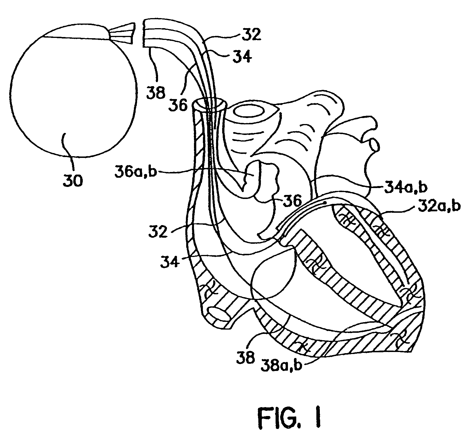
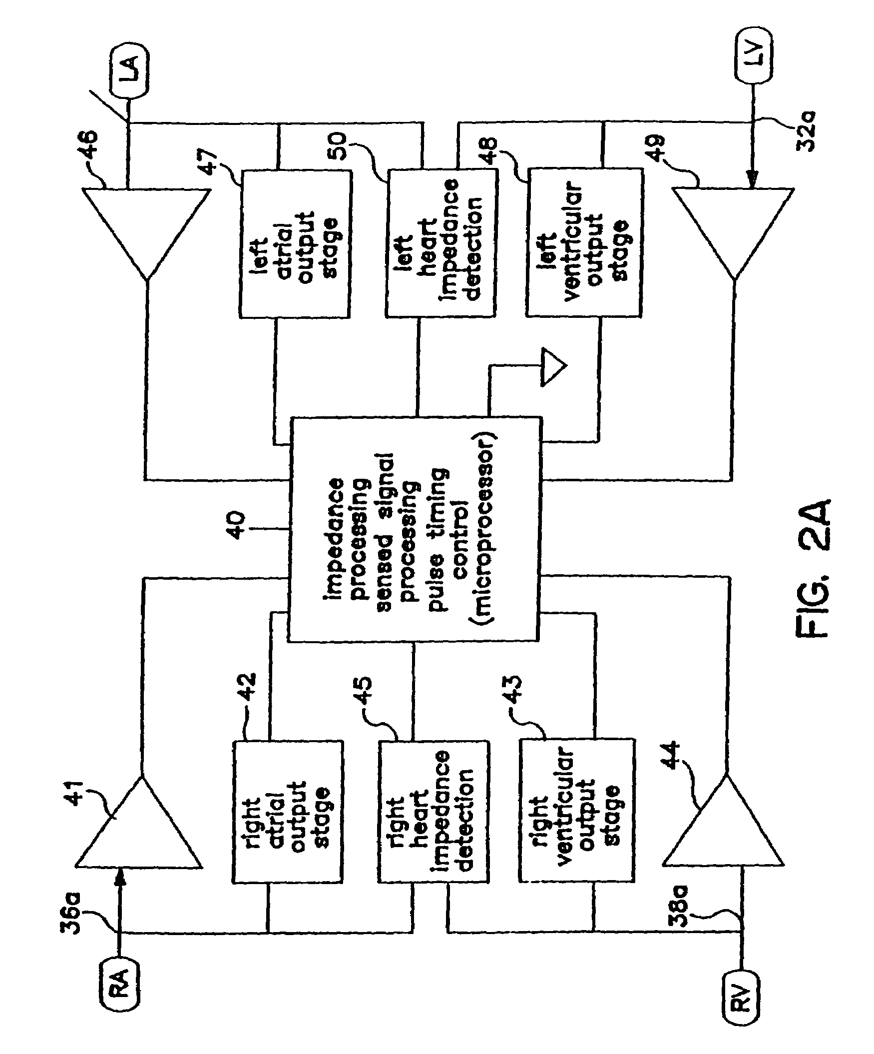
[0067]The impedance measurement of embodiment 300 and the other methods discussed in the present application, can be measured as a vector across the best available leads present in the heart. In a bi-ventricular implanted pacing device, the impedance can be measured between a right ventricular electrode and a left ventricular electrode disposed in a cardiac vein. In one example, the impedance signal is stimulated between the left ventricular ring and the right ventricular ring and detected by the left ventricular tip and the right ventricular tip. The electrode pairs can be selected to maximize the impedance changes caused by the ventricular contractions and expansions. In another embodiment, having a ventricular lead, different electrodes disposed along the ventricular lead may be used to stimulate and sense the impedance vector.

[0068]FIG. 5A illustrates another embodiment 320A for optimizing the AV interval. While method 300 attempted to maximize ventricular expansion as measured ...

embodiment 340

[0078]FIG. 6 includes a flow chart for embodiment 340, which varies the VV interval to find the maximum impedance. The maximum impedance corresponds to the maximum ventricular contraction and minimum ventricular volume. Method 340 optimizes the VV interval indicating the amount of time by which the right ventricle leads or lags the left ventricle. The VV interval may also be viewed as having a separate atrium to right ventricle delay and a separate atrium to left ventricle delay. The VV interval may also be referred to as the inter-ventricle time difference.

[0079]In step 341, the VV interval can be initialized in some methods to 0. In step 342, the AV interval can be initialized and maintained at a constant, preferably, optimal AV interval. In some methods, the optimal AV interval as determined by methods such as method 300, 320A, or 320B can be used to set the optimal AV interval.

[0080]In step 344, the atrium A-event occurrence can be paced or sensed, then the first ventricle paced...

embodiment 400

[0091]FIG. 8 illustrates an embodiment 400 for providing impedance data, to the interval optimization methods previously discussed. Method 400 is one example of a method for providing impedance data, which is preferably used in a clinical setting. In step 402, method 400 waits for a start signal. The patient is preferably supine, not moving, and has a regular breathing pattern. A treating physician can wait for the patient to be appropriately disposed then send a start signal from a programming unit, through a telemetry link, to the implanted pacing device. In step 402, the pacing device receives the start signal from the programmer, and proceeds to step 404.

[0092]In step 404, the implanted pacing device can automatically adjust pacing parameters as needed to carry out the interval optimization methods previously discussed to measure the impedance changes over one or more heartbeats. In the example illustrated, the impedance is measured over a single beat. With the impedance measure...


PUM


Abstract

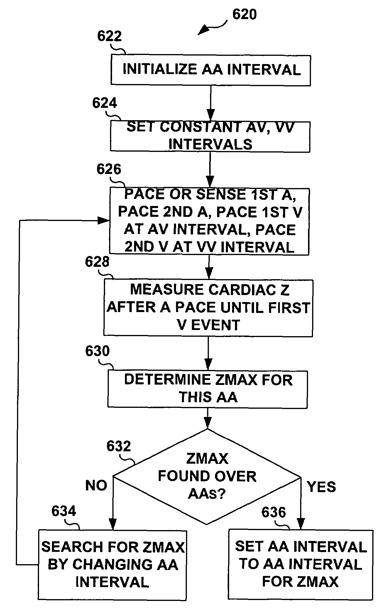
Impedance, e.g. sub-threshold impedance, is measured across the heart at selected cardiac cycle times as a measure of chamber expansion or contraction. One embodiment measures impedance over a long AV interval to obtain the minimum impedance, indicative of maximum ventricular expansion, in order to set the AV interval. Another embodiment measures impedance change over a cycle and varies the AV pace interval in a binary search to converge on the AV interval causing maximum impedance change indicative of maximum ventricular output. Another method varies the right ventricle to left ventricle (VV) interval to converge on an impedance maximum indicative of minimum cardiac volume at end systole. Another embodiment varies the VV interval to maximize impedance change. Other methods vary the AA interval to maximize impedance change over the entire cardiac cycle or during the atrial cycle.

Description

FIELD OF THE INVENTION[0001]The present invention is related generally to implantable cardiac pacemakers and cardioverter defibrillators. More specifically, the present invention includes apparatus and methods for using impedance measurements to set optimal pacing intervals.BACKGROUND OF THE INVENTION[0002]Congestive heart failure (CHF) is defined generally as the inability of the heart to deliver enough blood to the peripheral tissues to meet metabolic demands. Frequently CHF is associated with left heart dysfunction, but it can have a variety of other causes. For example, CHF patients may have any one of several different conduction defects. The natural electrical activation system through the heart involves sequential events starting with the sino-atrial (SA) node, and continuing through the preferred conduction pathways at the atrial level, followed by the atrio-ventricular (AV) node, Common Bundle of His, right and left bundle branches, and final distribution to the distal myoc...

Claims


Application Information

Patent Timeline
Patent Type & Authority Patents(United States)
IPC IPC(8): A61N1/362
CPCA61N1/36521
Inventor BURNES, JOHN E.IGEL, DAVID A.RUETER, JOHN C.CHO, YONG K.MONGEON, LUC R.STONE, HAROLD E.ZILINSKI, JODI
Owner MEDTRONIC INC
Learn More