
Biopsy method

a biopsy and method technology, applied in medical science, surgery, vaccination/ovulation diagnostics, etc., can solve the problem of inacceptable one-way biopsy

Inactive Publication Date: 2010-08-03
DEVICOR MEDICAL PROD
View PDF68 Cites 28 Cited by
  • Summary
  • Abstract
  • Description
  • Claims
  • Application Information

AI Technical Summary

Benefits of technology

Enables the collection of both FNA and core samples from the same tissue site using the same device, enhancing flexibility and safety by avoiding the need for separate procedures and devices, while maintaining the ability to perform either type of biopsy without removing the inner cannula.

Problems solved by technology

A surgeon may find one biopsy method to be unacceptable, necessitating a change to the alternative method.

Method used


Image

  • Biopsy method
  • Biopsy method
  • Biopsy method

Examples


Embodiment Construction

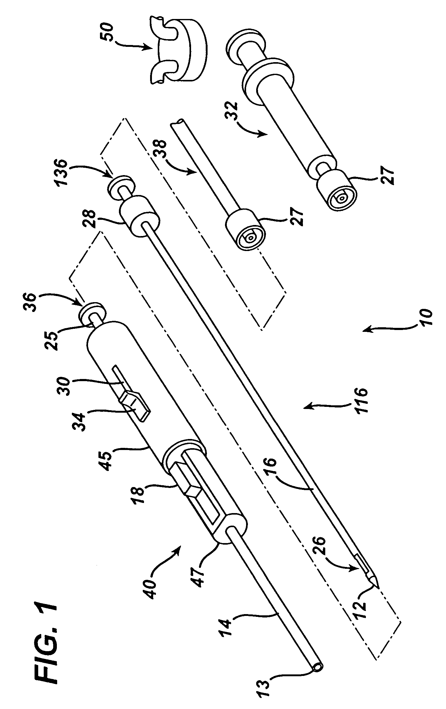
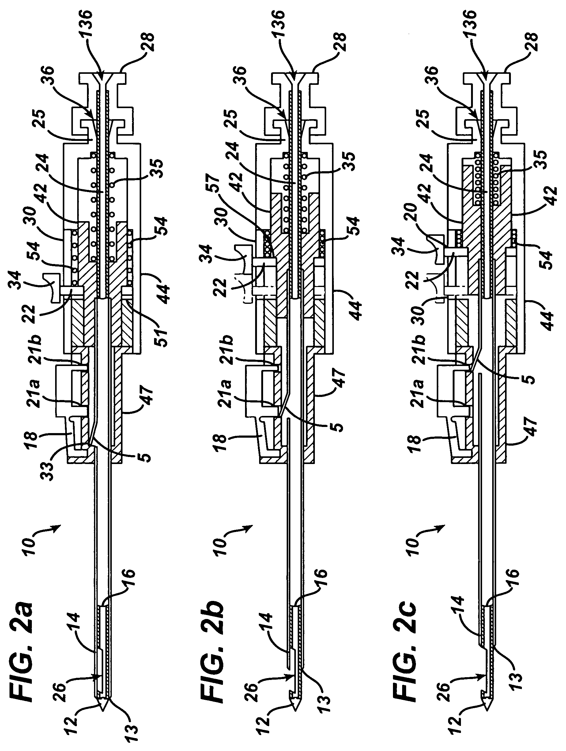
[0015]FIG. 1 illustrates a biopsy device 10 useful in the biopsy method according to one embodiment of the present invention. Biopsy device 10 can include an outer sheath assembly 40 and an inner cannula assembly 116. The outer sheath assembly 40 can include an outer cannula 14, a body 47, and a handle 45. A proximal opening 36 can be provided in the proximal end of handle 45.

[0016]The inner cannula assembly 116 can include an inner cannula 16 extending distally from an inner cannula locking hub 28. A proximal opening 136 can be provided in the proximal end of inner cannula assembly 116. Referring to FIG. 1, a vacuum tube 38, a syringe 32, a sample / fluid capture container 50, or other suitable device may be releasably attached to the proximal end of the inner cannula assembly 116, such as by using a locking hub 27 associated with the device.

[0017]Inner cannula 16 can include a closed, distal tissue penetrating tip 12 adapted for piercing tissue. Inner cannula 16 can also include a s...


PUM


Abstract

A biopsy method is disclosed. The biopsy method can be used to provide a fine needle aspiration sample and a core biopsy sample. The biopsy method can include adjusting a sample port length without removing the biopsy device from the patient.

Description

[0001]This patent application cross references and incorporates by reference the following copending, commonly assigned patent applications: U.S. patent application Ser. No. 11 / 072,719 filed Mar. 4, 2005 in the names of Weikel et al.; and U.S. patent application Ser. No. 11 / 222,575 filed Sep. 9, 2005 in the names of Weikel et al.[0002]This patent application cross references and incorporates by reference commonly assigned patent application “Biopsy Device” filed on the same day herewith and in the name of Voegele.FIELD OF THE INVENTION[0003]The present invention is directed to a biopsy method, and more particularly, to a biopsy method which can be used to obtain both fine needle aspiration and core samples.BACKGROUND OF THE INVENTION[0004]A biopsy may be performed in various ways, including by taking a fine needle aspiration (FNA) sample or, alternatively, a core sample.[0005]The diagnosis and treatment of tissue is an ongoing area of investigation. Medical devices for obtaining tis...

Claims


Application Information

Patent Timeline
Patent Type & Authority Patents(United States)
IPC IPC(8): A61B10/00
CPCA61B10/0275A61B2010/0208A61B10/0283
Inventor VOEGELE, JAMES W.
Owner DEVICOR MEDICAL PROD
Learn More