Methods and apparatus for computer automated diagnosis of mammogram images

a mammogram and automated diagnosis technology, applied in the direction of image enhancement, patient positioning for diagnostics, instruments, etc., can solve the problems of false positive findings, distorted summation artifacts, and inability to accurately interpret the information about some structures

Inactive Publication Date: 2011-01-04
CAROLINA IMAGING SPECIALISTS
View PDF4 Cites 19 Cited by
  • Summary
  • Abstract
  • Description
  • Claims
  • Application Information

AI Technical Summary

Benefits of technology

[0008]Among its several aspects, the present invention recognizes that false-positive findings associated with prior art approaches may result from the absence of the use of actual or derived points of reference from anatomic features such as the breast wall and the nipple. One aspect of the present invention relates to computer systems and methods for automated identification of diagnostically significant features, such as microcalcifications and masses, on film based or digital mammograms. Aspects of the invention include accurately determining if apparent clusters and focal densities on 2-D views are concordant or discordant in three dimensional space, specification of the location, of concordant lesions, in three dimensional space, by clock notation and distance from the nipple, and the assigning of a BIRADS classification to the mammographic or mammogram study as discussed further herein.
[0009]To such ends, the present invention comprises methods, procedures, systems, optical and magnetic media for computer automated diagnosis of screening and diagnostic mammography, especially to aid the physician in interpreting the mammogram and for application as an automated second read. Information from two mammographic or mammography views, such as cranio-caudal (CC) and medio-lateral oblique (MLO) or alternatively, cranio-caudal and medio-lateral (ML) is combined to reduce or eliminate false positive findings that result when mammographic findings, for example, microcalcifications, and focal densities, that are physically distant in three dimensional space, for example, greater than 1 cm apart, appear to be close together or mass-like in appearance, because of the collapsing of the third dimension on the 2-D image. This visual collapsing effect is analogous to the constellation effect and is known as a summation artifact, a pseudo-mass or pseudo cluster.

Problems solved by technology

If multiple x-ray absorbing structures lie in the same x-ray path, information about some of these structures are likely to be obscured, distorted or both.
Additionally, because mammography comprises two-dimensional imaging of a three-dimensional object, summation artifacts, that is the summation of the x-ray absorption of two or more, structures, may appear as false positive findings.
These subjective and qualitative criteria are only somewhat effective in correctly establishing the concordance or discordance of candidate lesions and thus decrease the accuracy of mammography.
Current methods including those described above are limited by the displacement of lesions due to the physical compression of the breast during the performance of the mammogram.
While existing solutions are sensitive, in other words, they identify almost all features of diagnostic interest, they are not sufficiently specific as they too often identify false positive findings.
Many false-positive findings associated with prior art approaches are constellation effect, summation artifacts, or the like, resulting from analyzing each 2-D mammogram view in isolation.
The absence of quantitative methods and automated tools for establishing the spatial concordance of a lesion or lesions between two or more mammographic or mammography views results in a higher than desired occurrence of false positive findings for both physician evaluated and computer evaluated mammography.
False positive findings add significant costs to the health care system and almost always result in added stress and anxiety for the patient.
False positive results can cause patients to be subjected to unnecessary follow-up testing which is costly, uncomfortable and emotionally stressful.
False positive findings may result in unnecessary medical intervention, including tissue sampling, such as cytology, biopsy, or surgical excision.
Furthermore, false positive CAD findings represent a potential for future litigation if the interpreting physician is not careful to adequately document his or her findings related to the CAD findings.
This extra documentation reduces physician productivity and profitability.
Since interpretation of mammograms requires the interpreting physician to use the breast imaging reporting and data system (BIRADS) lexicon, an incorrect BIRADS category may be assigned, resulting in an inappropriate follow-up interval, an unnecessary patient recall, or a missed or delayed diagnosis of breast cancer.

Method used


Image

  • Methods and apparatus for computer automated diagnosis of mammogram images
  • Methods and apparatus for computer automated diagnosis of mammogram images
  • Methods and apparatus for computer automated diagnosis of mammogram images

Examples


Embodiment Construction

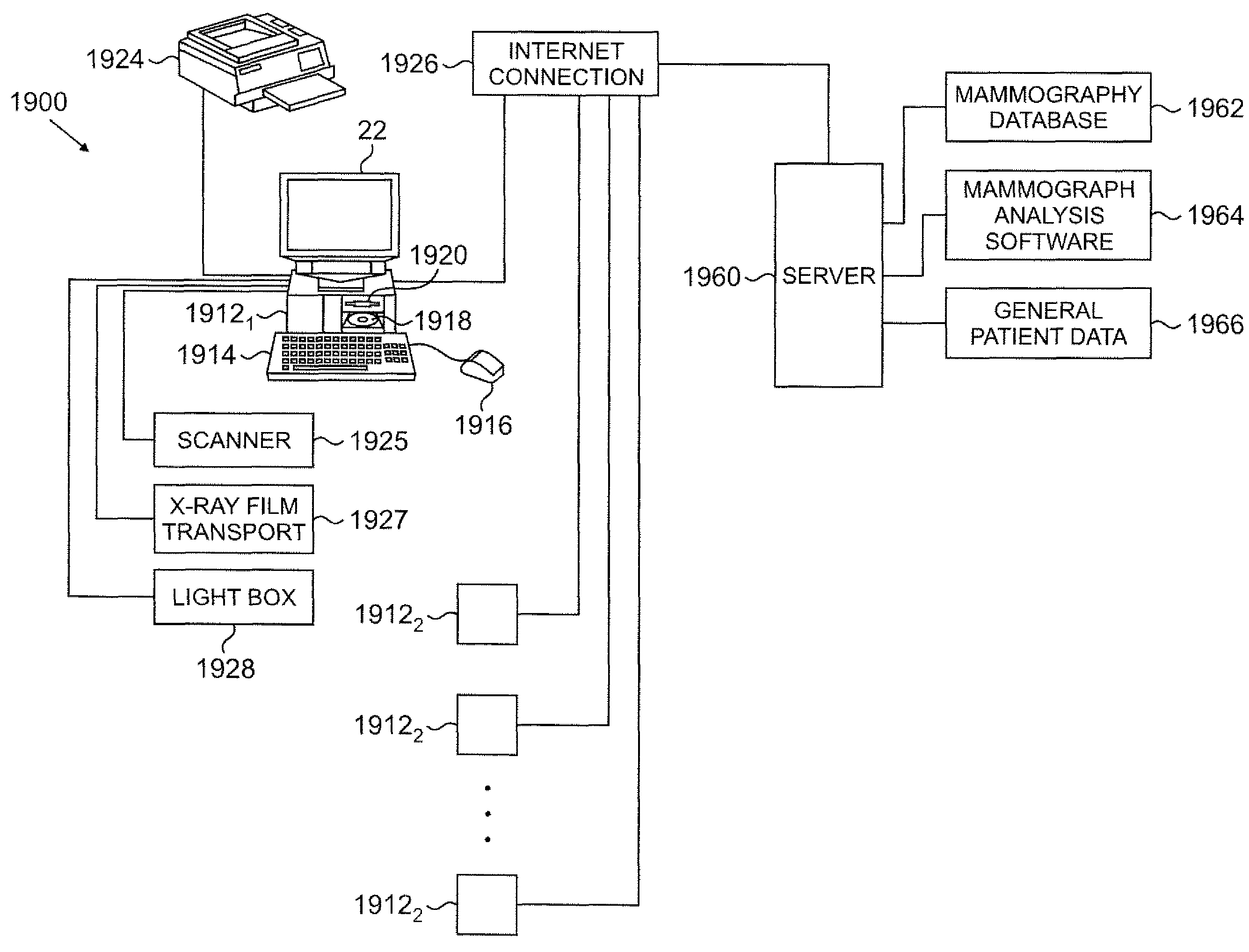
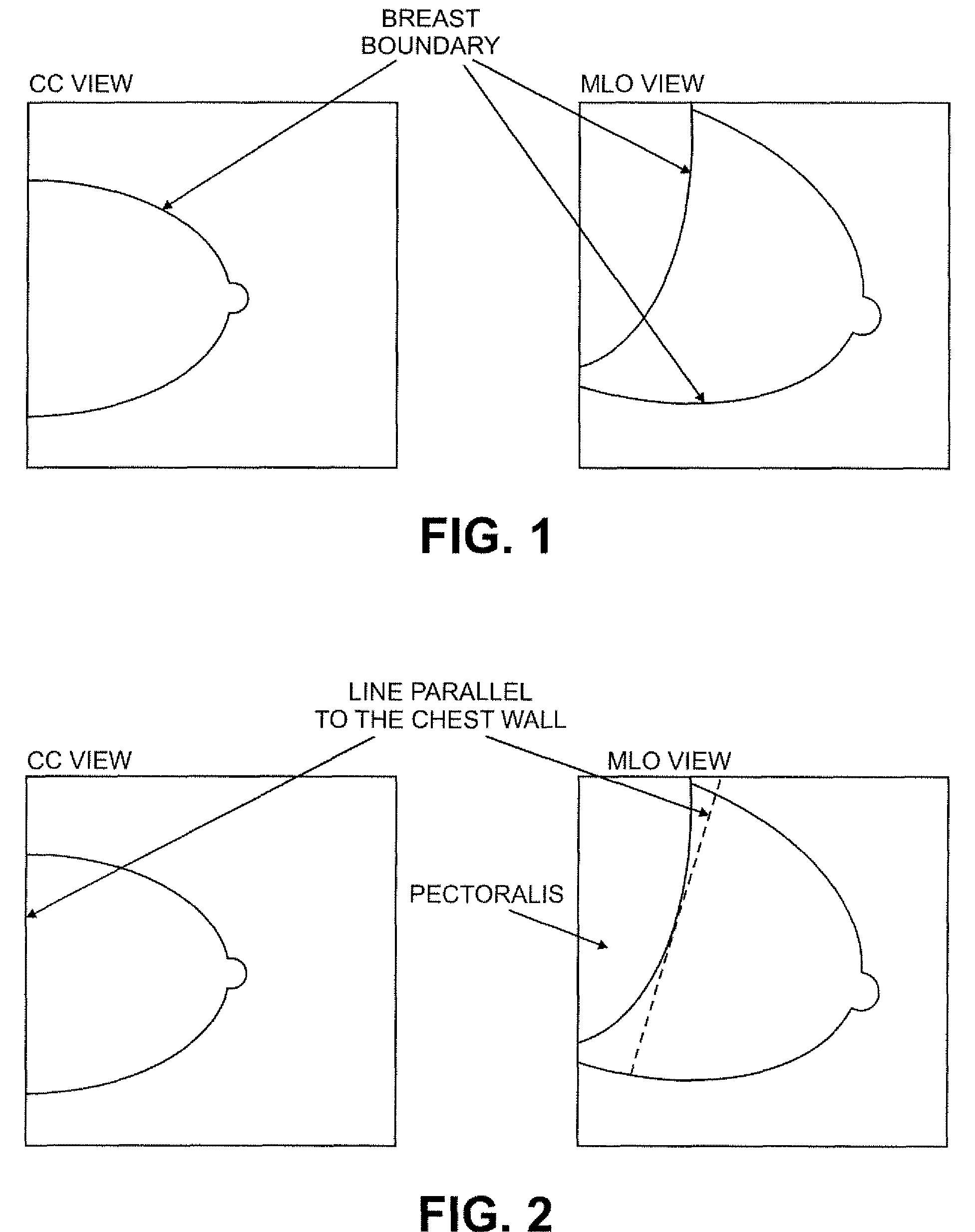
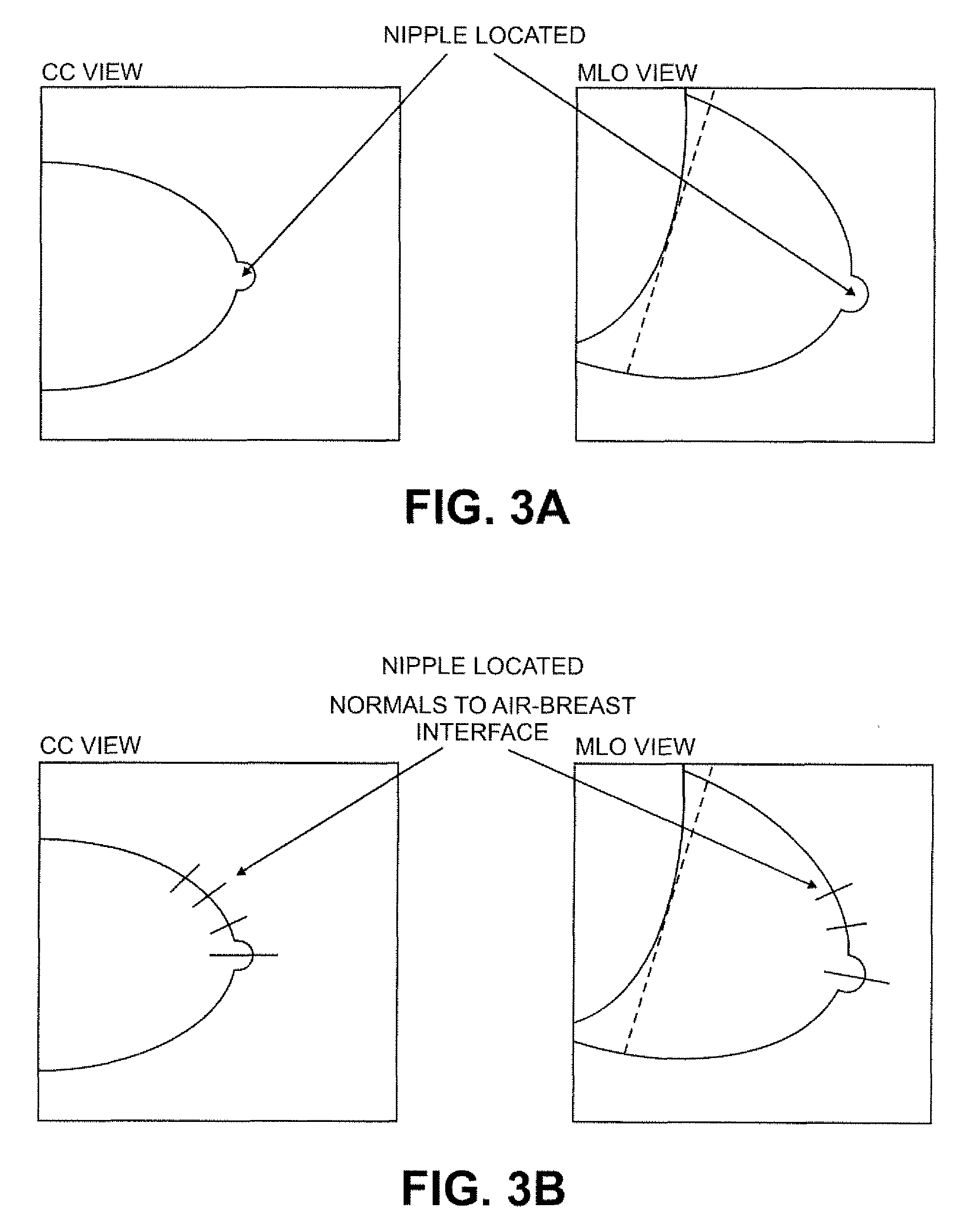
[0021]With reference to FIGS. 1-14 which show illustrative mammography views used to illustrate aspects of the present invention and FIGS. 15A and 15B which show a process 1500 in accordance with the invention, one embodiment of the invention starts with digital data from a standard screening mammography study comprising four views, two of each breast as follows:

[0022]left cranio-caudal (LCC) and left medio-lateral oblique (LMLO); and

[0023]right cranio-caudal (RCC) and night medio-lateral oblique (RMLO).

[0024]Medio-lateral (ML) views may be used instead of the MLO views. It is further noted that only two views are necessary, but where only two views are employed, they must be of the same breast. For example, left CC and left MLO views are acceptable, hut left CC and right MLO view are not an acceptable image pair.

[0025]In step 1502 of process 1500 of FIG. 15A, the output of a digital mammography system, or the output of a digitizer that has scanned a film mammogram, is converted to ...


PUM


Abstract

A system, method, and computer program product for computer analysis of lesions in digitized film-based and / or digital mammograms is described, wherein diagnostic information is combined from two different 2-D mammographic views with the information obtained from one view (or field of view) or mammographic position is processed with information obtained from a second (or a plurality of) related mammographic views to reduce false-positive findings (increase specificity) while preserving or improving diagnostic sensitivity. The digital mammograms or digitized film-based mammograms used, are those that are in current use, and those that conform to the requirements of the American College of Radiology and the Mammography Quality Standards Acts. In a preferred embodiment, a line constructed at the location of the chest wall (or parallel to the chest wall), the location of the nipple, and a line constructed perpendicular to the chest wall datum line and passing through the location of the nipple serve as reference datum across mammogram views. An algorithm locates suspicious lesions in each mammography view and evaluates the concordance of the 3-D spatial locations to rule out physically impossible false-positive findings, based on calculations of spatial relationships. Concordant findings are detected using anatomic landmarks and such findings are reported using terms that are currently in use by physicians and other health care providers in the field of mammography.

Description

[0001]The present application claims the benefit of U.S. Provisional Application Ser. No. 60 / 810,369 filed Jun. 2, 2006 which is hereby incorporated by reference in its entirety.FIELD OF THE INVENTION[0002]The present invention relates generally to improved techniques for the interpretation of mammogram images, and more particularly to advantageous techniques for computer automated analysis of diagnostic information from two or more different mammographic views.BACKGROUND OF THE INVENTION[0003]A standard mammogram consists of a set of two dimensional (2-D) x-ray images obtained in at least two different projections. The images formed using x-ray mammography are the result of differing x-ray absorption by the different tissues and structures of the body. The image formed is only two-dimensional. If multiple x-ray absorbing structures lie in the same x-ray path, information about some of these structures are likely to be obscured, distorted or both. Additionally, because mammography c...

Claims


Application Information

Patent Timeline
Patent Type & Authority Patents(United States)
IPC IPC(8): G06K9/00A61B6/04
CPCG06T7/0012G06T2207/10116G06T2207/30068
Inventor BASILICO, ROBERT F.SNYDER, WESLEYBASILICO, ALBERT
Owner CAROLINA IMAGING SPECIALISTS
Why Eureka
Try Eureka