

Non-invasive measurement of blood analytes using photodynamics
a technology of photodynamics and blood analytes, applied in the field of non-invasive in vivo measurement of blood analytes, can solve the problems of poor blood glucose control, risk of infection with repeated skin punctures, high cost of glucose testing supplies, etc., and achieve the effect of accurate blood glucose determination, non-invasive determination of glucose, and rapid repeatability
- Summary
- Abstract
- Description
- Claims
- Application Information
AI Technical Summary
Benefits of technology
Problems solved by technology
Method used
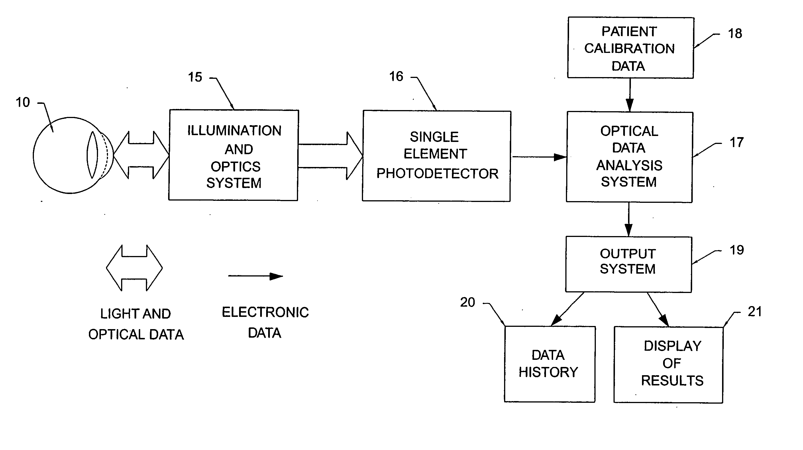
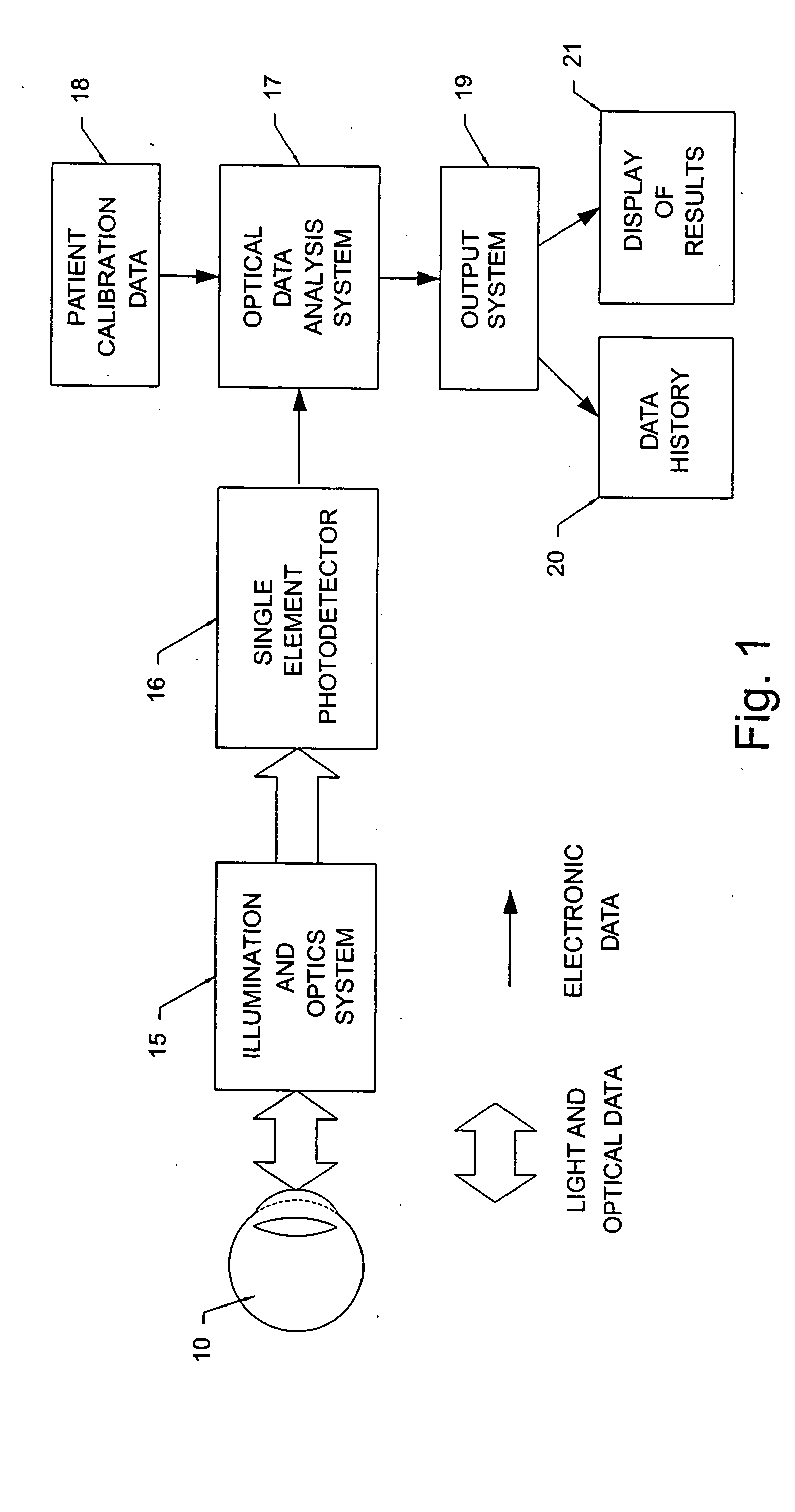
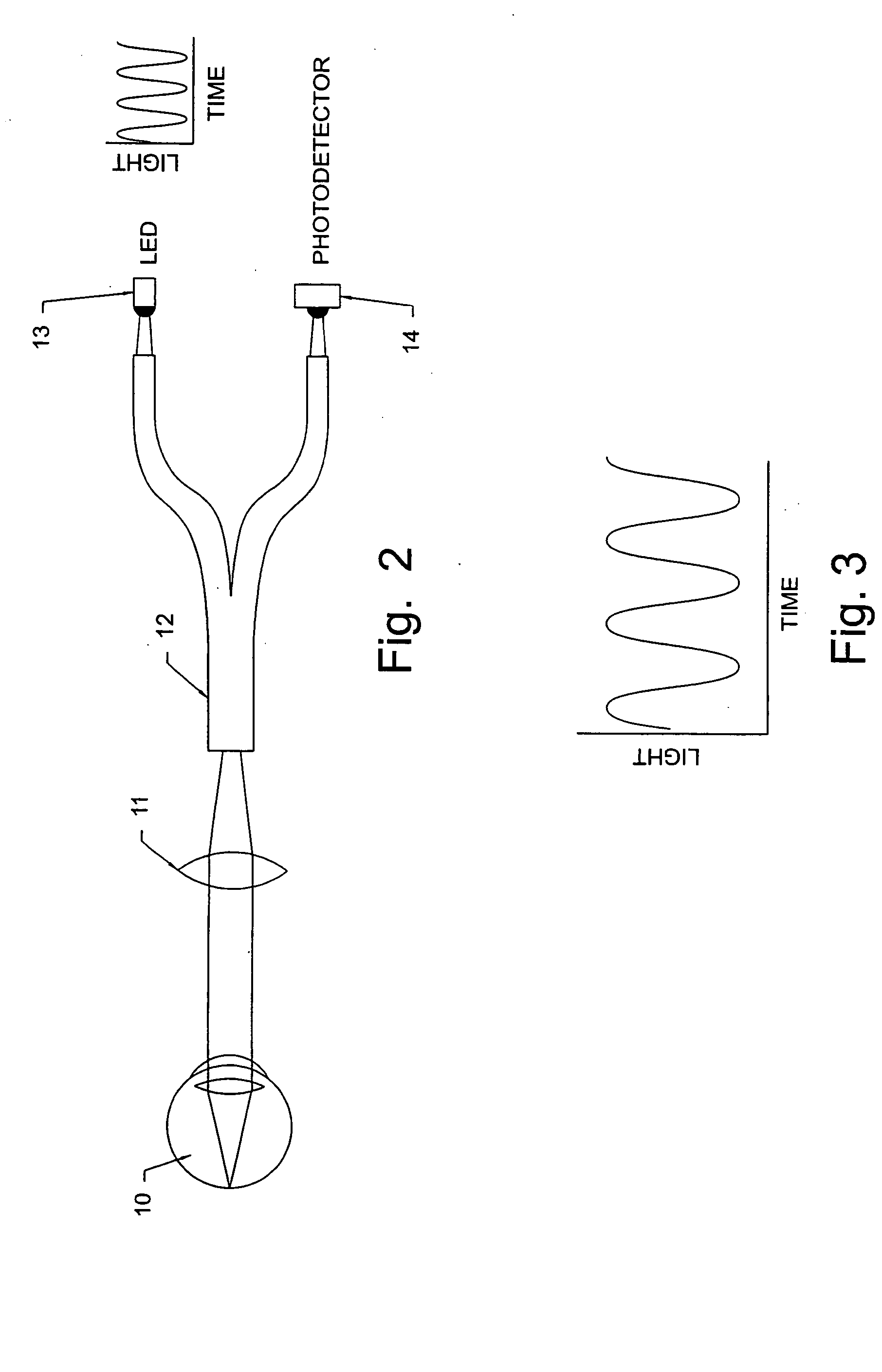
Image

Examples
Embodiment Construction
[0024] Rhodopsin is the visual pigment contained in the rods and cones of the retina. As this pigment absorbs light, it breaks down into intermediate molecular forms and initiates a signal that proceeds down a tract of nerve tissue to the brain, allowing for the sensation of sight. The outer segments of the rods and cones contain large amounts of rhodopsin, stacked in layers lying perpendicular to the light incoming through the pupil. There are two types of rhodopsin, with a slight difference between the rhodopsin in the rods (that allow for dim vision) and the rhodopsin in the cones (that allow for central and color vision). Rod rhodopsin absorbs light energy in a broad band centered at 500 nm, whereas there are three different cone rhodopsins having broad overlapping absorption bands peaking at 430, 550, and 585 nm.
[0025] Rhodopsin consists of 11-cis-retinal and the protein opsin, which is tightly bound in either the outer segment of the cones or rods. 11-cis-retinal is the photo...
PUM
 Abstract
Description
Claims
Application Information