

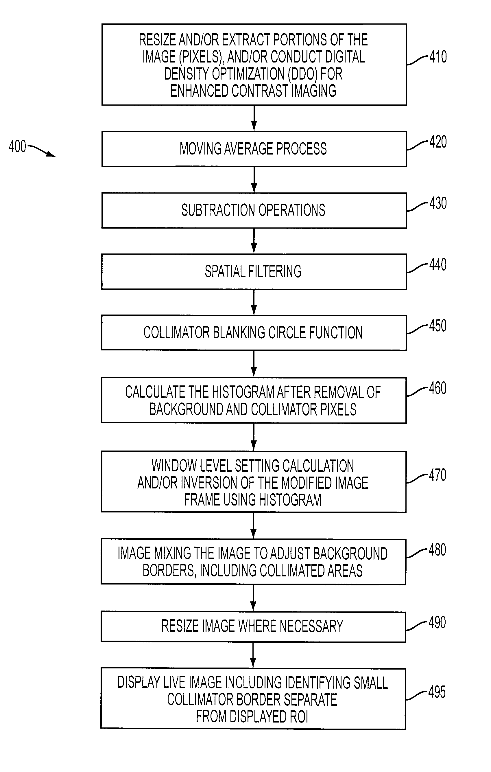
Histogram calculation for auto-windowing of collimated X-ray image
a collimated x-ray image and autowindowing technology, applied in the field of medical x-ray imaging, can solve the problems of cumbersome and time-consuming saving the settings for actual image acquisition, difficulty, and limited digital intensity values of pixels, and achieve the effect of improving image processing and improving contras
- Summary
- Abstract
- Description
- Claims
- Application Information
AI Technical Summary
Benefits of technology
Problems solved by technology
Method used
Image

Examples
Embodiment Construction
[0021]Reference will now be made in detail to embodiments of the present invention, examples of which are illustrated in the accompanying drawings, wherein like reference numerals refer to like elements throughout.
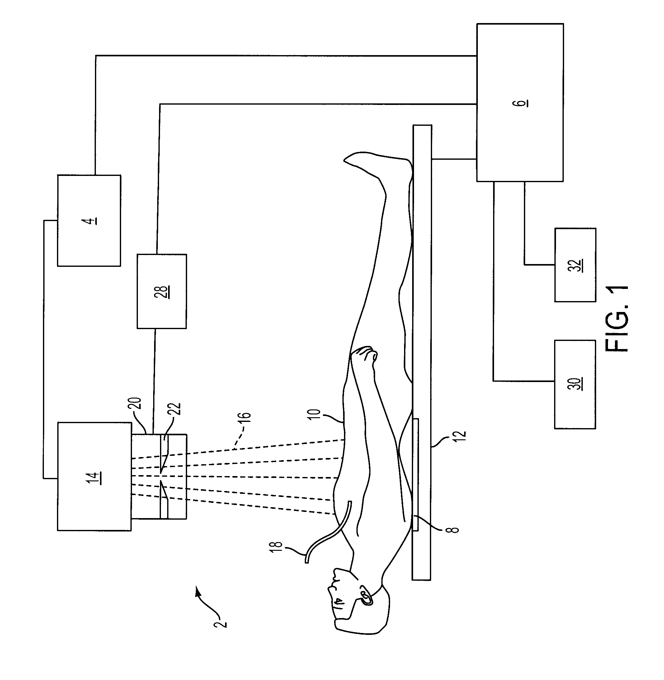
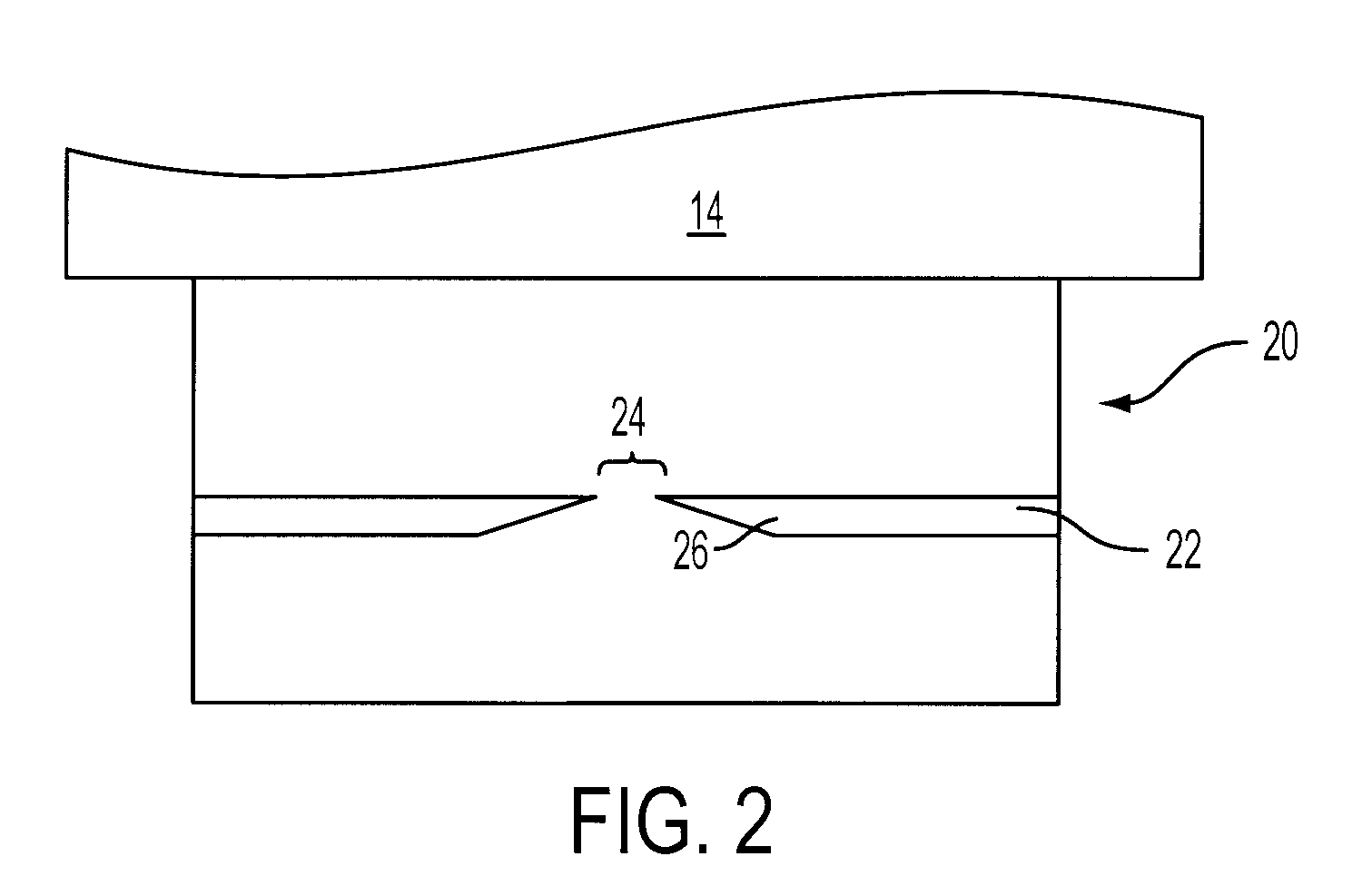
[0022]FIG. 1 represents an X-ray diagnostic imaging system 2 in accordance with an embodiment of the invention, under which is a patient 10 undergoing an X-ray fluoroscopic procedure. The X-ray system 2 is constructed to calculate a novel and improved histogram to support various post-acquisition processing such as window level setting, wherein pixels not included in the desired ROI, such as pixels located in collimated areas, and background pixels, are excluded from the histogram calculation. X-ray system 2 includes a high voltage transformer assembly 4, an X-ray source 14 with an X-ray tube, and a collimator assembly 20 including beam limiting filter plates 22. X-ray source 14 emits X-ray radiation 16 during a diagnostic or interventional procedure that is limited by the...
PUM
 Abstract
Description
Claims
Application Information