Human papilloma virus (HPV) detection using nucleic acid probes, microbeads and fluorescent-activated cell sorter (FACS)

a technology of human papilloma virus and nucleic acid probe, which is applied in the field of diagnostic and detection assays, can solve the problems of unsatisfactory assay for unequivocal determination, high labor intensity, and high cost of pap smear

Active Publication Date: 2011-03-08
INCITE HEALTH PTY LTD
View PDF31 Cites 6 Cited by
  • Summary
  • Abstract
  • Description
  • Claims
  • Application Information

AI Technical Summary

Problems solved by technology

Many of the existing tests for screening for the causes of infectious diseases are time consuming, labor intensive, expensive, often specific for only one specific pathogen and / or cannot differentiate between different strains of pathogens.
The Pap smear, however, is a somewhat unsatisfactory assay for unequivocally determining cervical cancer risk, as the technique has a false negative rate of approximately 20% and the technique cannot distinguish “high risk” and “low risk” taxon.

Method used


Image

  • Human papilloma virus (HPV) detection using nucleic acid probes, microbeads and fluorescent-activated cell sorter (FACS)
  • Human papilloma virus (HPV) detection using nucleic acid probes, microbeads and fluorescent-activated cell sorter (FACS)
  • Human papilloma virus (HPV) detection using nucleic acid probes, microbeads and fluorescent-activated cell sorter (FACS)

Examples


example 1

HPV Diagnosis—DNA Isolation and Amplification

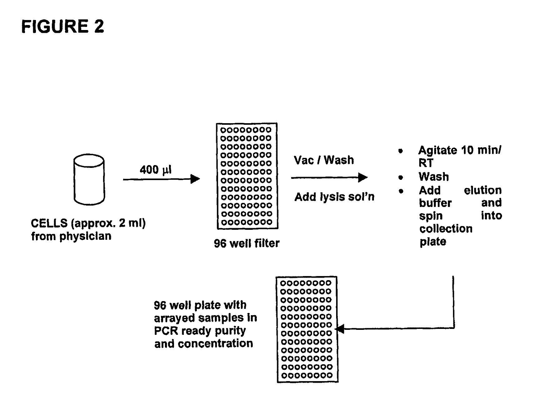
[0263]An overview of the DNA extraction protocol used to isolate DNA for the HPV diagnostic method is shown in FIG. 2.

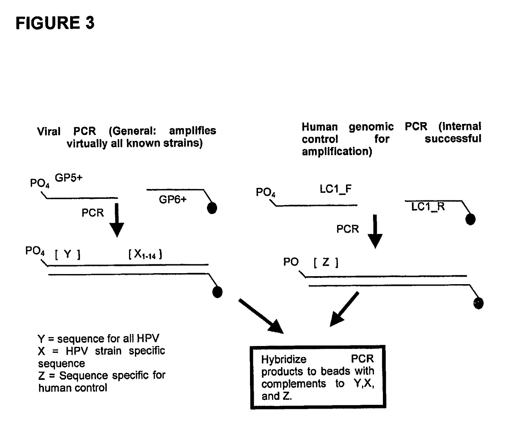
[0264]As shown in FIG. 3, PCR was used to amplify the DNA sample. The primers GP5+ and GP6+ were used to generate an amplicon for any HPV strain which was present in the DNA sample. Primer GP6+ comprised a fluorescent label, specifically Cy5 to allow later visualization of the amplicon binding to the binding agents. The viral amplicons generated comprised both a conserved region (Y) which is conserved among all the strains of HPV examined and a region which is variable (i.e. strain-specific) between HPV strains, Xn, wherein n represents a variable region associated with each HPV strain. The immobilized binding partners on beads specifically bind to the HPV strain-specific genome.

[0265]Also, an amplicon from the human subject genomic DNA was also generated using the LC1_F and LC1_R primers to serve as a control. In this cas...

example 2

HPV Diagnosis—Multiplex Detection

[0266]The amplicons generated in Example 1 were hybridized to an array of binding agents, each carrying a polynucleotide which is complementary to the variable region of the putative viral amplicon generated from each of HPV strains c, 11, 16, 18, 31, 33, 35, 42, 45, 51, 52, 56, 58, 59, 67 and 68 (X1 through X16). See FIG. 9 for the nucleotide sequences of the capture nucleic acids immobilized to the beads. Furthermore, the array comprises a binding agent which comprises a polynucleotide which is complementary to conserved region of the HPV viral amplicons (Y). Finally, a binding agent comprising a polynucleotide which is complementary to the sequence of the human control amplicon is included. The capture nucleic acid may be DNA or RNA. If RNA is used, a reverse transcriptase may be required to generate RNA from the DNA amplicon.

[0267]Each of the binding agents in the array is comprises a microsphere or bead with a distinct size and distinct intensit...

example 3

Comparison of the Multiplex Detection Method with Traditional HPV Diagnosis

[0271]Table 5, below, provides an overview comparing the multiplex HPV detection method of the present invention with the current histological method for HPV diagnosis.

[0272]

TABLE 5Comparison of HPV diagnostic methodsHPV DiagnosticMethodThroughputReportControlsPresent1600 per day,all 13 “high risk”internal control, positiveInventionper instrumentstrains individuallycontrol, human gDNA controlidentifiedHistological350 per day“high risk” classno internal control, “low risk”based methodgenerally identifiedpositive control, “high reiskpositive control


PUM

PropertyMeasurementUnit
bead sizesaaaaaaaaaa

Abstract

The present invention relates generally to the field of diagnostic and detection assays. More particularly, the present invention provides methods, and reagents including biochips for detecting the presence of, or distinguishing between, one or more analytes in a sample.

Description

[0001]This application is U.S. National Phase of International Application PCT / AU2005 / 001865, filed Dec. 9, 2005 designating the U.S., and published in English as WO 2006 / 060872 on Jun. 15, 2006, which claims priority to Australian Patent Application No. 2004907070 filed Dec. 10, 2004 and U.S. provisional application 60 / 704,974 filed Aug. 3, 2005.BACKGROUND OF THE INVENTION[0002]1. Field of the Invention[0003]The present invention relates generally to the field of diagnostic and detection assays. More particularly, the present invention provides methods, and reagents including biochips for detecting the presence of, or distinguishing between, one or more analytes in a sample.[0004]2. Description of the Prior Art[0005]Bibliographical details of references provided in the subject specification are listed at the end of the specification.[0006]Reference to any prior art in this specification is not, and should not be taken as, an acknowledgement or any form of suggestion that this prior...

Claims


Application Information

Patent Timeline
Patent Type & Authority Patents(United States)
IPC IPC(8): C12Q1/68
CPCC12Q1/708
Inventor POETTER, KARLGOULD, TOBY
Owner INCITE HEALTH PTY LTD
Why Eureka
Try Eureka