Bracket for mounting at least one position detecting sensor on an ultrasonic probe

a technology for ultrasonic probes and brackets, which is applied in the field of brackets for mounting at least one position detecting sensor on an ultrasonic probes, can solve the problems of not being suitable for retrofitting existing ultrasonic probes, increasing the cost and complexity of the probe, and not being desirabl

Active Publication Date: 2011-04-19
CIVCO MEDICAL INSTR CO
View PDF22 Cites 21 Cited by
  • Summary
  • Abstract
  • Description
  • Claims
  • Application Information

AI Technical Summary

Benefits of technology

[0012]In accordance with one aspect of this invention there is provided a bracket for use with an ultrasound transducer to releasably mount at least one sensor of a location / tracking system on the transducer. The at least one sensor has a body including an opposed pair of wall portions. The bracket comprises a member arranged to be releasably mounted on the transducer and includes at least one socket therein. The at least one socket has a pair of spaced-apart wall portions. The at least one socket and the at least one sensor are arranged to be coupled together via cooperating pins and slots to enable the at least one sensor to be oriented in a first position for insertion between the spaced-apart walls of the at least one socket to introduce said pins into respective ones of said slots so that the at least one sensor may be rotated to a second or locked position within the at least one socket. When the sensor is in the locked position, the wall portions of the at least one socket releasably engage, e.g., snap-fit, about a portion of the sensor to deter accidental displacement of the sensor with respect to the transducer.
[0014]In accordance with another aspect of this invention there is provided the combination of a bracket and at least one sensor of a location / tracking system. The bracket and sensor are arranged for enabling the location of an ultrasound transducer to be determined. The sensor forms a part of a location / tracking system. The bracket comprises a member arranged to be releasably mounted on the transducer and includes at least one socket in it. The at least one socket has a pair of spaced-apart wall portions. The at least one socket and the at least one sensor are arranged to be coupled together via cooperating pins and slots to enable the at least one sensor to be oriented in a first position for insertion between the spaced-apart walls of the at least one socket to introduce said pins into respective ones of said slots so that the at least one sensor may be rotated to a second or locked position within the at least one socket. When the at least one sensor is in the locked position, the wall portions of the at least one socket releasably engage, e.g., snap-fit, about a portion of the at least one sensor to deter accidental displacement of the at least one sensor with respect to the transducer.

Problems solved by technology

However, this approach may not be desirable if the probe is also intended to be used in applications wherein its position need not be determined, since the inclusion of such sensor(s) permanently on or in the probe will likely increase the cost and complexity of the probe.
Moreover, such an approach may not be suitable to retrofit existing ultrasonic probes.
While such brackets are suitable for their intended purposes, they are not designed for mounting one or more position sensors thereon.

Method used


Image

  • Bracket for mounting at least one position detecting sensor on an ultrasonic probe
  • Bracket for mounting at least one position detecting sensor on an ultrasonic probe
  • Bracket for mounting at least one position detecting sensor on an ultrasonic probe

Examples


second embodiment

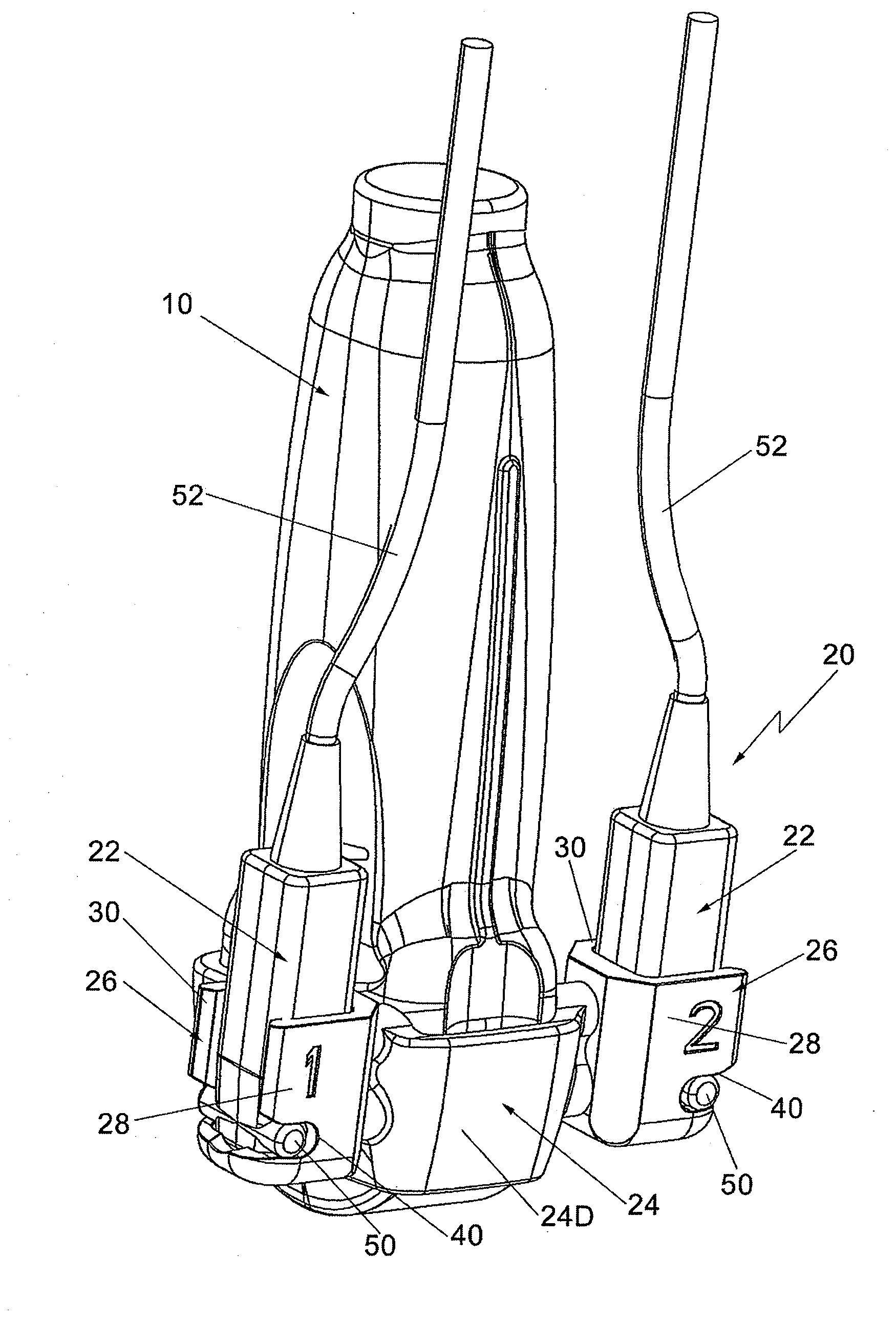
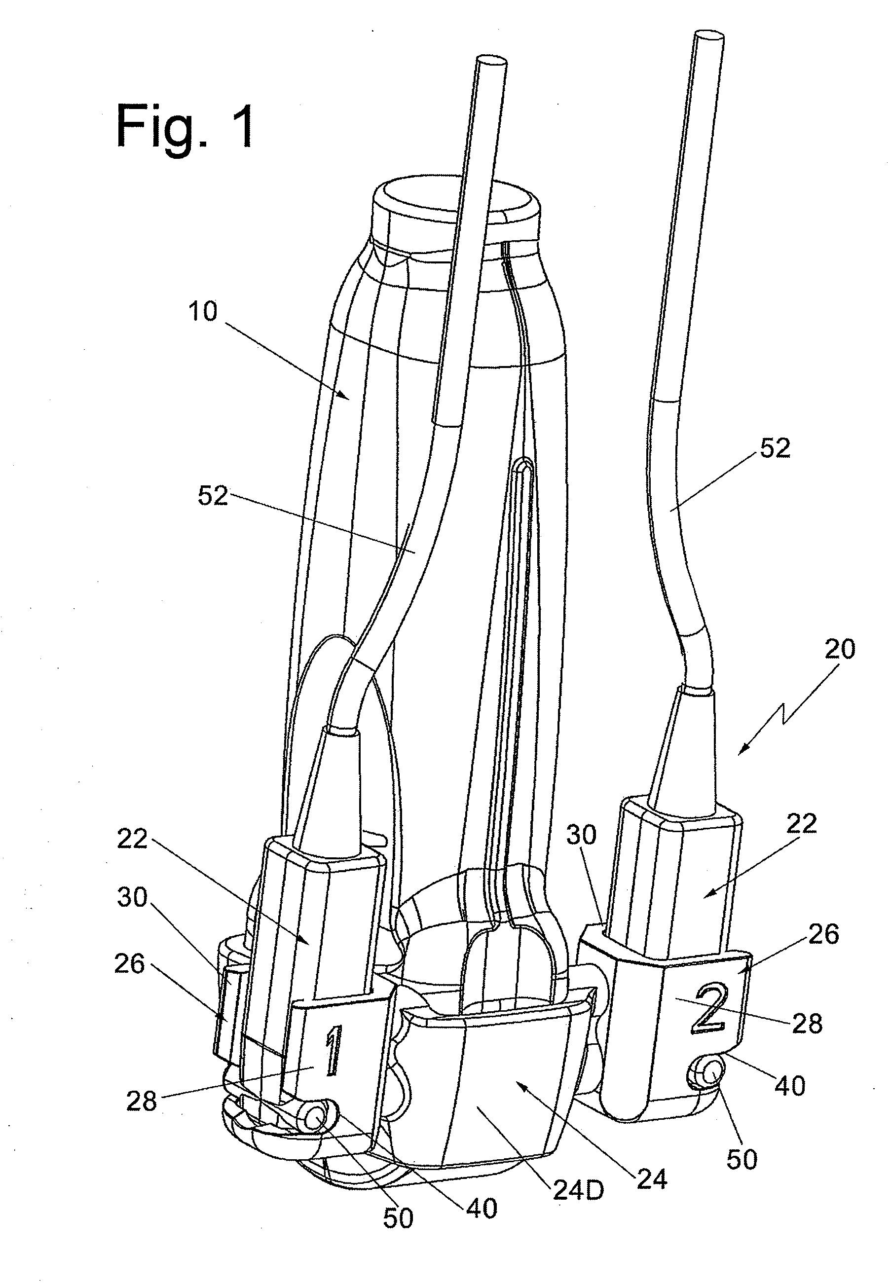
[0033]In FIG. 5 there is shown a bracket 20′ constructed in accordance with this invention. The bracket 20′ is identical in construction to bracket 20 except that it includes a conventional biopsy needle guide assembly 100 mounted on the rear wall 24D. The needle guide can be of any desired construction. In this embodiment the needle guide is a fixed angle needle guide, i.e., a guide wherein the angle that the biopsy needle is introduced into the patient's body via the transducer is a fixed, predetermined angle.

third embodiment

[0034]In FIG. 6 there is shown a bracket 20″ constructed in accordance with this invention. The bracket 20″ is identical in construction to bracket 20′ except that it includes an adjustable angle biopsy needle guide assembly 200 mounted on the rear wall 24D. The adjustable needle guide assembly 200 is preferably constructed in accordance with the teachings of my aforementioned U.S. Pat. No. 5,941,889, which as mentioned above is incorporated by reference herein, so that the biopsy needle can be introduced into the patient's body from any one of several predetermined angles. Other adjustable needle guides can be used in lieu of that adjustable needle guide.

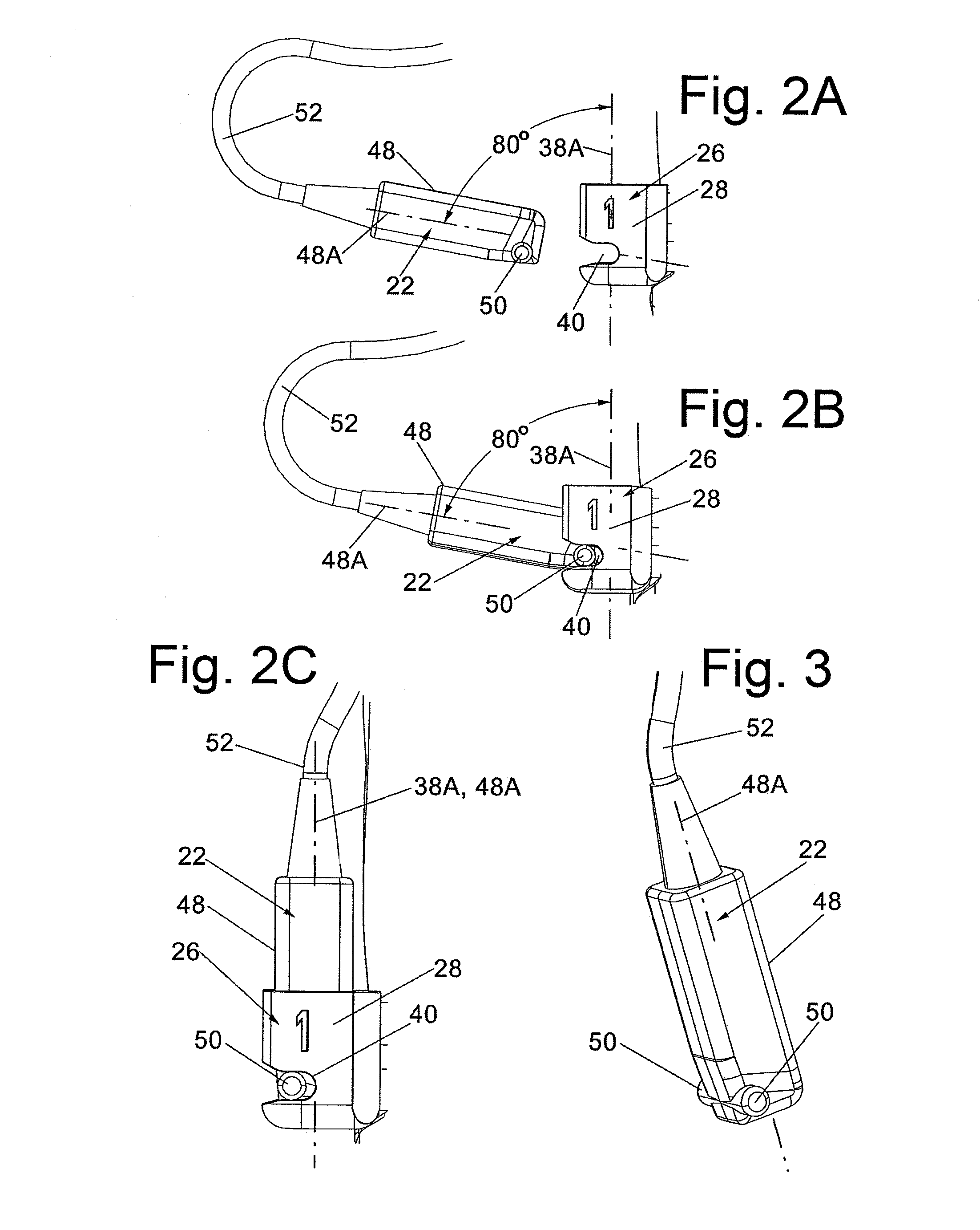
[0035]It should be pointed out at this juncture, that while the exemplary preferred embodiments disclosed above make use of the projecting pins being located on the sensor, it is contemplated that the pins form a portion of the sockets, e.g., extend inward into the cavity 38 from respective sidewalls 28 and 30, in which case the bo...


PUM


Abstract

A bracket for use on an ultrasound transducer to releasably mount at least one 3D tracking sensor on the transducer. Each sensor has a pair of axially aligned pins projecting outward from its body. The bracket includes at least one socket having a pair of spaced-apart wall portions, each of which includes a slot arranged to receive a respective one of the pins of the sensor when the sensor is in a first position. From this position the sensor's body can be rotated to a second position, whereupon the spaced apart wall portions releasably engage (snap-fit about) a portion of the body of the sensor to deter the accidental displacement of the sensor with respect to the transducer.

Description

CROSS-REFERENCE TO RELATED APPLICATIONS[0001]“Not Applicable”STATEMENT REGARDING FEDERALLY SPONSORED RESEARCH OR DEVELOPMENT[0002]“Not Applicable”INCORPORATION-BY-REFERENCE OF MATERIAL SUBMITTED ON A COMPACT DISK[0003]“Not Applicable”FIELD OF THE INVENTION[0004]This invention relates generally to medical devices and more particularly to devices for releasably mounting at least one sensor of a 3D tracking system on a ultrasound transducer or other hand-held medical instrument.BACKGROUND OF THE INVENTION[0005]Ultrasound has received widespread acceptance as a useful diagnostic tool by providing an image of the internal area of inquiry by emission of very high frequency sound waves from a transducer (commonly called a “probe”) placed in contact with the patient's skin adjacent that area of inquiry. Repeated arrays of ultrasonic beams scan that area and are reflected back to the transducer, where the beams are received and the data transmitted to a processing device. A processing unit, ...

Claims


Application Information

Patent Timeline
Patent Type & Authority Patents(United States)
IPC IPC(8): A47K1/08
CPCA61B8/4245A61B8/4281A61B8/4254A61B2017/00477
Inventor CERMAK, CRAIG J.
Owner CIVCO MEDICAL INSTR CO
Why Eureka
Try Eureka