Registering intra-operative image data sets with pre-operative 3D image data sets on the basis of optical surface extraction

a technology of optical surface extraction and image data, applied in the field of infraoperative registration of intra-operative image data sets with pre-operative 3d image data sets, can solve the problems of inability to obtain, extremely high cost of providing a modality combination, and localization of tumors, so as to improve the registration of intra-operative fluoroscopic images.

Inactive Publication Date: 2010-02-16
SIEMENS HEALTHCARE GMBH
View PDF5 Cites 24 Cited by
  • Summary
  • Abstract
  • Description
  • Claims
  • Application Information

AI Technical Summary

Benefits of technology

[0015]The object of the present invention is to further improve the registration of intra-operative fluoroscopic or functional image data with pre-operative morphological 3D image data.
[0028]With periodic detection in particular, in accordance with the invention, on the basis of the measurement results of the optical 3D sensor system, a movement field of the area under examination can be computed which is used to reduce movement artifacts in intra-operative recording or to improve their resolution.

Problems solved by technology

1. Intra-operative image data sets which are obtained as so-called real-time images during an intervention, usually with the aid of fluoroscopic imaging, are primarily used for navigation of surgical instruments (e.g. in the head or the heart). These types of fluoroscopic 2D images (recorded with a C-arm for example) are rapidly available, the imaging technology minimizes the radiation dosage for patient and doctor. Interventional equipment (operation instruments, catheters, guide wires etc.) are shown as high-resolution images almost in real time. However such fluoroscopy images—compared to images of 3D imaging modalities (e.g. CT, MRT, 3D angio)—do not show any spatial details. In the prior art the spatial information has been recovered by registering 3D images (for example from a CT, 3D angio or MR tomography) recorded pre-operatively with two-dimensional intra-operative radiological images and underlaid with these images, a process which is also referred to as co-registration. With this type of registration the direction from which a 3D volume must be projected so that it can be brought into alignment with the intra-operative 2D image must be determined.
2. Intra-operative image data sets can also be functional nuclear-medical 3D image data (e.g. PET or SPECT images) which supply a description of metabolic functions or processes of the body. One of the uses of such image data is to visualize anatomical regions which exhibit an abnormal metabolism, such as tumors for example, and to determine their size and activity. However these functional 3D images provide little information about the patient's anatomy in which the pathogenic region (e.g. the tumor) is embedded, which is a problem when such functional image data is used exclusively in respect of diagnostics, therapy planning and therapy, especially since the spatial resolution of this image data is greatly limited. It is therefore difficult to localize a tumor exactly solely using nuclear-medical 3D image data (SPECT, PET), since the exact spatial relationship to the patient anatomy is missing from this data. However this spatial relationship can also be re-established by registration (and fusion) of the functional 3D image data with high-resolution morphological 3D image data.
The problem from the medical technology standpoint—with 2D radiological images just as with 3D images—lies in the registration with the high-resolution morphological 3D images.
Such a software-based registration only operates reliably if enough common image information is available as regards functional and morphological image data, which is not always the case.
However the costs of providing a modality combination are extremely high and in addition it is not currently possible to obtain combinations which are more subject to failure but are still sensible and desired (e.g. PET+MRT, SPECT+MRT, ultrasound doppler+MRT, MEG+MRT etc.).
The disadvantage of the existing method is on the one hand a certain imprecision which is not to be tolerated and on the other hand the high time overhead sometimes involved in these methods.
Exact and fast registration, especially of functional with morphological 3D image data, turns out to be extremely difficult in practice however since only a small number of common factors exist between these two complementary image data categories.
However the scan duration of a laser scan is specified as around 30 seconds which makes the method susceptible to movement artifacts and it is unsuitable in particular for series of functional recordings or rapid instrument tracking.

Method used


Image

  • Registering intra-operative image data sets with pre-operative 3D image data sets on the basis of optical surface extraction
  • Registering intra-operative image data sets with pre-operative 3D image data sets on the basis of optical surface extraction
  • Registering intra-operative image data sets with pre-operative 3D image data sets on the basis of optical surface extraction

Examples


Embodiment Construction

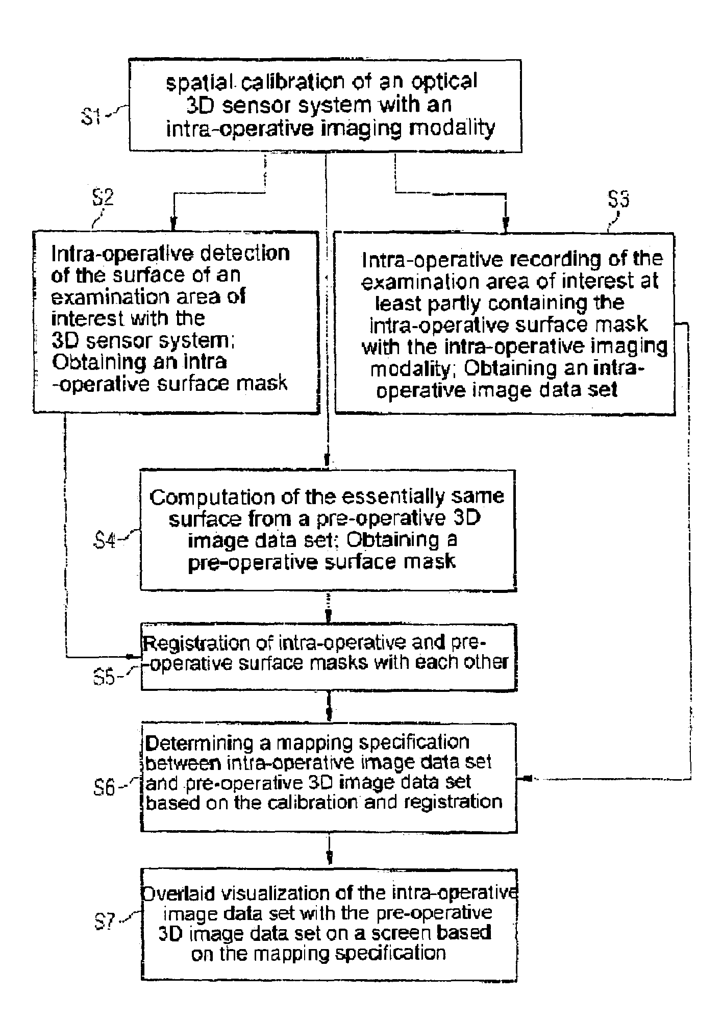
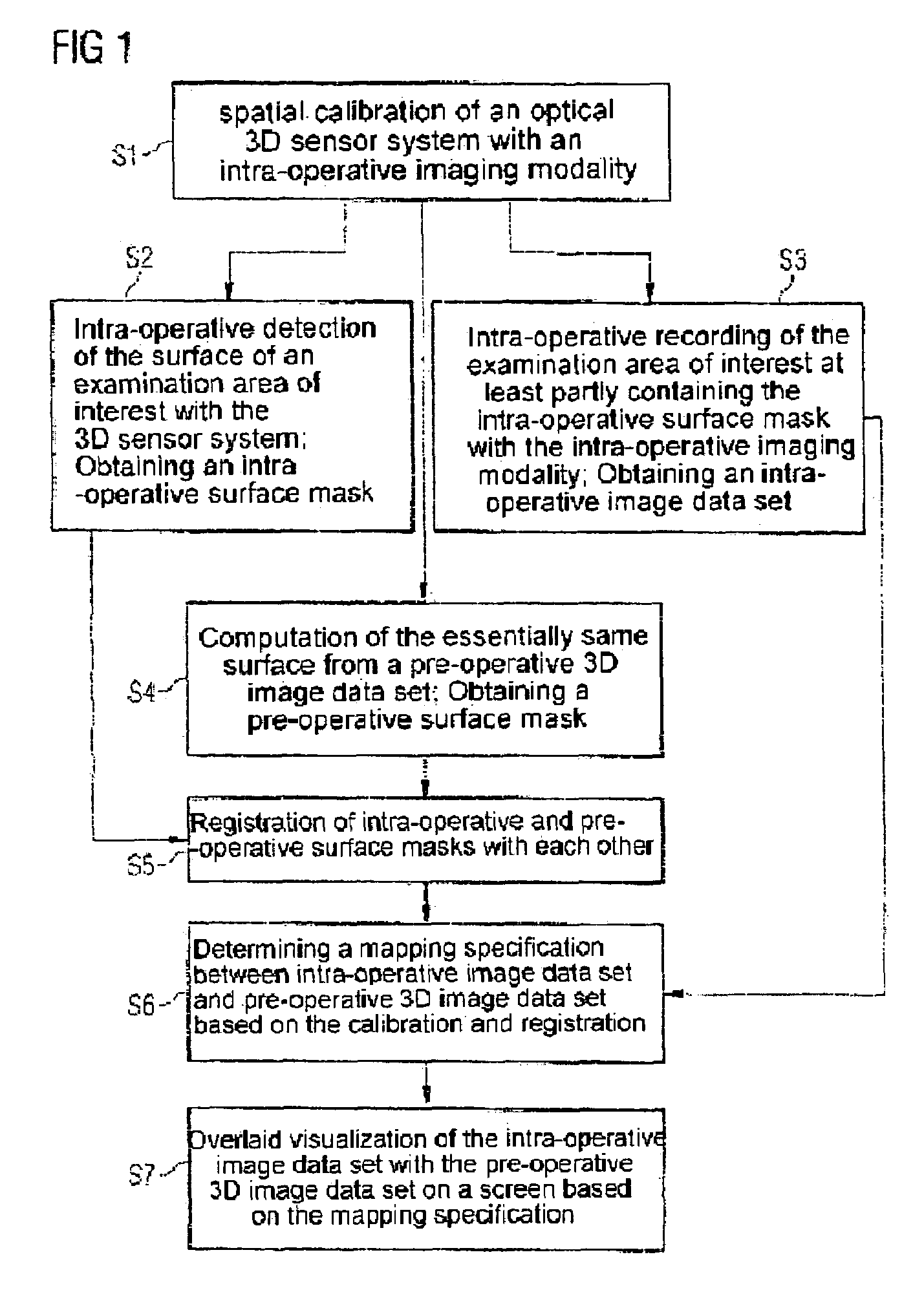
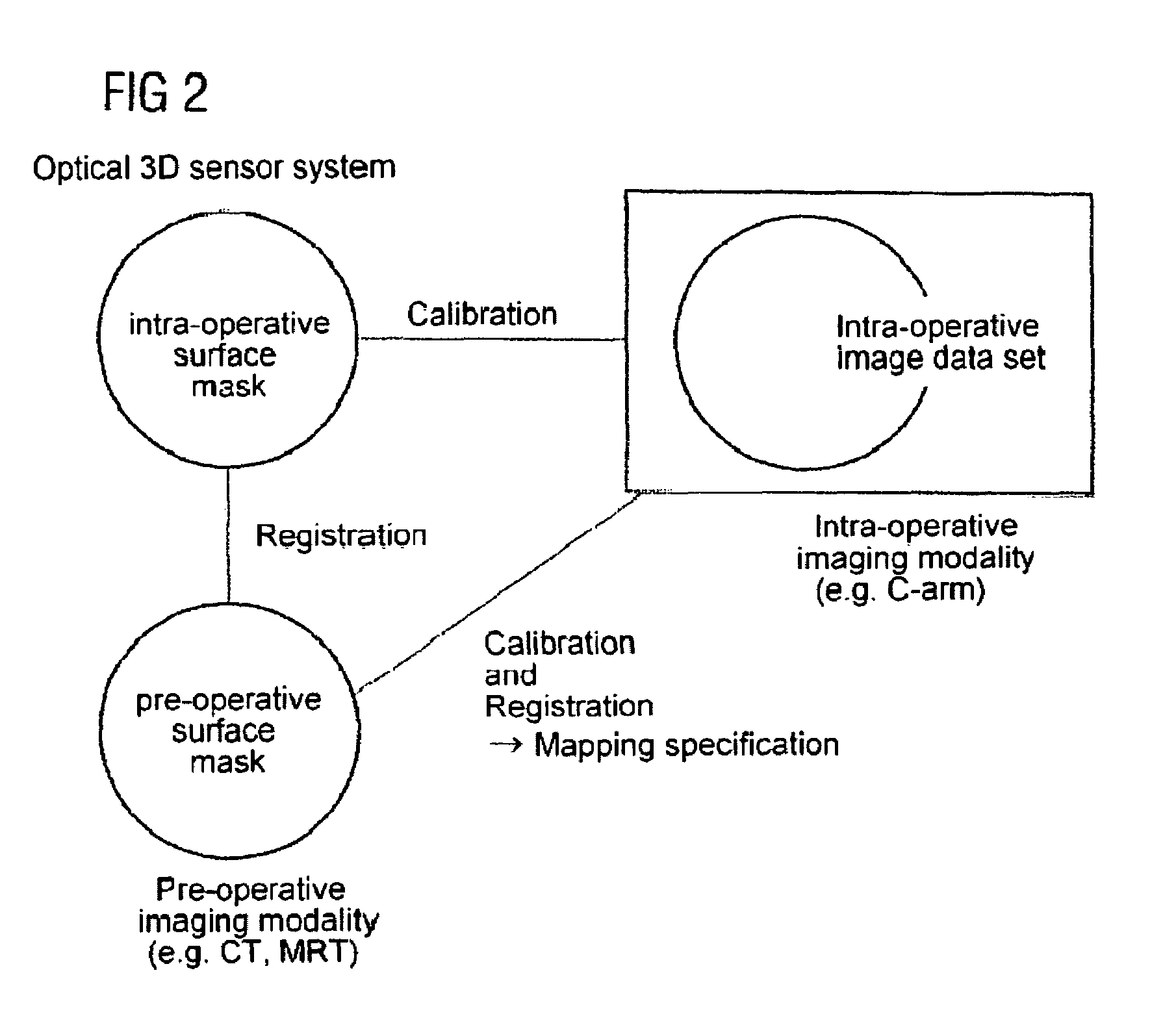
[0036]The present invention describes a method in which optical surface recognition is used to enable the position of the patient to be recorded precisely, rapidly and securely and thus an intra-operative 2D or 3D image data set can be quickly and thereby intra-operatively fused with a pre-operative 3D image data set.

[0037]Optical surface recognition is part of the prior art and is offered commercially.

[0038]The basis of this technology is an optical 3D sensor system which processes specific images of one or more 3D sensors in a suitable manner. These 3D sensors observe an object to be measured from the side. For measurement the surface of the measurement object is illuminated in accordance with a patentable method by means of white light projector with a pattern of stripes. From the displacement of the stripes produced in this observation from the side the surface form of the object is computed and for example is stored as a three-corner model for subsequent access. The small measu...


PUM


Abstract

The invention relates to a method for registering intra-operative image data set with pre-operative 3D image data set, including:spatially calibrating an optical 3D sensor system with an intra-operative imaging modality,intra-operatively detecting the surface of an examination area of interest with the 3D sensor system to produce an intra-operative surface mask,intra-operatively recording the area of interest for examination with the intra-operative modality at least partly containing the intra-operative surface mask to obtain an intra-operative image data set,computing the same surface from the pre-operative 3D image data set containing the detected surface to obtain a pre-operative surface mask,registering the intra-operative and pre-operative surface mask with each other,determining a mapping specification between pre-operative 3D image data set and intra-operative image data set based on the calibration and the registration, andoverlaying the intra-operative image data set with the pre-operative 3D data set based on the mapping specification.

Description

CROSS REFERENCE TO RELATED APPLICATIONS[0001]This application claims priority of German application No. 10 2005 036 322.9 filed Jul. 29, 2005, which is incorporated by reference herein in its entirety.FIELD OF THE INVENTION[0002]The present invention relates to a method for infra-operative registration of intra-operative image data sets with pre-operative 3D image data sets. In this regard the present invention relates especially to a method which is based on rapid optical surface recognition.BACKGROUND OF THE INVENTION[0003]In medical imaging a (frequent) problem which occurs is that of having to rapidly yet still precisely register intra-operative image data sets with pre-operative 3D image data sets; there being various reasons for this:[0004]1. Intra-operative image data sets which are obtained as so-called real-time images during an intervention, usually with the aid of fluoroscopic imaging, are primarily used for navigation of surgical instruments (e.g. in the head or the hear...

Claims


Application Information

Patent Timeline
Patent Type & Authority Patents(United States)
IPC IPC(8): A61B5/05A61B6/00
CPCG06T7/0038G06T7/0051G06T7/0065G06T2207/30004G06T7/38G06T7/50G06T7/55
Inventor BOESE, JANKLEEN, MARTINKLINGENBECK-REGN, KLAUSPFISTER, MARCUSRAHN, NORBERT
Owner SIEMENS HEALTHCARE GMBH
Why Eureka
Try Eureka