

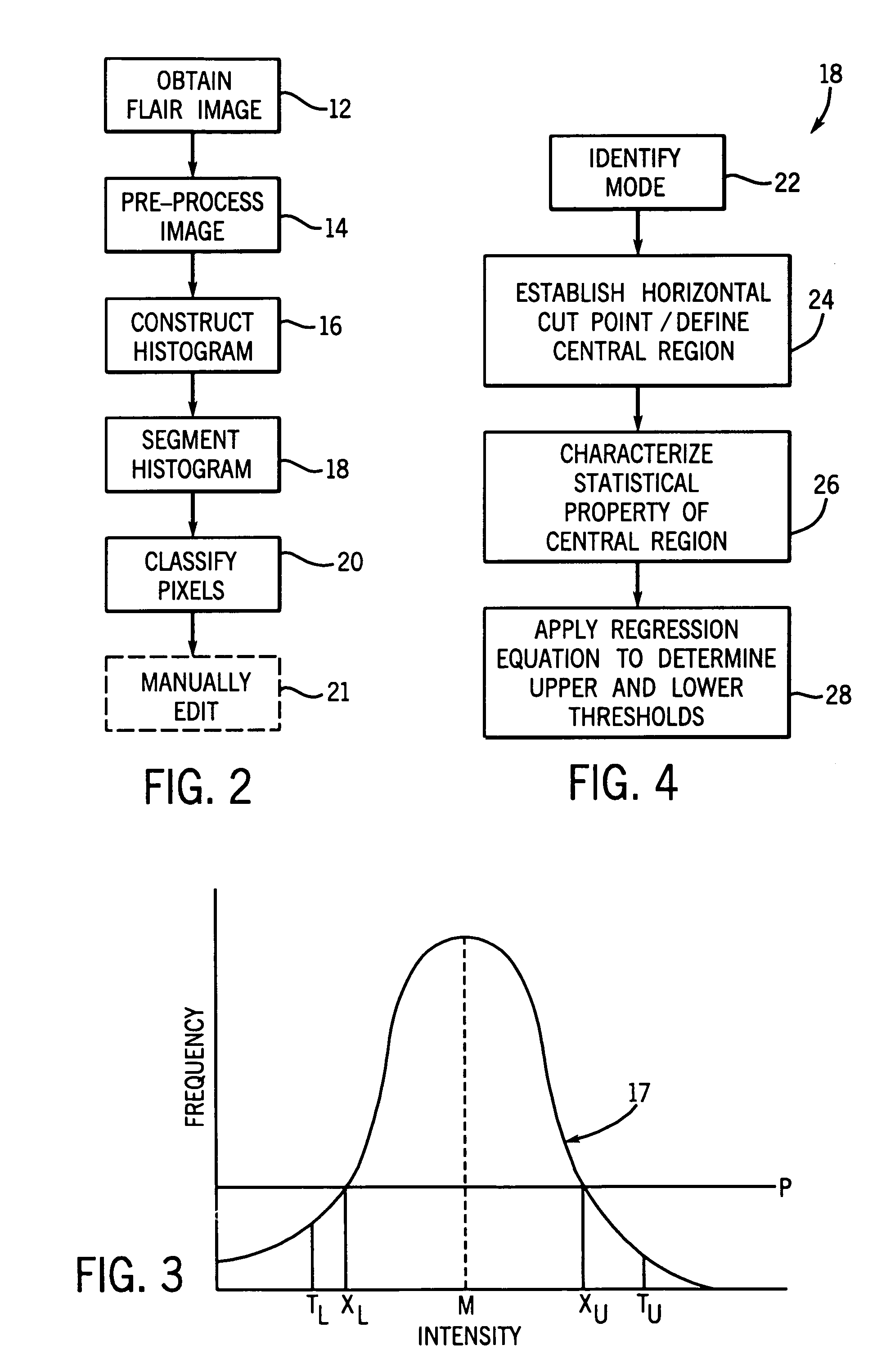
Histogram segmentation of FLAIR images
a technology of flair image and segmentation image, which is applied in the field of classifying and quantifying leukoaraiosis in the mri image of the brain, can solve the problems of inability to provide reproducible quantitative, difficult to accurately quantify leukoaraiosis tissue, and inability to automate methods
- Summary
- Abstract
- Description
- Claims
- Application Information
AI Technical Summary
Problems solved by technology
Method used
Image

Examples
Embodiment Construction
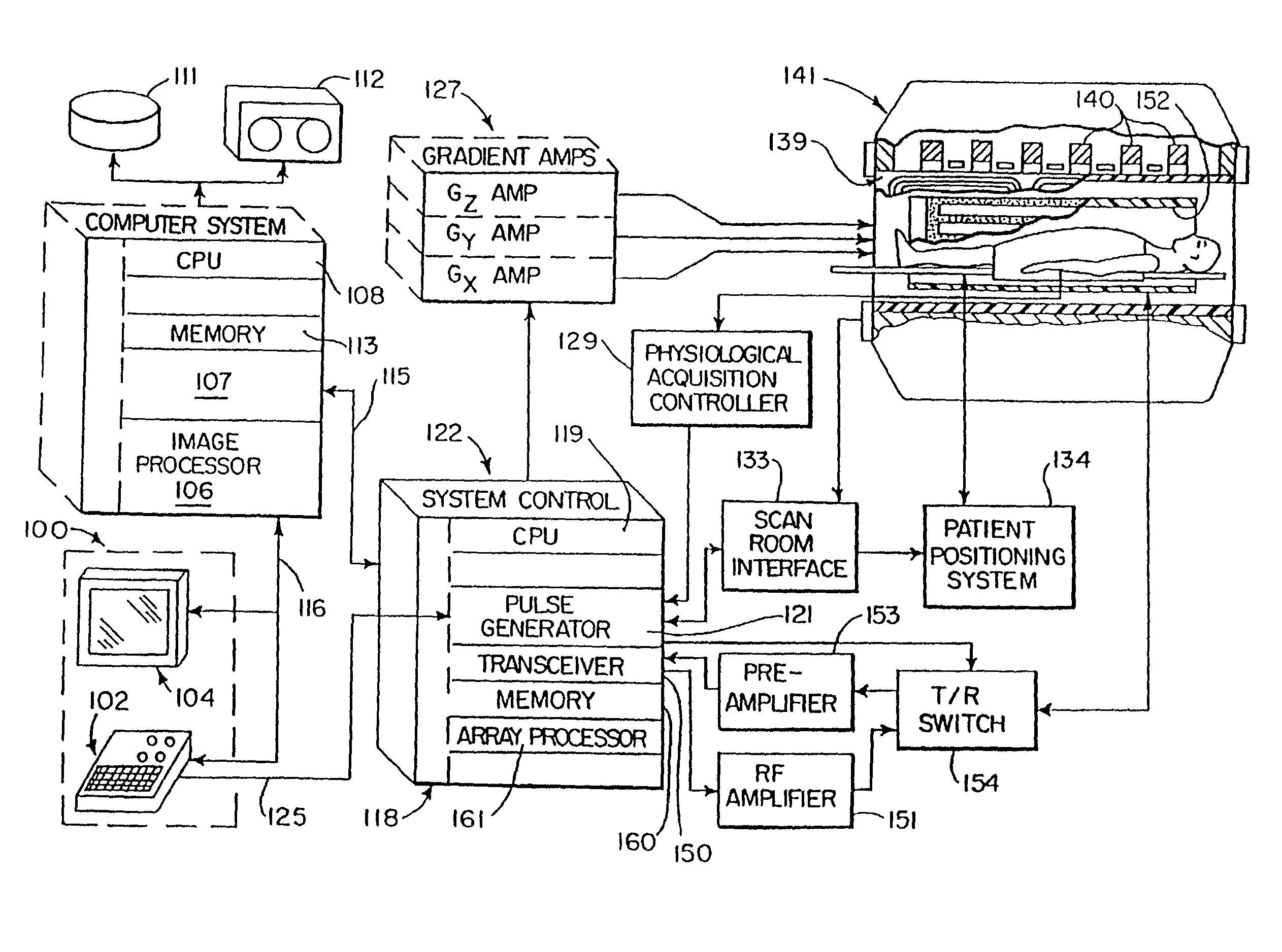
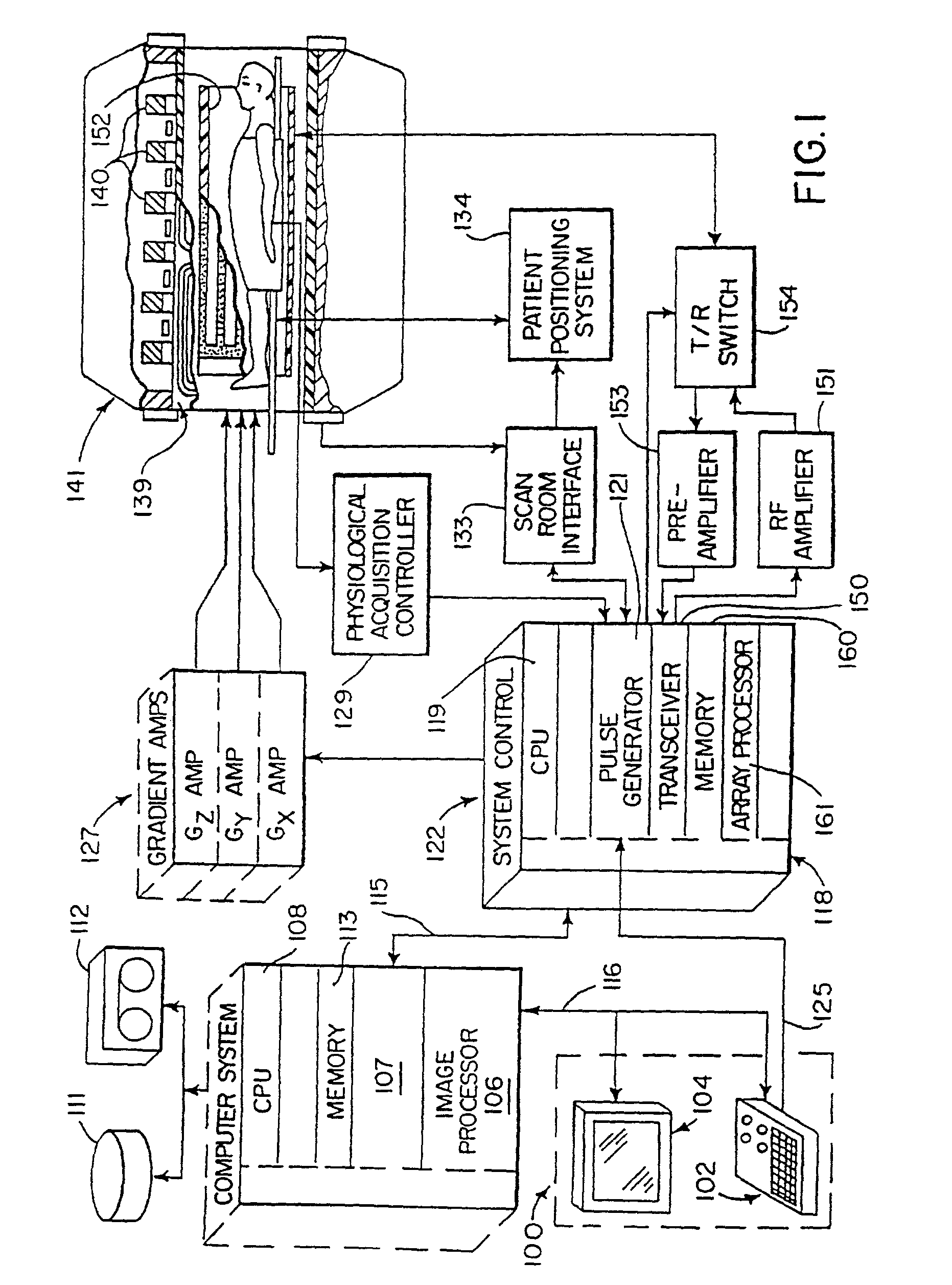
[0023]Referring first to FIG. 1, there is shown the major components of a preferred MRI system which incorporates the present invention. The operation of the system is controlled from an operator console 100 which includes a keyboard and control panel 102 and a display 104. The console 100 communicates through a link 116 with a separate computer system 107 that enables an operator to control the production and display of images on the screen 104. The computer system 107 includes a number of modules which communicate with each other through a backplane. These include an image processor module 106, a CPU module 108 and a memory module 113, known in the art as a frame buffer for storing image data arrays. The computer system 107 is linked to a disk storage 111 and a tape drive 112 for storage of image data and programs, and it communicates with a separate system control 122 through a high speed serial link 115.
[0024]The system control 122 includes a set of modules connected together by...
PUM
 Abstract
Description
Claims
Application Information