Assay for detecting tuberculosis in nonhuman primates

a technology for detecting tuberculosis and nonhuman primates, which is applied in the field of methods and compositions for detecting the presence of mycobacterial organisms, can solve the problems of limited methods, insensitive, inconvenient use, and significant problems for nonhuman primate populations and humans, and achieves the effect of convenient use and sensitiveness

Active Publication Date: 2012-03-06
CHEMBIO DIAGNOSTIC SYST +1
View PDF2 Cites 1 Cited by
  • Summary
  • Abstract
  • Description
  • Claims
  • Application Information

AI Technical Summary

Benefits of technology

[0014]The currently available methods for detecting TB in nonhuman primates, TST, are limited, for example, in that they are impractical, nonspecific, and insensitive. Nonspecific and insensitive nature of currently available tests leads to numerous false positives and false negatives, creating a significant problem for nonhuman primate populations and humans that come into contact with those populations. The present invention alleviates these problems by providing a diagnostic method for detecting TB in nonhuman primates that is advantageous, for example, in that it is easy to use, sensitive, and specific.
[0015]In contrast to TST, which requires intradermal injection of a crude assortment of mycobacterial antigens, the present invention requires only that one remove a small amount of a test sample from a test animal. Additionally, instead of a crude antigen mixture, the present invention utilizes defined mixtures of recombinant mycobacterial antigens. By utilizing these carefully selected mixtures of antigens, the present invention overcomes animal-to-animal variations in antigen recognition patterns among nonhuman primates. Additionally, the use of specific antigenic mixtures according to the present invention enables the production of a highly sensitive nonhuman primate test for TB. To achieve even greater specificity, various embodiments of the present invention utilize defined antigen mixtures that comprise polyfusion proteins.

Problems solved by technology

The currently available methods for detecting TB in nonhuman primates, TST, are limited, for example, in that they are impractical, nonspecific, and insensitive.
Nonspecific and insensitive nature of currently available tests leads to numerous false positives and false negatives, creating a significant problem for nonhuman primate populations and humans that come into contact with those populations.

Method used


Image

  • Assay for detecting tuberculosis in nonhuman primates
  • Assay for detecting tuberculosis in nonhuman primates
  • Assay for detecting tuberculosis in nonhuman primates

Examples


example 1

Selection of Antigens

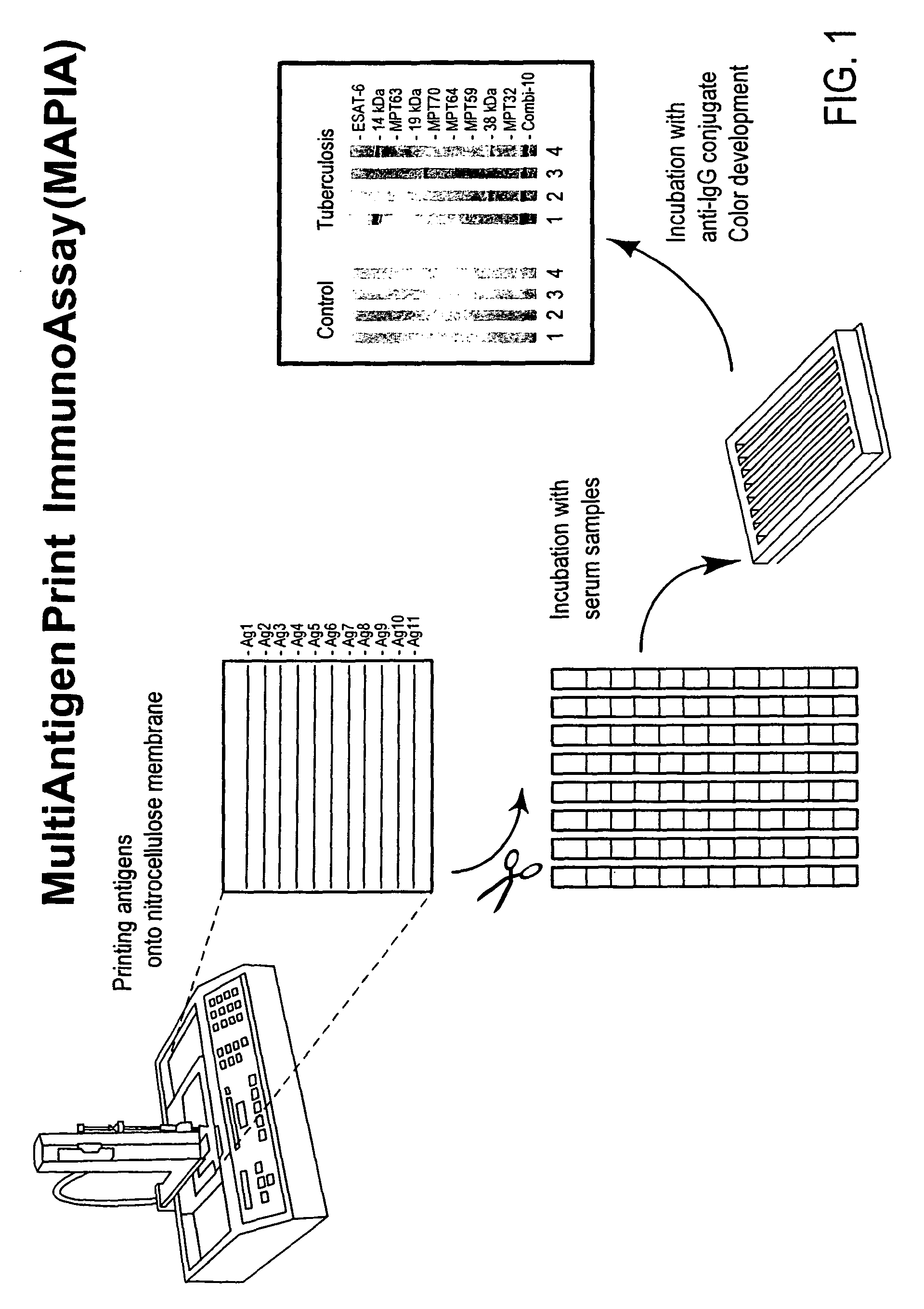
[0073]Mycobacterial antigens of the present invention were selected using Multi-Antigen Print ImmunoAssay (MAPIA) as described in Lyashchenko et al., 2000. Briefly, antigens were immobilized on a nitrocellulose membrane (Schleicher & Schuell BioScience, Inc. USA, Keene, N.H.) at a protein concentration of 0.05 mg / ml by using a semiautomated airbrush printing device (Linomat IV; Camag Scientific, Inc., Wilmington, Del.). The membrane was cut perpendicular to the antigen bands into 4-mm-wide strips. The strips were blocked for 1 h with 1% nonfat skim milk in PBS, pH 7.2, with 0.05% Tween 20 and then incubated for 1 h with serum samples diluted 1:50 in blocking solution. After being washed, the strips were incubated for 1 h with alkaline phosphatase-conjugated anti-human immunoglobulin G antibody (Sigma Chemical Co., St. Louis, Mo.) diluted 1:5,000 in blocking solution, followed by another washing step. Bound antibodies were visualized with 5-bromo-4-chloro-3-indol...

example 2

Serological Responses in Monkey Tuberculosis

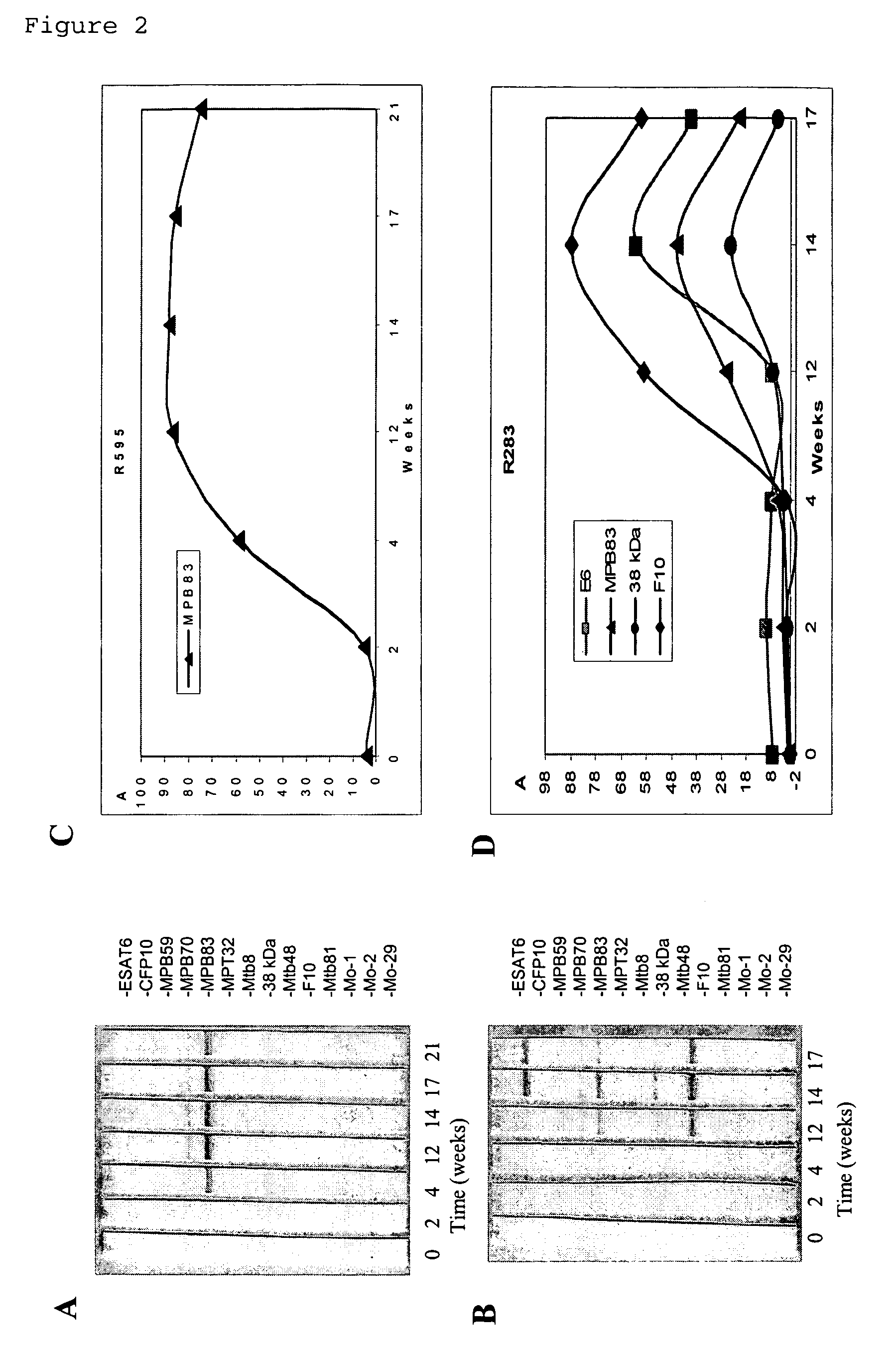
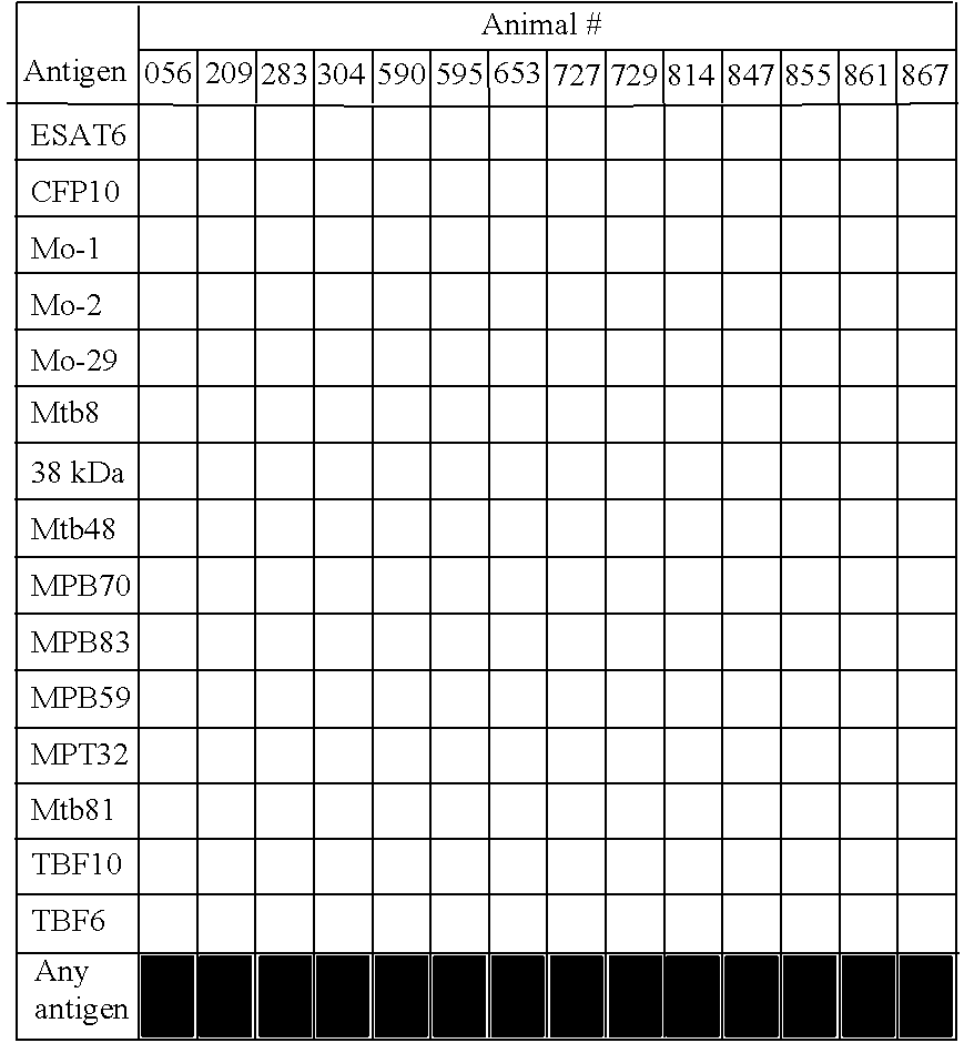
[0074]In MAPIA, 14 infected monkeys produced IgG antibodies of variable levels and starting at various time-points post-infection. FIG. 2. As shown in Table 2-4 below, antigen recognition patterns varied significantly from animal to animal. Three proteins, ESAT-6, MPB83, and TBF10 were most often recognized by post-infection serum samples.

[0075]

TABLE 2Antigen recognition patterns in M. tuberculosis infectionAntigen recognition is indicated by  cells.

[0076]

TABLE 3Antigens predominantly recognized in experimental TB**Antigens eliciting earliest antibody are shown in . Results are shown as negative (−) or Positive (+, weak; ++ moderate; +++, strong).

[0077]

TABLE 4Antigen recognition by serum IgG in monkey TBAntibodyreactorsAntigenNumber%ESAT-61071CFP10750Mo-100Mo-200Mo-29321Mtb821416 kDa64338 kDa17Mtb4817MPB70536MPB831179MPB5900MPT3200Mtb8100F10964CFP10 / ESAT-6107116 kDa / MPB831286

example 3

Specific Antigen Mixtures in Protein a Lateral Flow Assay

[0078]An antigen mixture consisting of the three selected proteins was formulated and evaluated in the lateral flow format. Printing conditions were optimized for ESAT-6, MPB83, and TBF10 separately by spraying each antigen at various concentrations onto the membrane, both singly and in mixtures, and selecting one concentration for each antigen that resulted in the strongest discrimination between selected TB positive and negative serum samples. A mixture of the antigens was then printed onto nitrocellulose membrane at concentration of 1 mg / ml. Protein A was printed onto the membrane as an additional line to produce the control line. Protein A was also conjugated to colloidal gold particles following routine procedures known in the art and was used as the nonhuman primate IgG antibody-detecting molecule in the lateral flow immunoassay. Test strips were laminated, such that a conjugate pad was placed between the sample pad and ...


PUM

PropertyMeasurementUnit
pore sizesaaaaaaaaaa

Abstract

Systems, methods, and compositions for a diagnostic method for detecting TB in non-human primates that is easy to use, sensitive, and specific. The method utilizes recombinant mycobacterial antigens, such as polyfusion proteins. The method utilizes an antigen-antibody-antigen arrangement to detect TB infection in nonhuman primates. The method can detect IgM antibodies to TB, in addition to IgG antibodies, providing ability to detect TB earlier in nonhuman primate TB infection as compared to conventional TB tests.

Description

[0001]This application claims the benefit of provisional patent application No. 60 / 605,304, filed on Aug. 26, 2004.FIELD OF INVENTION[0002]The present invention relates to methods and compositions for detecting the presence of mycobacterial organisms, and in particular to methods and compositions for rapidly detecting tuberculosis infections in nonhuman primates.BACKGROUND[0003]Tuberculosis (TB) is one of the most important bacterial diseases of nonhuman primates (Adams et al., Biosafety in Nonhuman Primates in Biomedical Research: Biology and Management, (eds) Bennett, B. T., Abee, C. R. and R. Henrickson, Academis Press, San Diego, 1995). This is particularly true for captive nonhuman primate colonies, in which close quarters among colony members allow this highly contagious disease to spread rapidly. As in humans, most reported cases of this disease are caused by the acid-fast bacillus Mycobacterium tuberculosis or Mycobacterium bovis. And because these organisms are communicable...

Claims


Application Information

Patent Timeline
Patent Type & Authority Patents(United States)
IPC IPC(8): A61K39/04A61K39/02G01N33/53
CPCG01N33/5695G01N2469/20
Inventor LYASHCHENKO, KONSTANTINESFANDIARI, JAVANBAKHSHMCCOMBS, CANDACE
Owner CHEMBIO DIAGNOSTIC SYST
Why Eureka
Try Eureka