
Method and apparatus for removing tissue autofluorescence

a tissue and autofluorescence technology, applied in the field of tissue autofluorescence removal methods and apparatuses, can solve the problems of reducing the specificity of detection, affecting the analysis of images obtained of these cells, and affecting the detection accuracy of images, so as to remove autofluorescence and remove dark current noise.

Active Publication Date: 2012-03-13
LEICA MICROSYSTEMS CMS GMBH
View PDF9 Cites 0 Cited by
  • Summary
  • Abstract
  • Description
  • Claims
  • Application Information

AI Technical Summary

Benefits of technology

This method effectively reduces autofluorescence and dark current noise, enhancing the sensitivity of detection and preserving signal fluorescence, making it adaptable for various biological samples and fluorescence imaging applications.

Problems solved by technology

This autofluorescence can interfere with the analysis of images obtained of these cells.
For example, autofluorescence may reduce signal detection sensitivity by masking the fluorescence of the probe of interest.
In addition, autofluorescence may also decrease the specificity of detection by providing false positive results.
The extent of autofluorescence may be limited by treating the sample with certain dyes or chemicals prior to image acquisition.
However, these treatment techniques involve further manipulation of the biological sample, which may degrade the quality of the sample itself.
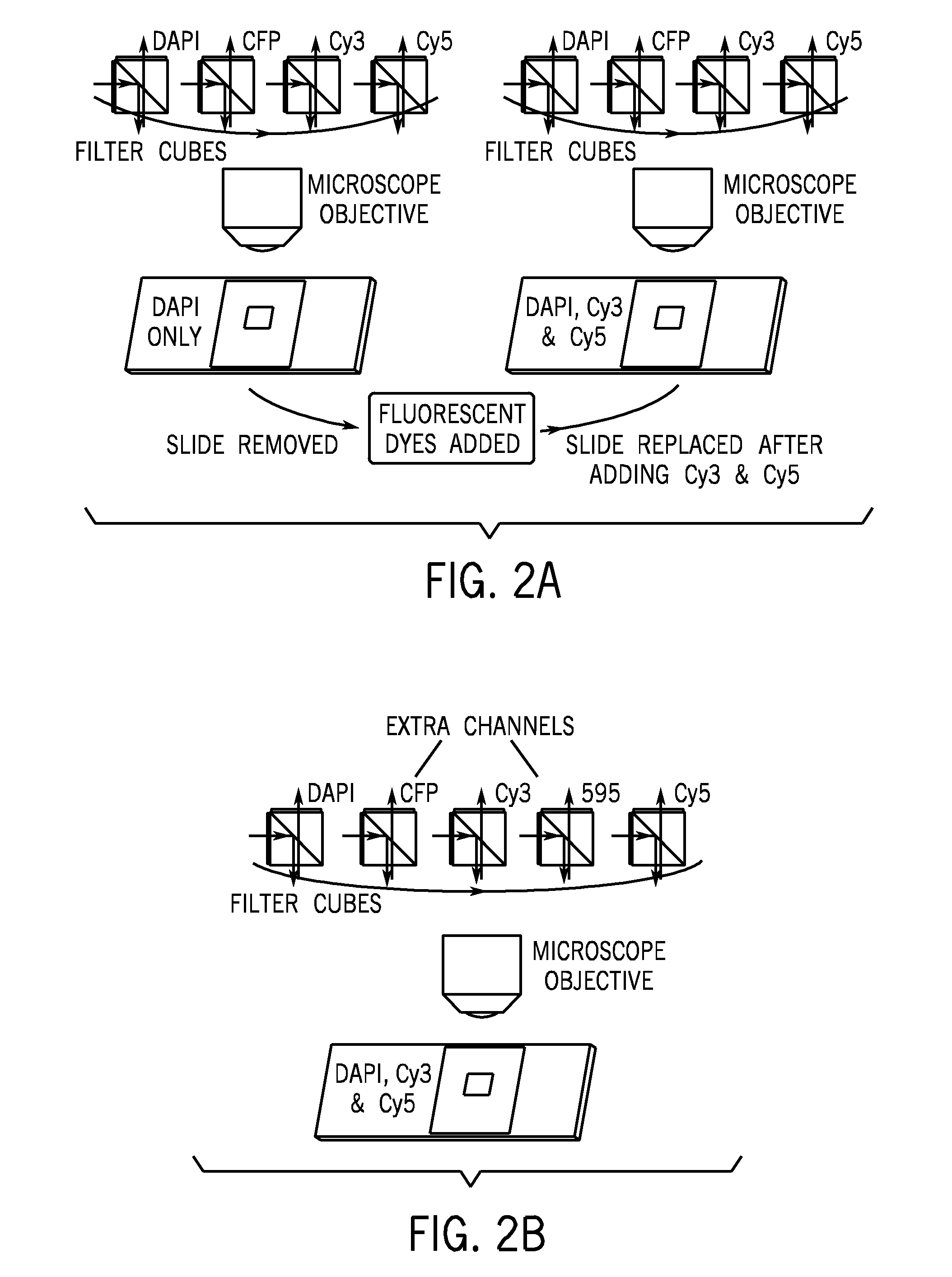
However, these techniques may not be suitable with standard fluorescent microscopes in widespread use.
While these techniques may be more cost effective, they may not be able to completely remove the autofluorescence component from fluorescence microscopy images.
For example, spectral unmixing methods often require prior knowledge of the amount of autofluorescence in the sample.
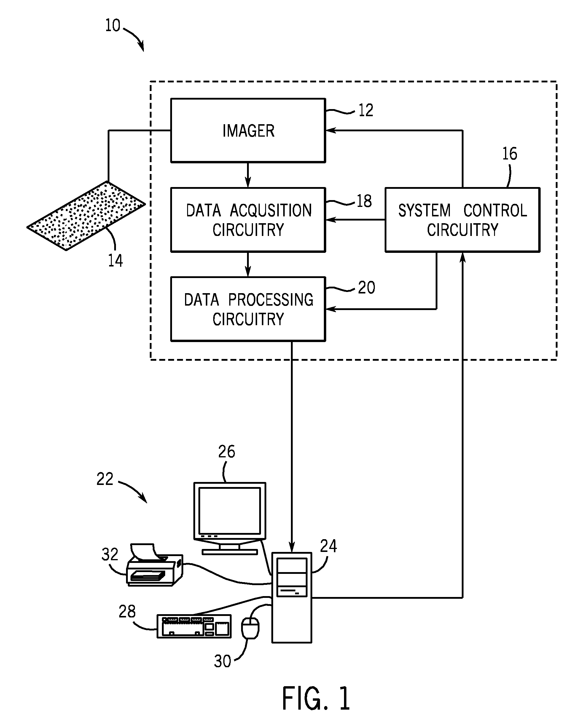
In addition, cell or tissue images may also include a certain amount of noise contributed by the acquisition system itself.
However, there is no image processing method that deals with both autofluorescence and dark current.

Method used


Image

  • Method and apparatus for removing tissue autofluorescence
  • Method and apparatus for removing tissue autofluorescence
  • Method and apparatus for removing tissue autofluorescence

Examples


Embodiment Construction

[0021]The methods and systems of the present techniques may provide the advantage of improving image analysis for fluorescently stained images. Such techniques may preserve signal fluorescence while reducing or eliminating autofluorescence, as well as dark current. The techniques increase the resulting signal-to-autofluorescence ratio and the overall sensitivity of detection. These fully automatic systems and methods operate without user intervention and complicated and expensive instrumentation. The methods and systems are adaptable to a wide array of biological samples, including tissue micro arrays. However, they are not limited to use with tissue micro arrays and may be applied to any fluorescence imaging application.

[0022]The present techniques provide an automatic autofluorescence removal method for fluorescent spectroscopy, based on a realistic physical model. In one embodiment, the image processing technique provided herein uses a non-negative matrix factorization algorithm ...


PUM

PropertyMeasurementUnit
wavelengthsaaaaaaaaaa
lengthaaaaaaaaaa

Abstract

Techniques for removing image autoflourescence from fluorescently stained biological images are provided herein. The techniques utilize non-negative matrix factorization that may constrain mixing coefficients to be non-negative. The probability of convergence to local minima is reduced by using smoothness constraints. The non-negative matrix factorization algorithm provides the advantage of removing both dark current and autofluorescence.

Description

BACKGROUND[0001]The invention relates generally to methods and systems for removing inherent autofluorescence and the contribution of dark current from images of biological materials.[0002]Recent advances in imaging and microscopy technologies combined with the development of fluorescent probes have allowed researchers to study variations in movement, distribution, and concentration of various molecular markers. In many such studies, fluorescent probes with a binding specificity to one or more cellular molecules are introduced into a sample. Images are obtained of the sample, for example with fluorescence microscopy, and these images may be processed and further analyzed to determine the amount of fluorescence in the image, which may provide information about the cellular marker. Accurate detection and analysis of the contribution of fluorescence from the fluorescent probes may be critical for many microscopy applications, such as molecular pathology imaging. For example, the fluore...

Claims


Application Information

Patent Timeline
Patent Type & Authority Patents(United States)
IPC IPC(8): G06K9/00A61B1/06
CPCG01N21/6458G06K9/0014G06V20/695
Inventor CAN, ALIGERDES, MICHAEL JOHNBELLO, MUSODIQ OLATAYOTAO, XIAODONGPAVAN-WOOLFE, FRANCIS EDWARDBHAGALIA, ROSHNI
Owner LEICA MICROSYSTEMS CMS GMBH
Learn More