
Arrangement and method for assessing tissue qualities

a tissue quality and arrangement technology, applied in the direction of optical radiation measurement, diagnostics using spectroscopy, instruments, etc., can solve problems such as methods which disrupt the cartilage layer

Active Publication Date: 2012-05-01
ARTHREX
View PDF11 Cites 7 Cited by
  • Summary
  • Abstract
  • Description
  • Claims
  • Application Information

AI Technical Summary

Problems solved by technology

These authors all question the methods which disrupt the cartilage layer because of the thickness changes caused by dehydration or hydrophilic swelling.

Method used


Image

  • Arrangement and method for assessing tissue qualities
  • Arrangement and method for assessing tissue qualities
  • Arrangement and method for assessing tissue qualities

Examples


Embodiment Construction

Tissue Layer Thickness Measurement—Materials and Methods

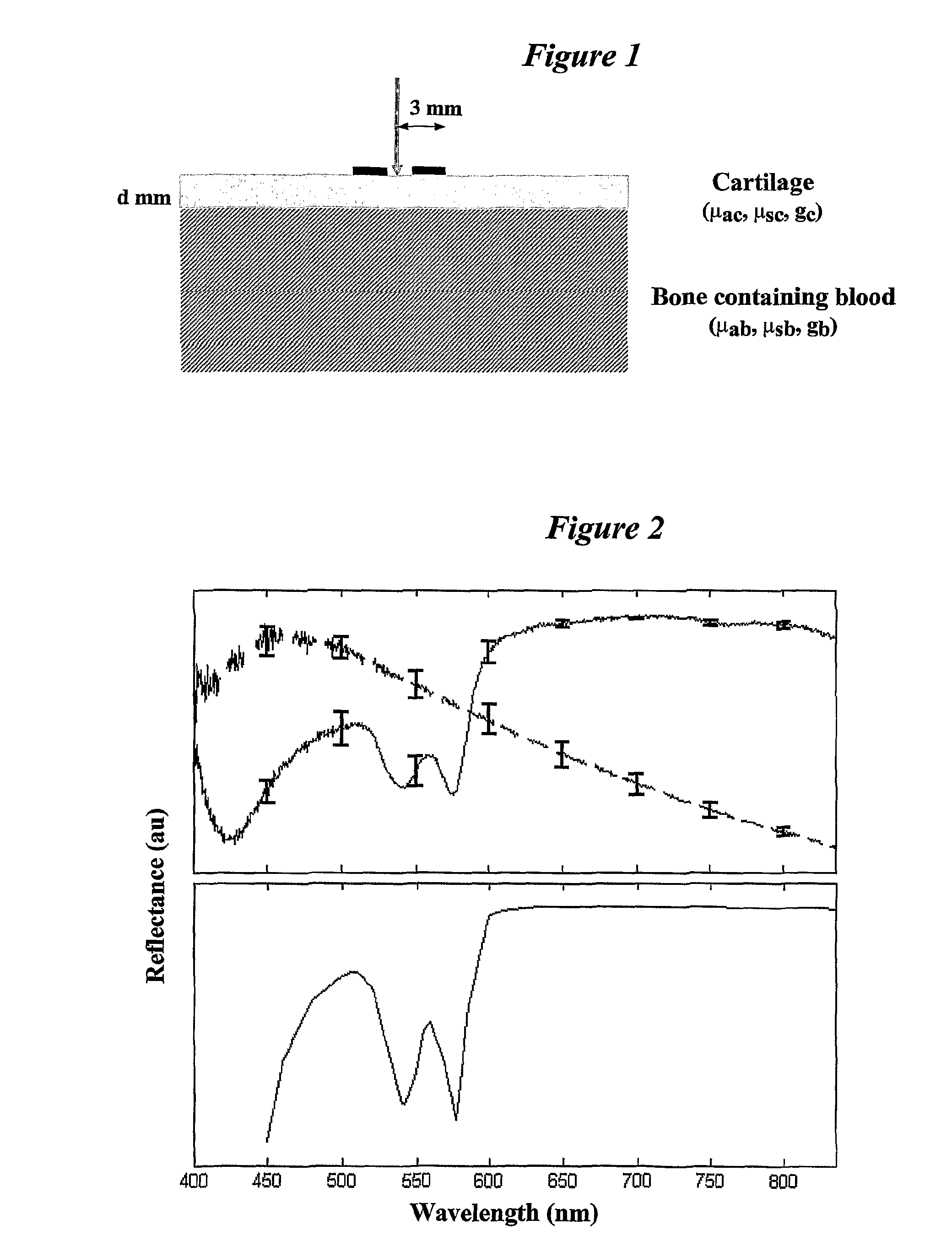
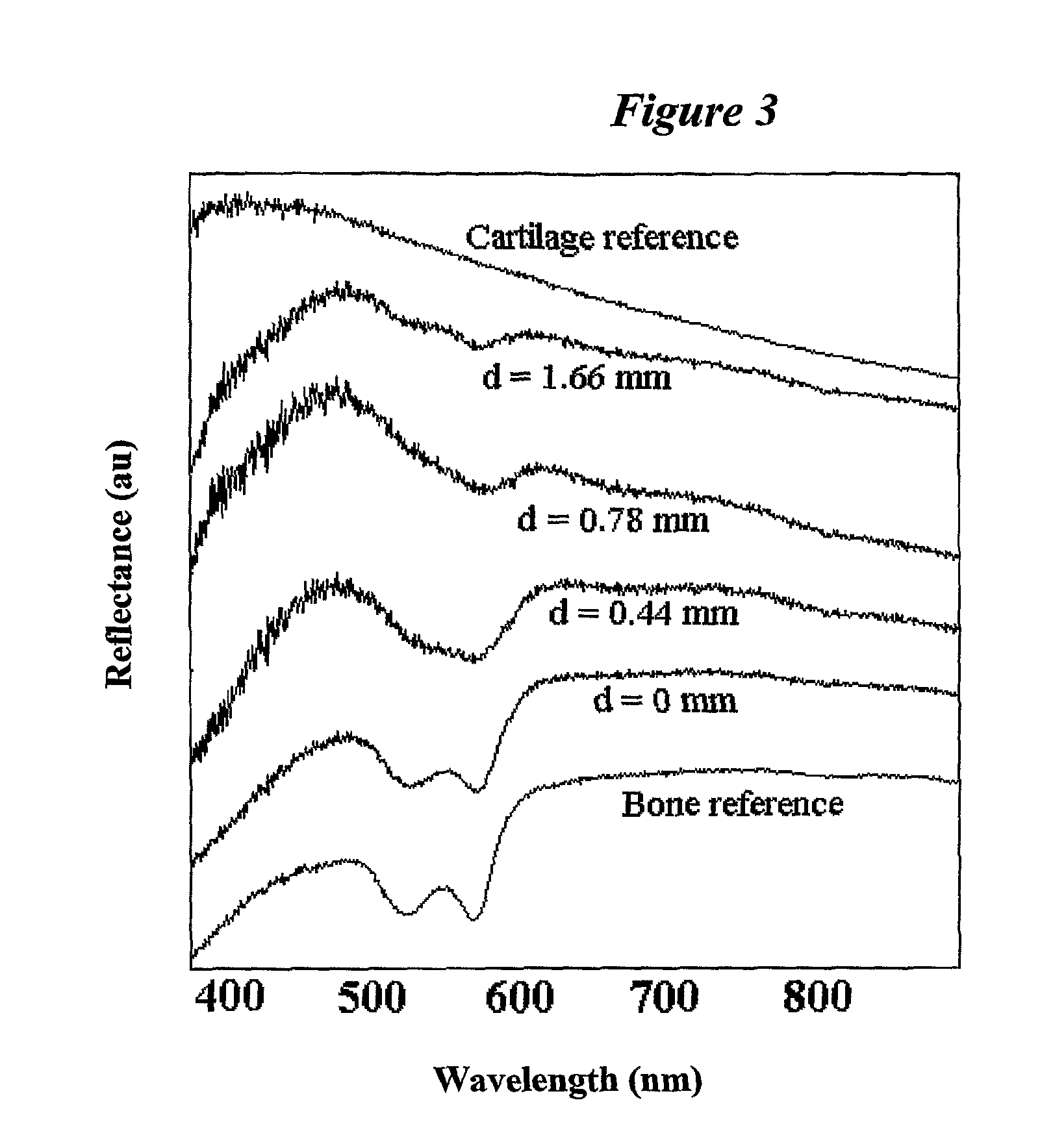
[0021]Twelve hip joint condyles from bovine calves were obtained from a local slaughterhouse less than 24 hours after sacrifice. Two of the condyles were used for reference measurements and the other ten for thickness experiments. The condyles were stored in saline in a refrigerator and prepared for cartilage measurements through the removal of soft tissues and tendons surrounding the joint. Three sites on each condyle surface were used for the measurements. A handheld, rotating, grinding machine was used to reduce the cartilage layer thickness. Sandpaper with the roughness P100 was used for grinding. Care was taken to grind in short episodes (5-15 s) so as not to increase the temperature of the cartilage. Thickness measurement of the cartilage layer was done with a high-resolution ultrasound scanner (B-mode 20 MHz, Dermascan 3v3, Cortex Technology, Hadsund, Denmark). The probe scanned over the measurement site and an image of ...


PUM

PropertyMeasurementUnit
wavelength rangeaaaaaaaaaa
wavelengthaaaaaaaaaa

Abstract

An arrangement and method for imaging and / or measuring tissue qualities, such as tissue thickness, tissue surface roughness and degree of tissue fiber linearization. The arrangement includes at least one light generating element, at least one light detecting element, a probe with an extension, and possibly a control apparatus including a signal processor for processing the detected signals and / or images. The extension is designed to convey light from the light generating element to the tissue for visualization and / or measurement. The extension is also designed to convey light back-scattered from the tissue to the light detecting element. The detecting element is designed to measure the intensity and / or spatial distribution of light back-scattered from the tissue.

Description

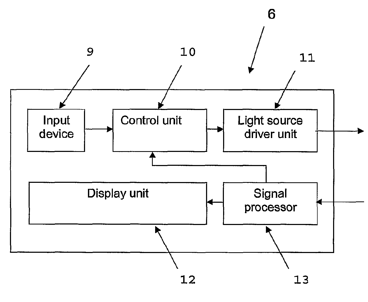
TECHNICAL FIELD[0001]The present invention generally relates to an arrangement and a measurement method for assessing tissue qualities. More specifically, the present invention relates to an arrangement and a method for measuring tissue thickness, tissue surface roughness and degree of tissue fiber linearization. The arrangement comprises a probe and means for light source driving / control, light detection, signal processing and presentation.PRIOR ART[0002]Endoscopic techniques have been used for the diagnosis and therapy of disorders since the beginning of the twentieth century. One typical example is arthroscopy, where the interior of a joint is visualized. Takagi (TAGAKI, 1918, in ALTMAN and KATES, 1983) modified a pediatric cytoscope to fit the problem area of knee joint visualization, which became the birth of this dynamic and rewarding field. The arthroscope is primarily a diagnostic device but therapeutic variants allow the removal of adhesions, intra-articular debris and meni...

Claims


Application Information

Patent Timeline
Patent Type & Authority Patents(United States)
IPC IPC(8): A61B6/00A61B5/00A61B5/103A61B5/107G01B11/06G01N21/21G01N21/47
CPCA61B5/0059A61B5/0084A61B5/0086A61B5/1075A61B5/4514G01B11/0625G01N21/21G01N21/274G01N21/474A61B5/0075A61B5/441A61B5/4504A61B5/4528G01N2021/4709G01N2021/4735G01N2021/4792
Inventor JOHANSSON, ANDERSSUNDQVIST, TOMMYOBERG, .ANG.KE
Owner ARTHREX
Learn More