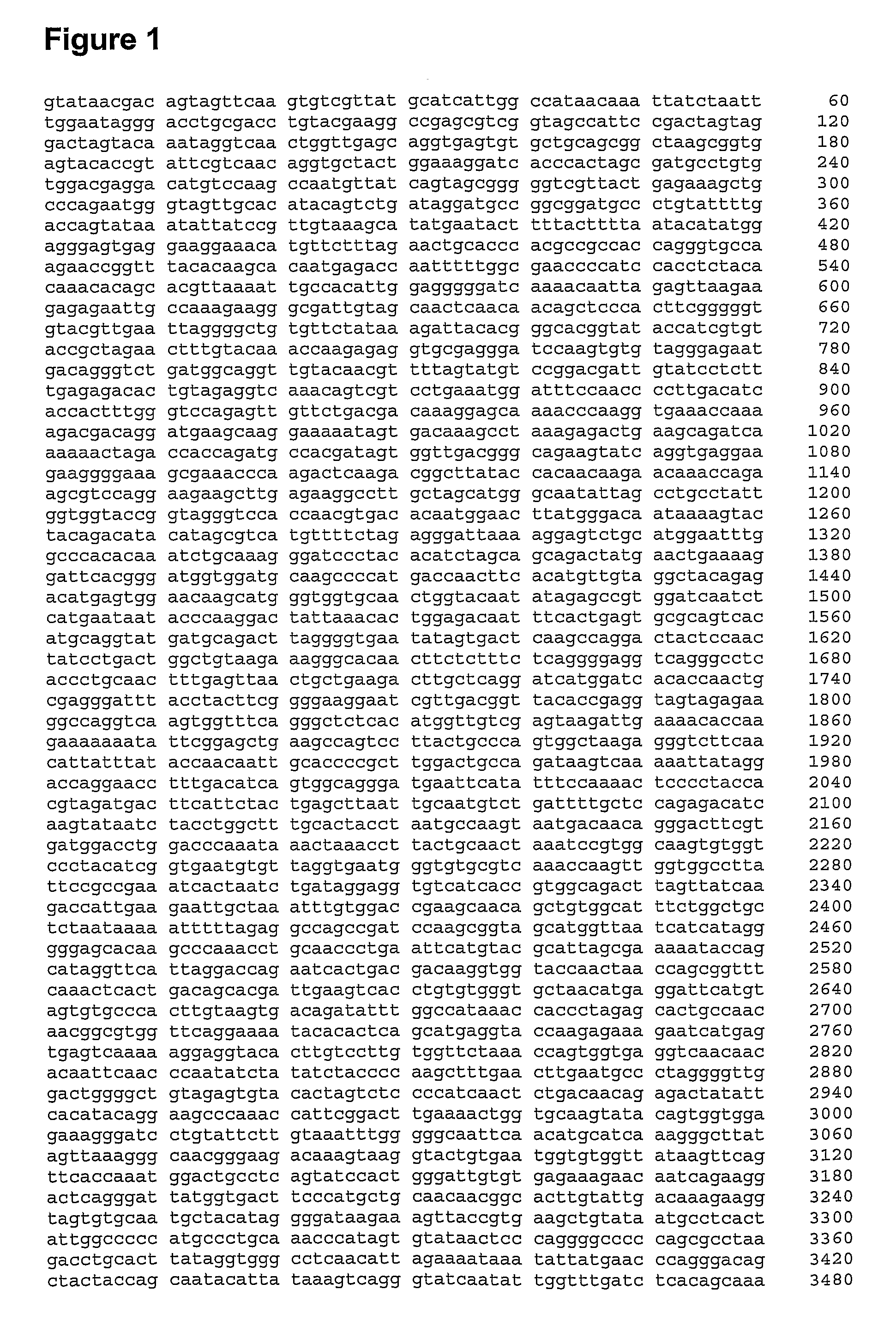
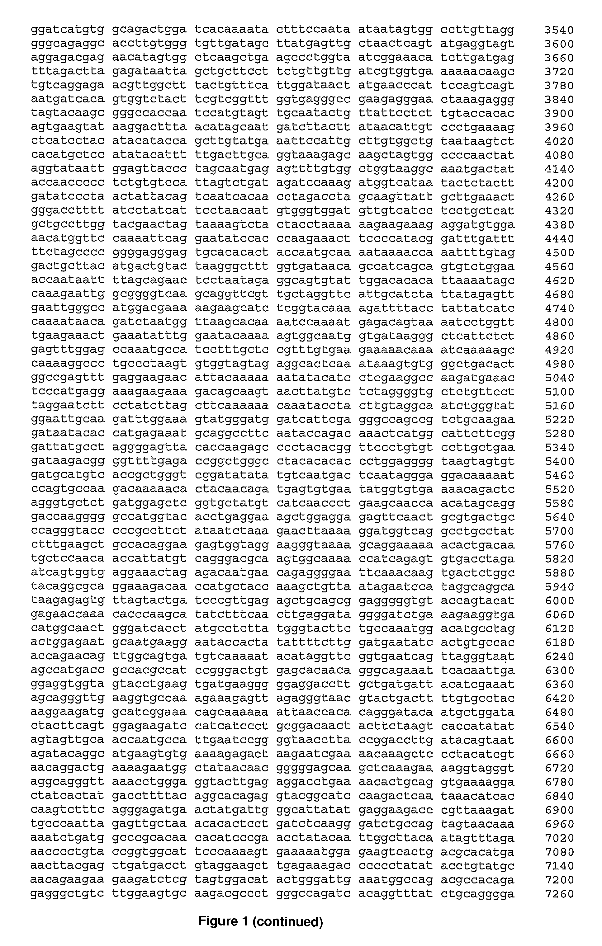
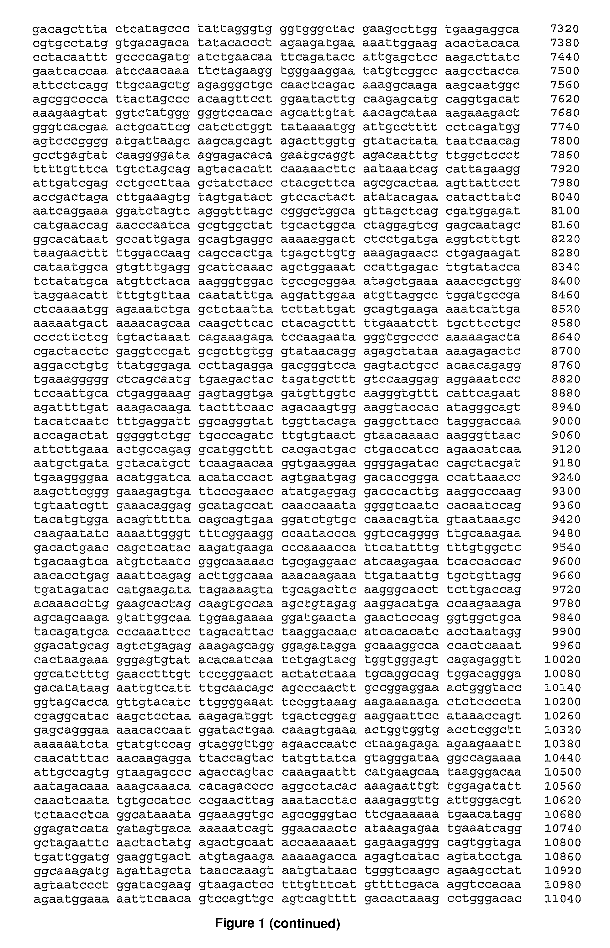
Pestivirus species
a pestivirus and pestivirus technology, applied in the field of new viruses, can solve the problems of large economic losses, congenital defects and the birth of persistently infected animals, high contagious and often fatal diseases, etc., and achieve the effect of enhancing the immunocompetence of the host individual and eliciting specific immunity
- Summary
- Abstract
- Description
- Claims
- Application Information
AI Technical Summary
Benefits of technology
Problems solved by technology
Method used
Image
Examples
example 1
Sample Preparation
[0458]Tissue samples were extracted and prepared using a method whose main basis was derived from Allander et al (2001) “A virus discovery method incorporating DNase treatment and its application to the identification of two bovine parvovirus species.” Proc Natl Acad Sci USA. 98(20): 11609-14, with some modifications to improve the efficiency from Baugh et al (2001) “Quantitative analysis of mRNA amplification by in vitro transcription.” Nucleic Acids Res. 29(5): E29. However, the methods were modified to improve efficiency.[0459]1. Preparation of Serum Samples:[0460]a) Obtain at least 240 μL of supernatant from a tissue homogenate or serum and divide into 2×120 ul lots[0461]b) To each 120 ul of sample add 240 ul of PBS or H2O (or take 50 ul sera+100 ul PBS)[0462]c) Filter diluted sample through two separate 0.2 um filters by centrifuging at 2000×g (wash top of filter and keep at −20° C.)[0463]d) Add 25 ul DNASE I (250 U) to each tube of filtered sample and incubat...
example 2
Enzyme Linked Immunosorbent Assay to Detect Antibodies to PMC Virus
[0605]1. Clone and express the PMC virus protein of interest (eg E2, NS3) in baculovirus and purify the expressed protein. This purified protein can be used as an antigen to detect specific antibodies to the PMC virus proteins of interest.[0606]2. Coat ‘medium binding’ 96 well microplates (50 uL per well) with antigen diluted in carbonate buffer (0.05M Carbonate buffer 1× (pH 9.6): Na2CO3 (1.59 gm); NaHCO3 (2.93 gm) water to 1 L). Hold overnight at room temperature (18-25° C.).[0607]3. Dilute samples and controls (Negative, High and Low Positive) 1 / 100 in sample diluent (phosphate buffered saline (pH 7.3) solution containing 1% skim milk powder and 0.05% Tween 20).[0608]4. Wash plates 5 times with PBS-Tween and tap to dry.[0609]5. Transfer diluted samples and controls to the ELISA plate in duplicate: 50 uL to each well.[0610]6. Incubate at 37° C. for 1 hr in a humidified container.[0611]7. Wash plates 5 times with PB...
example 3
Enzyme Linked Immunosorbent Assay to Detect Antigens of PMC Virus
[0619]It should be noted that working solutions of the detector reagent and enzyme conjugate reagents should be made within approximately 1 hour of anticipated use and then stored at 4° C.[0620]Materials
[0621]ELISA Wash Buffer—10× concentrate: 1 M Tris; HCl (6.25 Normal) for pH adjustment; 0.01% Thimerosal; and 5% Tween 20.
[0622]Detector Reagent—10× concentrate: 25% Ethylene Glycol, 0.01% Thimerosal, approximately 5% biotinylated goat anti-PMC virus antibody, and 0.06% yellow food colouring in PBS (pH 7.4). The working Detector Reagent is prepared by mixing 1 part of the Detector Reagent—10× concentrate, 1 part of NSB Reagent 10× concentrate, and 8 parts of Reagent Diluent Buffer. This working agent should be prepared within approximately 1 hour of anticipated use.
[0623]NSB Reagent—10× concentrate: 25% Ethylene Glycol, 0.01% Thimerosal, 0.2% Mouse IgG, 0.06% red food colouring in PBS (pH 7.4).
[0624]Reagent Diluent Buff...
PUM
| Property | Measurement | Unit |
|---|---|---|
| temperatures | aaaaa | aaaaa |
| temperatures | aaaaa | aaaaa |
| temperatures | aaaaa | aaaaa |
Abstract
Description
Claims
Application Information
 - R&D
- Intellectual Property
- Life Sciences
- Materials
- Tech Scout
- Unparalleled Data Quality
- Higher Quality Content
- 60% Fewer Hallucinations
Browse by: Latest US Patents, China's latest patents, Technical Efficacy Thesaurus, Application Domain, Technology Topic, Popular Technical Reports.
© 2025 PatSnap. All rights reserved.Legal|Privacy policy|Modern Slavery Act Transparency Statement|Sitemap|About US| Contact US: help@patsnap.com


