
Endoscope disposable sheath

a technology for endoscopy and cover, which is applied in the field of medical devices, can solve the problems of not being able to disinfect with steam, not being able to be used only once, and not being able to save water and electricity, and achieves the effects of convenient fixing of the cover, improved disinfection reliability, and easy installation and detachmen

Active Publication Date: 2012-08-14
SHENYANG SHENDA ENDOSCOPE
View PDF10 Cites 13 Cited by
  • Summary
  • Abstract
  • Description
  • Claims
  • Application Information

AI Technical Summary

Benefits of technology

[0015]3. the one-way valve disposed on the water air tube and outside the disposable sheath prevents backstreaming in the water air tube, and thus cross infection among different patients;
[0018]a. The invention is coronary, and can cover the outer surface of the operating portion and other portions near the operating portion from the top to the bottom and has an opening facing downwards, in a manner of wearing a cap; while in the FIGS. 1-13 of the U.S. Pat. No. 6,852,077 B2, it covers the operating portion from the bottom to the top, that is, from 102 to the top, in a manner of wearing trousers, and the big opening 103a covers the light guide portion and the doctor's left hand. In addition, in the FIGS. 15, 16, 29, and 30, the cover covers the outer surface of the endoscope operating portion, and closes it, and does not protect the operator's hand.
[0019]b. The invention is made of transparent film and easy for observation, and features light weight, low cost and so on. These materials enable the heat seal forceps to align with the hole of the three-way sealing cap, and to cut off the disposable forceps channel; while in the U.S. Pat. No. 6,852,077 B2, it uses high polymer annular elastic materials that do not have the above advantages of the invention, in addition, it is not elastic.
[0020]c. The cover is fixed on the endoscope by connecting the instrument insertion opening to the three-way sealing cap, which makes it easy for installing and detaching. While in FIGS. 22 and 23 of U.S. Pat. No. 6,852,077 B2, the water air valve connects the suction valve; the position of 441 and 442 is near, but the connection method is different. The instrument insertion opening of the invention is a component with protection film, while in U.S. Pat. No. 6,852,077 B2, the cover is disposed on the endoscope 441 first, then, 442 is disposed on it, obviously it has essential difference from the invention. As for FIGS. 27, 28 in the FIG. 27, the connection has no feasibility; in addition, as shown in FIG. 13, the operator's left hand is not in the cover, so it cannot be protected and is unsafe.
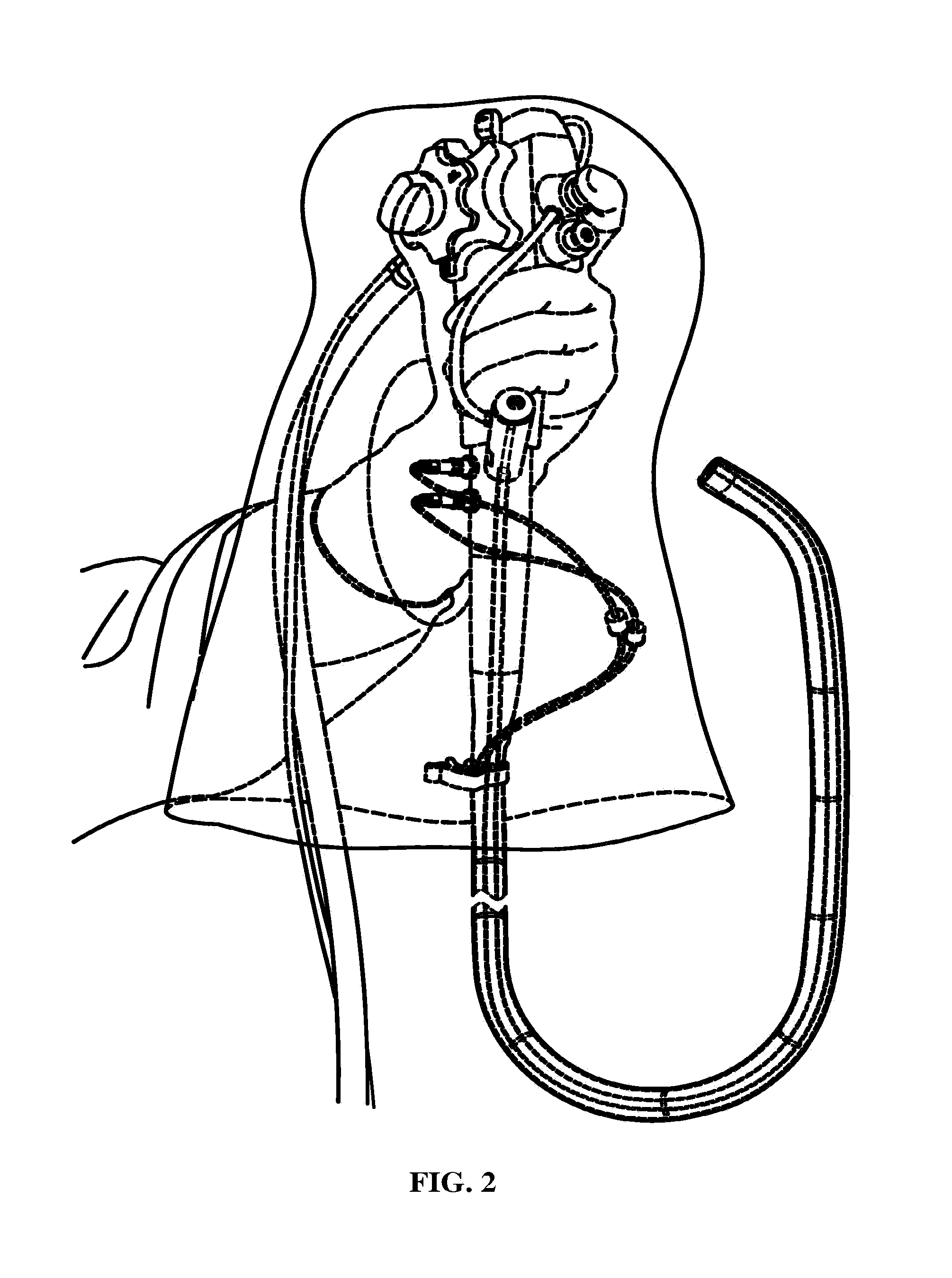
[0021]d. The instrument insertion opening has two purposes: firstly makes it convenient for fixing the cover on the endoscope. The instrument insertion opening is elastically connected to the three-way sealing cap, so installation and detachment are very easy, in a manner of pressing and releasing a button on clothes, and it is better than the method in U.S. Pat. No. 6,852,077 B2. Secondly, because of the arched endoscope and close cut in the instrument insertion opening, as the instruments is inserted or taking out, the contaminant cannot be discharged from the three-way sealing cap. After use, the disposable suction tube, disposable valve disk, coronary transparent film protection cover and the three-way sealing cap can be removed as a whole part. This is very convenient, and improves the reliability of disinfection. In addition, the invention features low cost, and is easy to operate and promote.
[0016]4. the disposable protecting cover of the invention enables a reusable endoscope to have reliable disinfecting effect, reduces disinfecting time, increases work life of the endoscope of the endoscope, saves a large amount of water and electricity, and cost spent on processing medical sewage, implements energy conservation and emission reduction, reduces the number of the endoscopes used in hospitals and corresponding cost, and protects environment.

Problems solved by technology

Due to materials and structure of the endoscope, it cannot be disinfected by steam at high temperature and pressure, and as the high cost thereof, it cannot be used for only one time like an injector or a cardiac catheter.
To facilitate disinfection, washing via disinfectant solution and dipping not only cost large amount of water and electricity, but also require sewage treatment, and disinfectant effect thereof is not stable enough.
However, there are several disadvantages with the endoscope: firstly, a suction tube is disposed in a endoscope of the endoscope and cannot be changed after use, which makes it the most polluted and unsafe; secondly, there is no fixing device between the front of a sheath and a head of the endoscope, which may cause one of them to detach from the other; thirdly, no one-way valve is disposed on a water air tube to prevent backstreaming, which causes water or air to flow reversely as pressure in a cavity of a patient varies; fourthly, an operating portion of the endoscope is not covered and protected, and may be contaminated by hands of a doctor; fifthly, though a prior art (such as U.S. Pat. No. 6,852,772 B2) discloses a method for protecting the operating portion, materials and structure of the endoscope are not practical, and production cost thereof is high.

Method used


Image

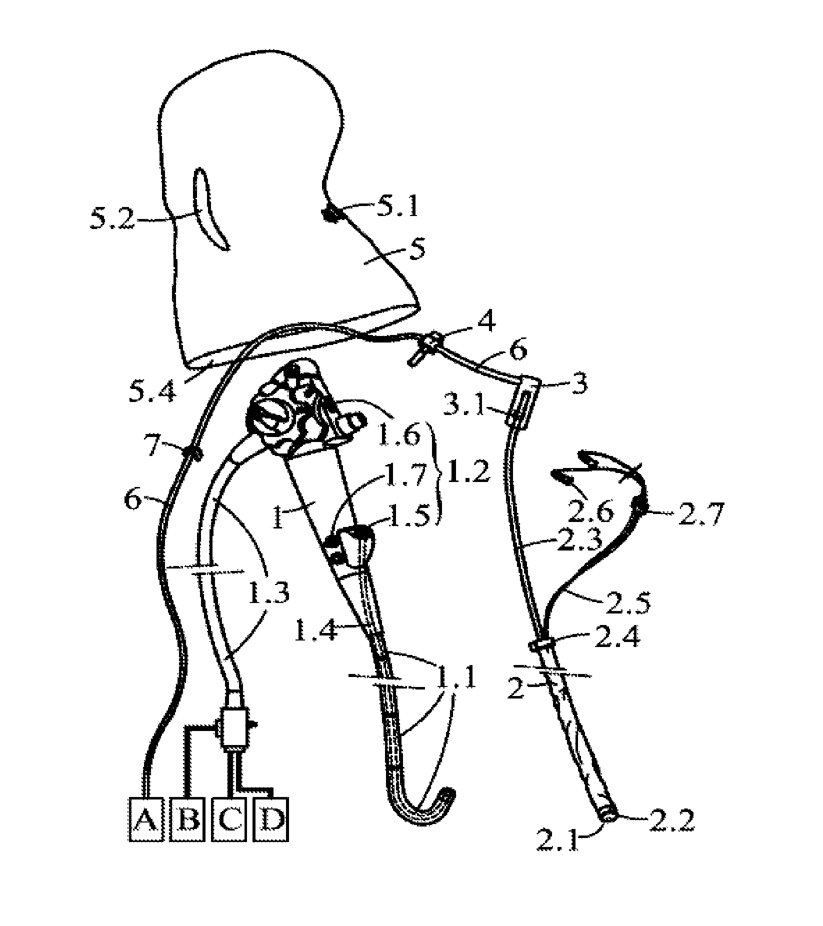
  • Endoscope disposable sheath
  • Endoscope disposable sheath
  • Endoscope disposable sheath

Examples


Embodiment Construction

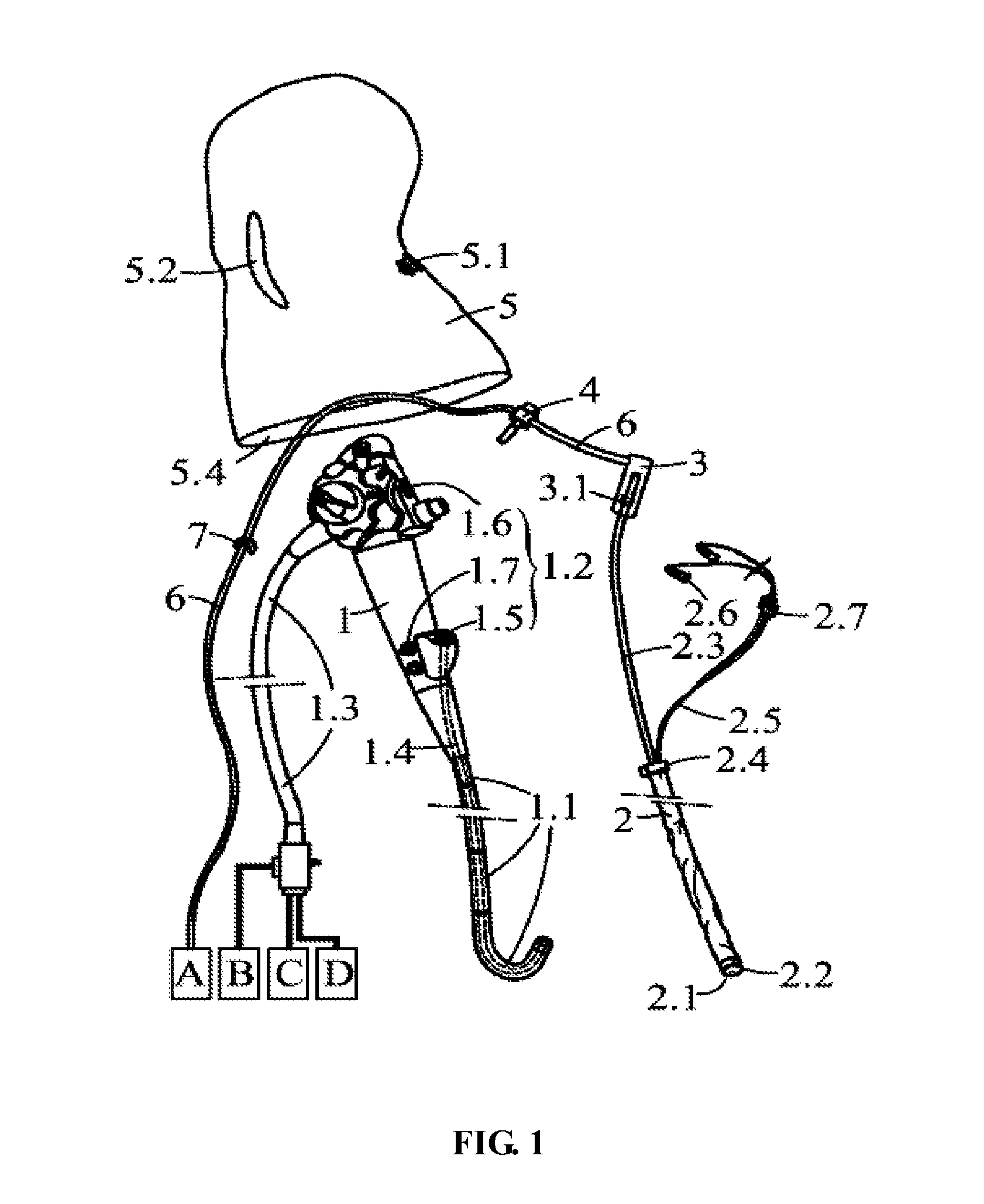
[0032]Further description of the invention will be given below in conjunction with accompanying drawings and specific embodiments.

[0033]As shown in FIG. 1, a disposable protecting cover of the invention operates to protect a endoscope 1 of an endoscope, and comprises a sheath 2 operating to protect an endoscope insertion portion 1.1 of the endoscope 1. The sheath 2 is made of elastic materials.

[0034]A diameter of the sheath 2 is greater than that of the insertion portion.

[0035]After the sheath 2 covers the endoscope insertion portion 1.1, a locking ring 2.4 disposed at the back of the sheath 2 is pulled up, at this time a diameter of the sheath 2 is decreased and thickness of a wall thereof is reduced, and the sheath 2 is tightly disposed on outer surface of the endoscope insertion portion 1.1.

[0036]A support ring 2.1 is disposed on a front end of the sheath, and a convex portion and a concave portion are respectively disposed on both sides of the support ring 2.1 and fit with a con...


PUM


Abstract

A disposable protecting cover for an endoscope, the endoscope comprising a endoscope and a disposable valve disk, and the disposable protecting cover comprising a disposable sheath, a clamping tube, a disposable suction tube, a three-way sealing cap, a water air tube, and a protecting cover comprising a hand entrance and an instrument insertion opening. A support ring is disposed in the front of the disposable sheath, a convex portion and a concave portion are respectively disposed on both sides of the support ring and fit with a concave portion and a convex portion on a head of the endoscope, the water air tube is connected to the disposable sheath, a one-way valve is disposed on the water air tube and outside the disposable sheath, the protecting cover is in the shape of a cap and made of transparent film, the protecting cover operates to cover an operating portion of the endoscope of the endoscope, the instrument insertion portion is elastically fit with an upper opening of the three-way sealing cap, a suck-in connector and a suck-out connector are disposed on a valve seat of the disposable valve disk, and connected to the disposable suction tube, and the suck-in connector, the suck-out connector, and the disposable suction tube are dispose outside the endoscope.

Description

CROSS-REFERENCE TO RELATED APPLICATIONS[0001]Pursuant to 35 U.S.C. §119 and the Paris Convention Treaty, the invention claims the benefit of Chinese Patent Application No. 201010162668.8 filed on Apr. 9, 2010, the contents of which are incorporated herein by reference.BACKGROUND OF THE INVENTION[0002]1. Field of the Invention[0003]The invention relates to a medical device, and more particularly to a disposable protecting cover for an endoscope.[0004]2. Description of the Related Art[0005]An endoscope is widely used to go deeply into a cavity of a patient for examination and operation in medical field. The endoscope comprises an insertion portion disposed in the front thereof, comprising a viewing system, an illumination system, an operating system, a biopsy channel, a water air tube and a suction tube, the insertion portion having a 1-2 meter flexible cylinder, an operating portion disposed in the middle thereof, and a light guide portion disposed at the back thereof and connected t...

Claims


Application Information

Patent Timeline
Patent Type & Authority Patents(United States)
IPC IPC(8): A61B1/12A61B1/04
CPCA61B1/00068A61B1/00135A61B1/00142
Inventor JIANG, KERANGJIANG, SHOUMEI
Owner SHENYANG SHENDA ENDOSCOPE
Learn More