
Method and system for segmentation of tubular structures in 3D images

a tubular structure and image technology, applied in the field of image segmentation, can solve the problems of difficult to fully automatic segmentation methods to yield robust, tubular structures that are typically too tedious to be practical in clinical applications, and the runtime of segmentation techniques is too slow for use, so as to achieve robust results

Active Publication Date: 2012-10-16
SIEMENS HEALTHCARE GMBH +1
View PDF12 Cites 3 Cited by
  • Summary
  • Abstract
  • Description
  • Claims
  • Application Information

AI Technical Summary

Benefits of technology

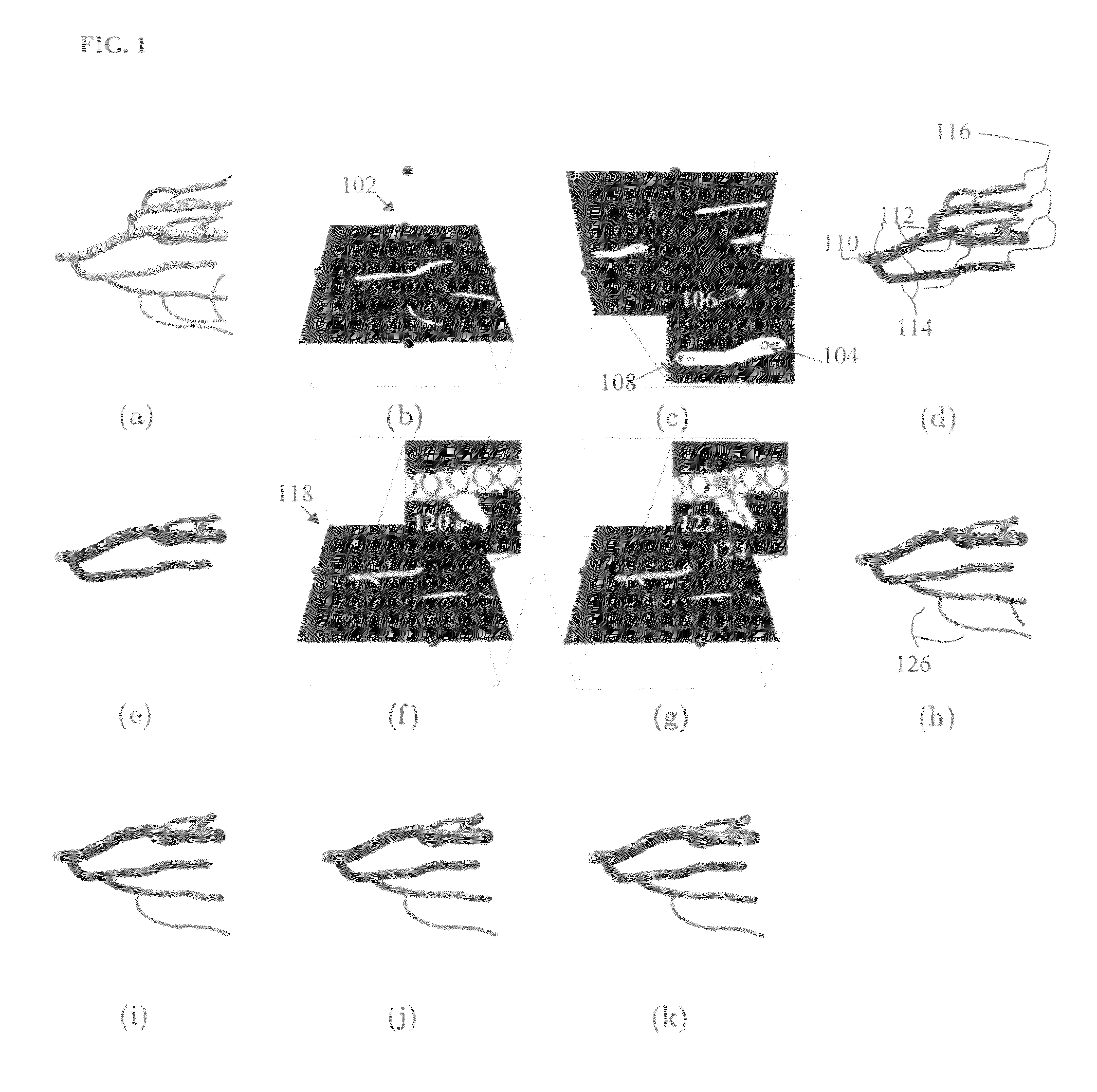
[0005]The present invention provides a method for segmenting tubular structures in 3D images. Embodiments of the present invention are directed to segmenting tubular structures in 3D images using a method referred to herein as pearling. Pearling generates an ordered series of pearls, which are 3D spheres of possibly different radii, as representation of a tubular structure. Pearling is robust to fluctuations in image intensities (due to noise, etc.) as the forces acting on a pearl are integrated over the region inside the pearl. Pearling is computationally efficient and well suited to user interactivity. Such interactivity can allow operator guidance of the segmentation in a particular direction, as well as operator correction of errant or undesired segmentation results.

Problems solved by technology

Accordingly, segmentation, or extraction, of 3D tubular structures from 3D image data is a fundamental problem in medical imaging, and is an important component of clinical applications involving diagnosis (e.g., stenosis, aneurysm, etc.), surgical planning, anatomical modeling and simulation, and treatment verification.
Using manual segmentation techniques to segment 3D tubular structures typically requires too much tedious labor to be practical in clinical applications.
However, due to poor contrast, noise, and clutter that is common to medical images, it is often difficult for fully automatic segmentation methods to yield robust results.
Typical conventional automatic segmentation techniques have runtimes that are too slow for use in such an interactive segmentation method.

Method used


Image

  • Method and system for segmentation of tubular structures in 3D images
  • Method and system for segmentation of tubular structures in 3D images
  • Method and system for segmentation of tubular structures in 3D images

Examples


Embodiment Construction

[0016]The present invention is directed to a method for segmenting tubular structures in 3D images. Embodiments of the present invention are described herein to give a visual understanding of the segmentation method. A digital image is often composed of digital representations of one or more objects (or shapes). The digital representation of an object is often described herein in terms of identifying and manipulating the objects. Such manipulations are virtual manipulations accomplished in the memory or other circuitry / hardware of a computer system. Accordingly, it is to be understood that embodiments of the present invention may be performed within a computer system using data stored within the computer system.

[0017]Embodiments of the present invention are directed to segmenting tubular structures in 3D images using pearling. Pearling allows a user to guide the automated construction of a 3D model of a tubular structure directly from 3D volume data, without going through slice cont...


PUM


Abstract

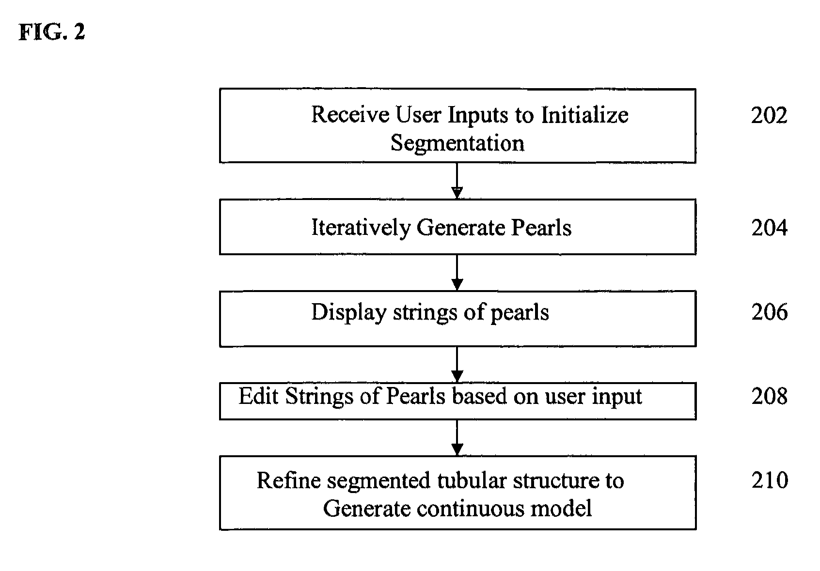
A method and system for segmenting tubular structures in 3D images is disclosed. User inputs identifying a first region on the image inside of a tubular structure and a second region of the image outside of the tubular structure are received. Based on this information, an ordered series of pearls are generated along the tubular structure. Pearls are spheres, each having a center location and a radius determined based on the center locations and radii of previous pearls and on local voxel intensities in the image. A continuous model of the tubular structure can be generated by interpolating the center locations and radii of the ordered series of pearls. The ordered series of pearls can be displayed and easily edited in response to user input, thus providing an efficient and flexible method for interactive segmentation of a potion of interest in a tubular structure.

Description

[0001]This application claims the benefit of U.S. Provisional Application No. 60 / 938,239, filed May 16, 2007, the disclosure of which is herein incorporated by reference.BACKGROUND OF THE INVENTION[0002]The present invention relates to image segmentation, and more particularly, to segmentation of tubular structures in 3D images.[0003]Computed tomography (CT) and magnetic resonance imaging (MRI) scans are frequently used to support diagnosis and analysis of cardiovascular and pulmonary anatomic structures. Such structures can appear as tubular structures in CT and MR images. Accordingly, segmentation, or extraction, of 3D tubular structures from 3D image data is a fundamental problem in medical imaging, and is an important component of clinical applications involving diagnosis (e.g., stenosis, aneurysm, etc.), surgical planning, anatomical modeling and simulation, and treatment verification.[0004]Using manual segmentation techniques to segment 3D tubular structures typically requires...

Claims


Application Information

Patent Timeline
Patent Type & Authority Patents(United States)
IPC IPC(8): G06K9/00
CPCG06K9/342G06T7/0081G06T2200/04G06T2207/10072G06T2207/20101G06T2207/30101G06T7/11G06V10/267
Inventor SLABAUGH, GREGORY G.UNAL, GOZDEFANG, TONGWHITED, BRIANROSSIGNAC, JAROSLAW
Owner SIEMENS HEALTHCARE GMBH
Learn More