Devices and processes for analysing individual cells

a technology of individual cells and devices, applied in the field of cell analysis, can solve the problems of inability to provide information, loss of subtle but important variations between cells within experimental noise, etc., and achieve the effects of low contrast in images, enhanced contrast in images, and flexible selection of field of view

Inactive Publication Date: 2013-02-12
OXFORD GENE TECH IP
View PDF31 Cites 12 Cited by
  • Summary
  • Abstract
  • Description
  • Claims
  • Application Information

AI Technical Summary

Benefits of technology

[0119]A major advantage of the invention is its capacity to analyse the contents of a cell even though the analytes can range from zero to many thousand copies per cell. As mentioned above, the invention provides improvements in fluorescence detection where signal strengths span several orders of magnitude, and one aspect of the invention is an integrated fluorescence detector for implementing this method. Thus the invention provides an apparatus for detection of fluorescence on a reaction substrate, comprising a light source, a fluorescence detector, a receptacle for a substrate of interest, and a computer programmed to select between an integrating detection mode and a counting detection mode. The light source will typically be a laser. The fluorescence detector will typically be a fluorescence microscope. The substrate of interest will typically be a device of the invention, or a portion thereof (e.g. a lid member). Selection between integrating and counting modes may be made manually, but is preferably made automatically depending on one or more pre-selected criteria e.g. signal intensity, etc. The apparatus will usually be able to move the substrate, the light source and / or the detector relative to each other e.g. in a way that is adapted to the specific format of the substrate and the disposition of the probes. For example, ignoring the empty regions between lines of probes will speed up the reading process.
[0120]The fluorescence being detected preferably results from specific binding of two biological molecules e.g. two nucleic acids, an antibody & antigen, etc.
[0121]When analysing results in a single channel, this apparatus can operate in various ways. For instance, it can move along the channel scanning individual patches in an integration mode, and then return to patches that did not reach a signal threshold in order to analyse them in a counting mode. As an alternative, it can make this selection at each patch along the channel, rather than making a crude ‘first sweep’ and requiring a second measurement of some patches. Similarly, the apparatus can operate in either of these ways, but scanning orthogonally to the channels. Further variations will be apparent.Common Analytical Components
[0122]As mentioned above, a powerful aspect of the invention is to perform identical individual analysis in parallel on different cells, and the invention provides a device for individually analysing a plurality of cells, comprising a plurality of channels, each of which is for receiving the contents of an individual cell, wherein each channel contains a series of sequential analytical components along the channel, and wherein the sequence of analytical components in one channel is the same as in another channel.
[0123]Thus a cell will experience a common series (e.g. A, B, C, D, E, F, G, . . . ) of analytical components regardless of which channel it enters. This common arrangement of analytical components within multiple channels means that each cell being analysed experiences the same analytical reagents, meaning that the results for one cell can readily and directly be compared to the results for another cell.
[0124]Preferably, at least 10 (e.g. 10, 50, 100, 250, 500, 1000 or more) analysis channels, and more preferably all of the analysis channels, contain a common sequence of analytical components.

Problems solved by technology

Analyses are most often carried out after lysing cells to release their contents, and it is usually necessary to use a large number of cells, because it is difficult to isolate single cells and because normal methods of detection are not sensitive enough to measure the contents of single cells.
Analysis of a mixture of cells thus masks heterogeneity within the mixture, and fails to provide information which is likely to be important for understanding the disease state.
Subtle but important variations between cells are lost within experimental noise.

Method used


Image

  • Devices and processes for analysing individual cells
  • Devices and processes for analysing individual cells
  • Devices and processes for analysing individual cells

Examples


Embodiment Construction

Microfabricated Device

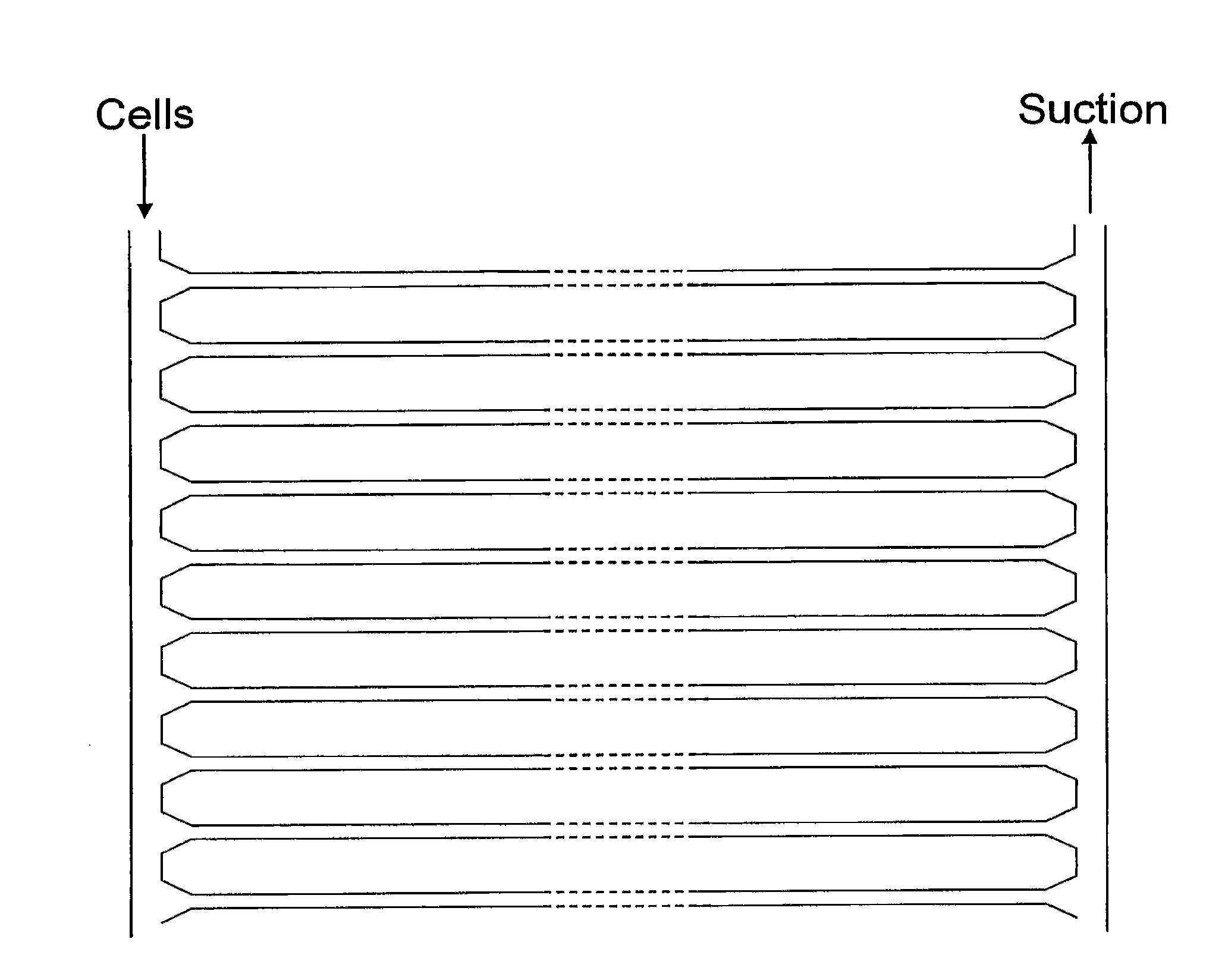
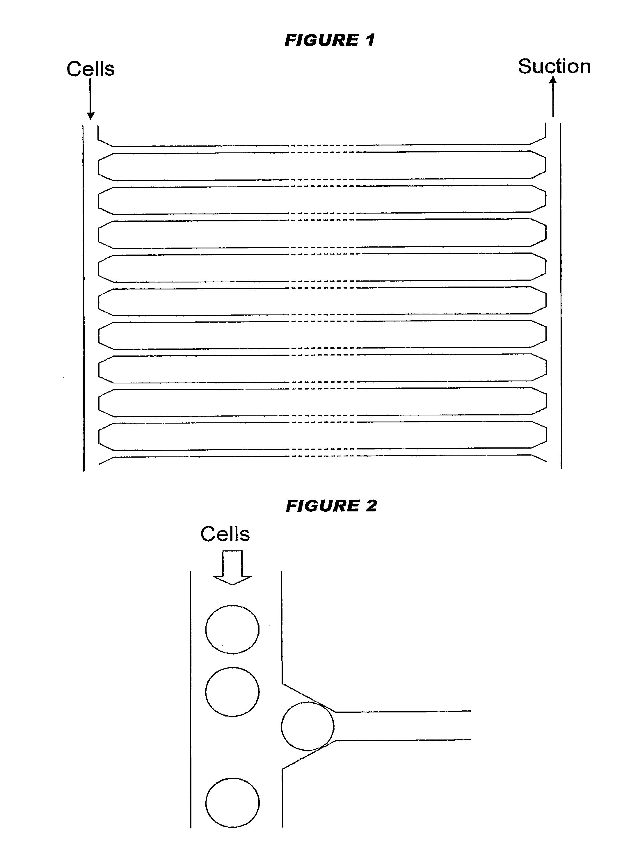
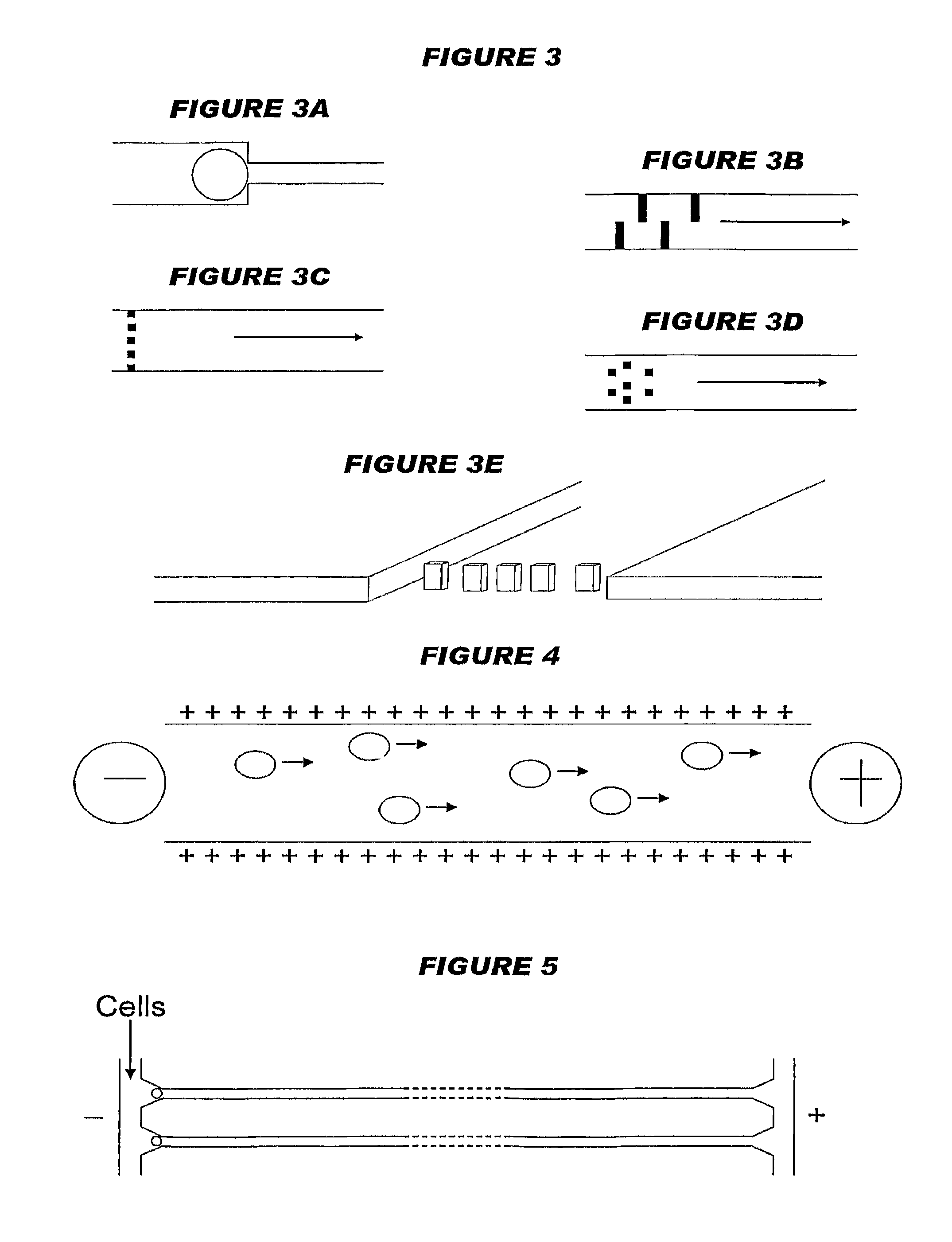
[0271]A microfluidic network (1) with a plan view as shown in FIG. 33 was made within a PDMS slab. Cells enter at the top of the device, along delivery line (10). Single cells are trapped at the tapered entrances (20) of multiple parallel channels (30) and their contents travel in the direction shown by the arrow. After travelling along the channels (30), reagents leave the output ends (40) of the device (1) by exhaust (50). An enlarged view of the output ends (40) in the final PDMS device (1) is shown in FIG. 34. An enlarged view of the tapered entrances (20) in the final PDMS device (1) is shown in FIG. 35. Detail of the regions downstream of entrances (20), including various arrangements of projections (60) for trapping cells, is shown in FIG. 36. The channels (30) have a rectangular cross-section, being 10 μm wide and, depending on the thickness of PDMS used, from 2-20 μm high. Adjacent channels are separated by 60 μm. The entrances (20) taper from 50 μm to...


PUM

PropertyMeasurementUnit
heightaaaaaaaaaa
areaaaaaaaaaaa
volumeaaaaaaaaaa

Abstract

A device for individually analysing cells of interest, comprising (a) a channel for receiving the contents of a cell of interest, wherein the channel has an input end and an output end, and (b) a cell trapping site in proximity to the input end of the channel, wherein (i) the input end of the channel is adapted such that an intact cell of interest cannot enter the channel; and (ii) the channel contains one or more analytical components for analysing the contents of the cell of interest. In use, a cell is applied to the device, where it is trapped by the cell trapping means. The cell cannot enter the channel intact, but its contents can be released in situ to enter the channel's input end. The contents can then move down the channel, towards the output end, and they encounter the immobilised reagents, thereby permitting analysis of the cell contents.

Description

[0001]This application is a U.S. national stage of International Application No. PCT / GB2006 / 001593 filed May 3, 2006.[0002]All documents and on-line information cited herein are incorporated by reference in their entirety.TECHNICAL FIELD[0003]This invention is in the field of cell analysis, and in particular the analysis of individual cells.BACKGROUND ART[0004]There are many methods for biochemical characterisation of cells and tissues. Methods such as electrophoresis, chromatography, mass spectrometry, microarrays, etc. are used to analyse the molecular composition of cells or tissues. The results of such analyses may indicate a disease state, for example. Analyses are most often carried out after lysing cells to release their contents, and it is usually necessary to use a large number of cells, because it is difficult to isolate single cells and because normal methods of detection are not sensitive enough to measure the contents of single cells.[0005]It is rare, however, to find a...

Claims


Application Information

Patent Timeline
Patent Type & Authority Patents(United States)
IPC IPC(8): C12M3/00C12M1/00C12M1/22B01L3/00C12M1/33
CPCB01L3/502753C12Q1/6876C12M47/06B01L3/5025B01L3/502715B01L2200/027B01L2200/0668B01L2300/0816B01L2300/0864B01L2400/0415G01N2015/1006C12M1/00C12M1/33
Inventor SOUTHERN, EDWINMEULEMAN, WOUTERLUEERSSEN, DIETRICH WILHELM KARLMILNER, NATALIE
Owner OXFORD GENE TECH IP
Why Eureka
Try Eureka