
Subretinal access device

a subretinal access and device technology, applied in the field of eye surgery instruments, can solve the problems of reducing visual acuity, eventually blindness, and difficult to perform interventional procedures targeting tissues beneath the sensory retina, and achieve the effect of stabilizing the device location and facilitating imaging

Inactive Publication Date: 2013-04-23
ISCI INTERVENTIONAL CORP
View PDF158 Cites 37 Cited by
  • Summary
  • Abstract
  • Description
  • Claims
  • Application Information

AI Technical Summary

Benefits of technology

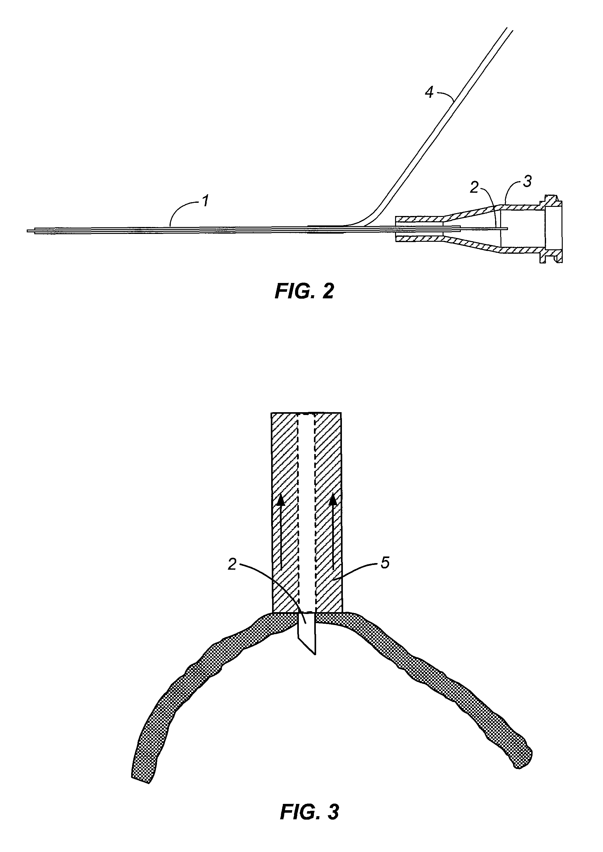
The invention provides devices that can access the space beneath the retina to deliver devices, materials, energy, or substances. These devices can hold the retina in place while therapies are performed underneath it. One device uses vacuum pressure to hold the retina and can also infuse fluids or gases. Another device uses a pointed tip to pierce the retina and access the subretinal space without damaging the underlying tissues. These devices can be introduced into the eye through a sclerostomy port. The technical effects of these devices include improved access to the retina for therapeutic purposes.

Problems solved by technology

There are many diseases and conditions that affect the posterior segment of the human eye which can lead to a decrease in visual acuity and eventually blindness.
Interventional procedures targeting tissues beneath the sensory retina are difficult to perform due to limited accessibility and the delicate structure of the retina which can be easily damaged during surgical manipulation.

Method used


Image

  • Subretinal access device
  • Subretinal access device
  • Subretinal access device

Examples


example 1

Access Device

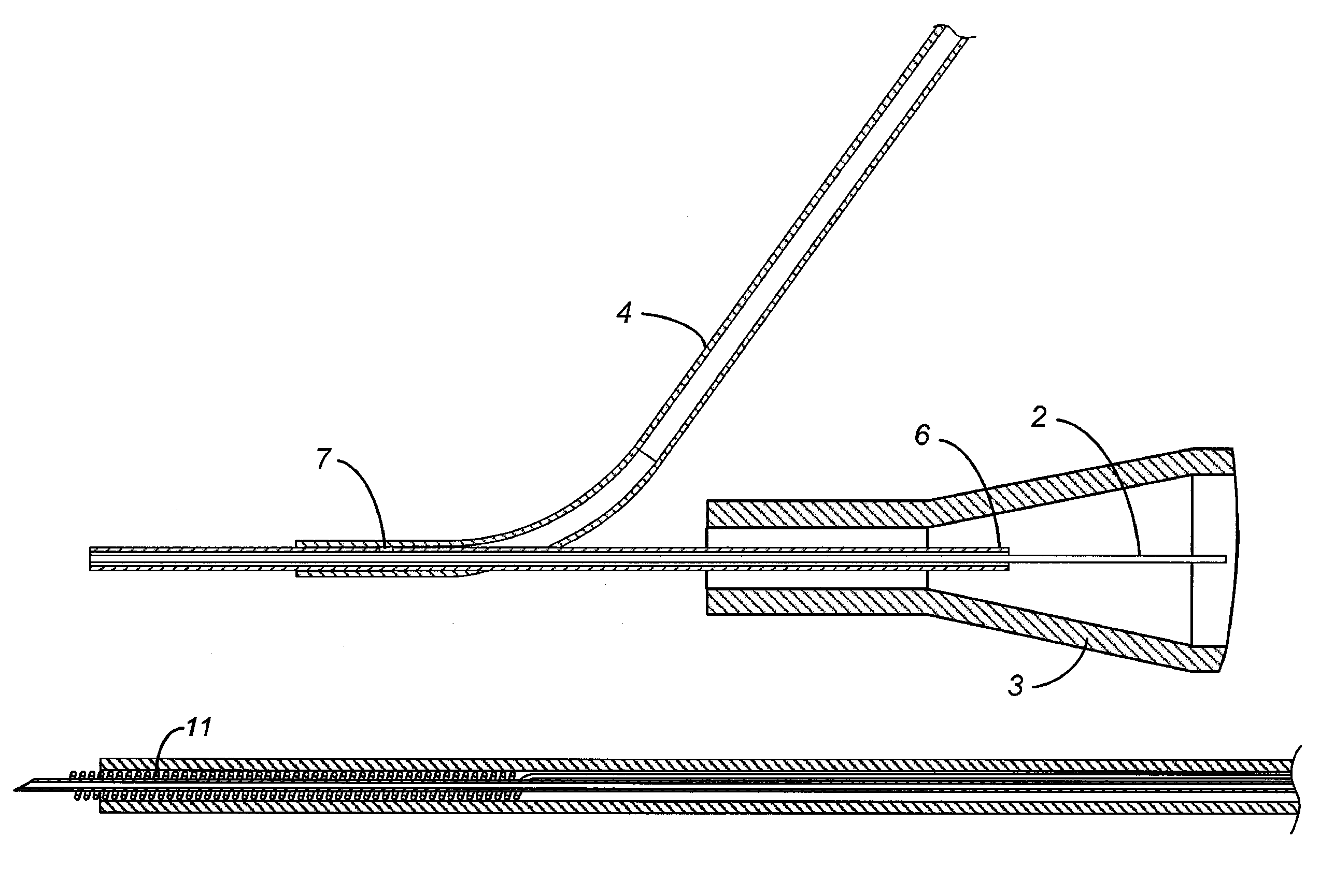
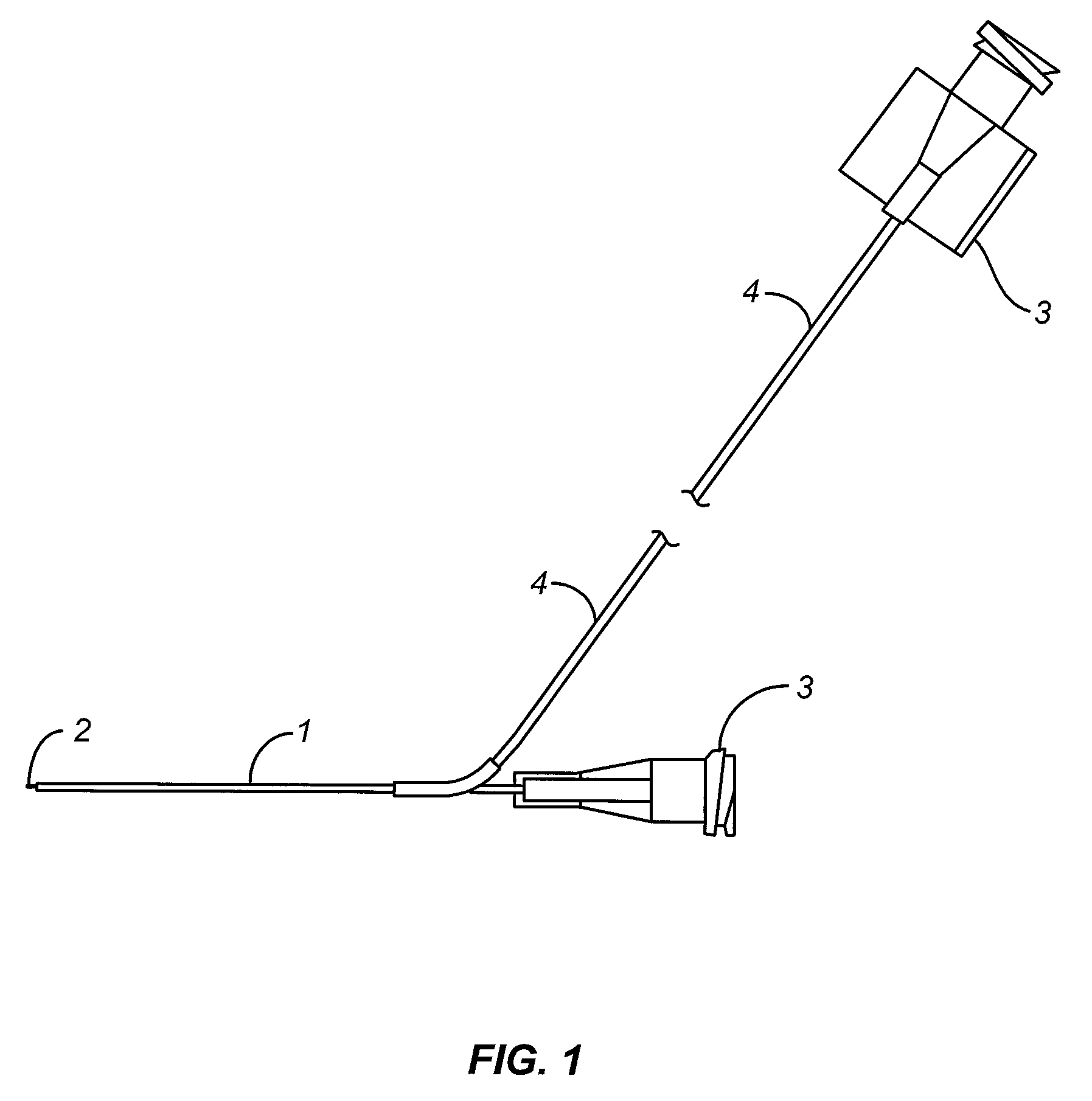
[0056]Approximately 2″ of thin walled 25 Gauge stainless steel hypotube, 0.020″ by 0.012″, (MicroGroup, Inc) was used as the main shaft. A single hole was drilled approximately 1.25″ from the distal edge of the hypotube. A skive was created approximately 0.010″ from one end of a 3″ length of 0.020″ by 0.060″ Tygon tubing. The main shaft was inserted into the Tygon tubing through the skive until the hole of the main shaft was in communication with the Tygon tubing. UV cure cyanoacrylate adhesive (Loctite 4305, Loctite, Inc.) was applied at the proximal and distal interfaces between the Tygon tubing and the main shaft to create a seal, such that communication existed between the Tygon tubing branching from the main shaft and the lumen of the main shaft.

[0057]A 0.028″ stainless steel mandrel was heated with the proximal end of the Tygon tubing, and then fed into the proximal end of the Tygon tubing in order to flare the inner diameter of the Tygon tubing from 0.020″ to 0.0...

example 2

Laboratory Testing with the Access Device

[0061]A human cadaver eye was obtained from an eye bank. The cornea, the iris, natural lens, and the vitreous were removed, providing access to the retina from the interior of the globe without significantly damaging the retina tissue. The open globe was filled with phosphate buffered saline.

[0062]An access device as described in Example 1 was set-up as follows. The side port of the device was connected to a vacuum source to provide aspiration in the outer annulus. The vacuum source was capable of providing vacuum levels from 300 to 600 mm Hg. A 6 inch long extension tube was attached to Luer fitting in communication with the access shaft distal tip. A syringe containing 0.1% Alcian Blue dye was attached to the extension line.

[0063]In a first trial, the device tip was placed against a portion of the cadaver eye retina that had detached from the underlying RPE during preparation. Vacuum aspiration was applied to the outer annulus and its attac...


PUM


Abstract

The invention provides surgical devices that provide access to the sub-retinal space using delicate traction to hold the sensory retina to create and maintain a patent sub-retinal space of sufficient size to introduce and perform treatments on the eye. Such treatments may include the introduction of illumination or imaging agents or tools, surgical tools, the infusion of pharmaceutical or biological agents, and the placement of grafts, transplants or implants and the closure of the site through the delivery of a sealant.

Description

CROSS-REFERENCE TO RELATED APPLICATION[0001]This application is related to co-pending, commonly assigned Ser. No. 12 / 359,169, filed on an even date herewith, entitled “Device for Aspirating Fluids” in the names of Chang, Tom S.; Ho, Friedrich; Conston, Stanley R. and Yamamoto, Ronald.FIELD OF THE INVENTION[0002]The present invention relates to surgical instruments for use in the eye. More particularly, the invention relates to instruments that can provide access to the sub-retinal space using delicate traction to hold the sensory retina to create and maintain a patent sub-retinal space of sufficient size to introduce and perform treatments on the eye. Such treatments may include the introduction of illumination or imaging agents or tools, surgical tools, the infusion of pharmaceutical or biological agents, and the placement of grafts, transplants or implants.BACKGROUND OF INVENTION[0003]There are many diseases and conditions that affect the posterior segment of the human eye which c...

Claims


Application Information

Patent Timeline
Patent Type & Authority Patents(United States)
IPC IPC(8): A61M25/00A61M1/00A61M5/32A61M31/00A61M35/00
CPCA61F9/007A61F9/008A61B1/313A61B3/102A61B3/12A61B5/0066A61B17/3421A61F2009/00874A61F9/0017A61F2009/00863A61B2017/306A61M25/01A61M31/00A61L29/04
Inventor HO, FRIEDRICHCONSTON, STANLEY R.YAMAMOTO, RONALD K.
Owner ISCI INTERVENTIONAL CORP
Learn More