Method of predicting responsiveness to autologous adoptive cell transfer therapy
a technology of autologous adoptive cells and predicting responsiveness, applied in the field of predicting responsiveness to autologous adoptive cell transfer therapy, can solve the problems of toxic side effects of the current generation of chemotherapeutic drugs utilized in cancer treatment, inability to cure, and inability to cur
- Summary
- Abstract
- Description
- Claims
- Application Information
AI Technical Summary
Benefits of technology
Problems solved by technology
Method used
Image

Examples
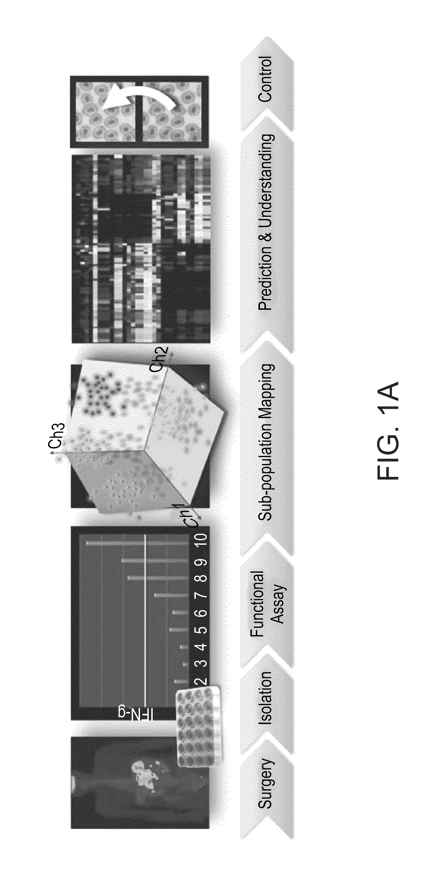
example 1
Determination of Immune Subpopulation Composition of IFN-γ Secreting TILs (Tumor Infiltrating Lymphocytes)
[0133]Materials and Methods
[0134]Measurement of IFN-γ Secretion:
[0135]IFN-γ secretion was measured after co-incubation of 105 TIL cells with 105 viable autologous melanoma cells for an overnight period. The amount of IFN-γ secretion in the culture supernatant was detected using standard sandwich ELISA protocol.
[0136]Flow Cytometry:
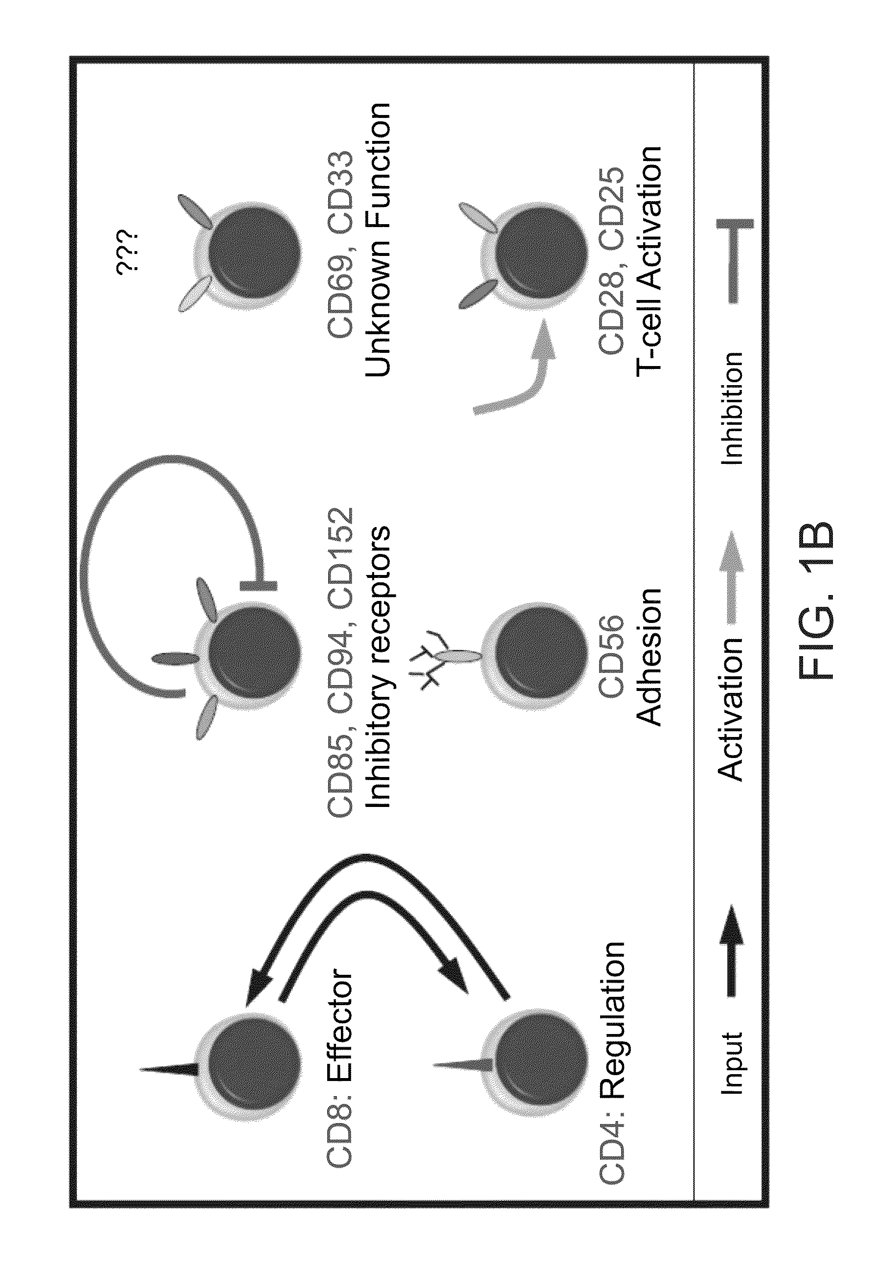
[0137]The markers used for subpopulation mapping included combinations of triple staining from the pool of the following surface receptors: CD3, CD4, CD8, CD25, CD28, CD33, CD56, CD69, CD85, CD94, CD152 (FIG. 1B) and the intracellular cytotoxic proteins perforin and granzyme B.
[0138]The following antibodies (Abs) were purchased from DakoCytomation: CD4, CD25, CD28, CD56, CD69, CD85, CD94, CD152. The following Abs were purchased from BD Pharmingen: CD3, CD8, CD33. Perforin and Granzyme B antibodies were purchased from eBioscience.
[0139]For flow cytometr...
example 2
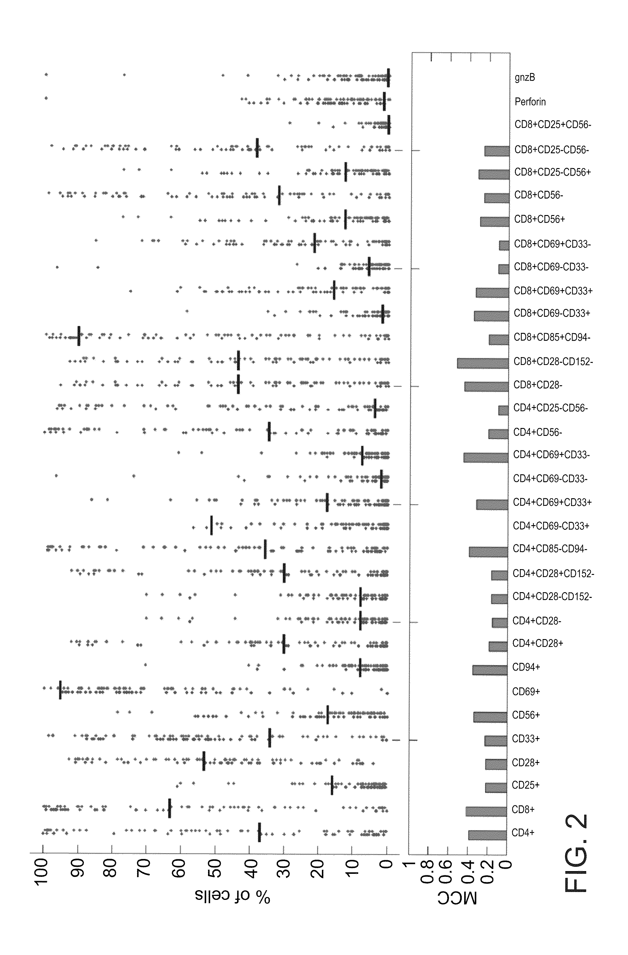
Comparison Between Individual Subpopulation Fractions and Use Thereof to Predict Reactive and Nonreactive TILs
[0142]Materials and Methods
[0143]SVM Classification:
[0144]SVM classifications were performed with the gist-train-svm software www.bioinformatics.ubc.ca / gist / . All classifications were performed with a linear kernel and input data was normalized by rescaling the columns to values between −1 and 1. All tests were conducted by applying a ‘leave three out’ procedure. SVM performance was evaluated by the ROC (receive operating characteristics) analysis which calculates the true positive rate versus True negative rate for different cutoffs. The ROC value namely the area under A ROC curve was reported for each test. In addition, the total accuracy (TA), sensitivity (SN), specificity (SP) and the Matthews correlation coefficient (CC) were calculated.
[0145]The present inventors compared individual subpopulation fractions and used them to predict reactive and nonreactive TILs (FIG. 2)...
example 3
Use of Subpopulation Signatures to Predict TIL Reactivity
[0154]Results
[0155]Since the SVM model does not lend itself easily to biological interpretation, the present inventors decided to investigate the underlying biological rational that governs TIL reactivity. The usage of differential expression signatures has become a well established method for distinguishing between various cellular states and different pathological conditions. This concept was applied to cell populations, by using a similar notion of “subpopulations signature” that can be used to differentiate between reactive and nonreactive TILs (see FIG. 3A and FIG. 11). Each column corresponds to a TIL culture and the rows represent subpopulations. Two significant clusters emerge, each representing a profile of CD4+ and CD8+ enriched subsets. These two markers represent regulatory and cytotoxic T-cell subpopulations respectively (FIG. 1B). Interestingly, the two clusters also separate between nonreactive and reactive TILs...
PUM
| Property | Measurement | Unit |
|---|---|---|
| frequency | aaaaa | aaaaa |
| affinity | aaaaa | aaaaa |
| degree of TIL reactivity | aaaaa | aaaaa |
Abstract
Description
Claims
Application Information
 - R&D
- Intellectual Property
- Life Sciences
- Materials
- Tech Scout
- Unparalleled Data Quality
- Higher Quality Content
- 60% Fewer Hallucinations
Browse by: Latest US Patents, China's latest patents, Technical Efficacy Thesaurus, Application Domain, Technology Topic, Popular Technical Reports.
© 2025 PatSnap. All rights reserved.Legal|Privacy policy|Modern Slavery Act Transparency Statement|Sitemap|About US| Contact US: help@patsnap.com