System and method of guided treatment within malignant prostate tissue

a prostate cancer and guided treatment technology, applied in the field of systems and methods of planning treatments, can solve the problems of radical intervention using standard treatments that might offer more harm than good, treatment is not completely curative or devoid of side effects, and cannot be completely correct, so as to improve the accuracy of imaging and localization, prevent unnecessary damage to non-malignant tissue, and improve the delivery of ablative therapy

Active Publication Date: 2013-10-01
TRACHTENBERG JOHN +2
View PDF11 Cites 14 Cited by
  • Summary
  • Abstract
  • Description
  • Claims
  • Application Information

AI Technical Summary

Benefits of technology

[0007]A system and method to identify the malignant tissue region and a method to focally and selectively destroy the tumor tissue is disclosed for the diagnosis of malignant tissue and prevention of unnecessary damage to non-malignant tissue in the delivery of ablation. The enabled technology is achieved through convergence of technologies that include accurate imaging to detect and target the malignant tissue within the prostate, an appropriate tissue removal systems such as automated (e.g., robotic) sectioning devices or an ablative device and energy source or any other appropriate surgical device, guided delivery of activity in the automated device or energy from the ablative device, the use of software being optional but preferred in planning, manipulation, and imaging following ablation such that only the target malignant is destroyed as may be confirmed by subsequent imaging of the target. The components may be employed sequentially over short or long time span. Advantages of the invention may include at least some of the following: a) improved accuracy in imaging and localization of the tumor (malignant tissues) within the prostate is a result of a novel magnetic resonance imaging-based technique or other contrast-enhancing imaging modalities; b) improved planning for optimizing delivery of therapy to the focal malignant tissue with minimal damage outside the focal volume, based on pre-treatment imaging with or without biopsy; c) improved delivery of ablative therapy to the malignant tissue, such ablative therapy comprising any of thermal therapy (using laser, ultrasound, radiofrequency or microwave energy sources); photodynamic therapy (using a combination of a
[0009]a) improved safety of the system and method through use of minimally-invasively delivery of treatment based on the planning, with or without on-line sensing and / or imaging of the treatment delivery and tissue response; and
[0013]b) a surgical system (e.g., an energy source and an ablative device for removing tissue such as cutting devices, sectioning devices, ablative devices for deposition of energy into the malignant prostate tissue; and means for quantifying a surgical procedure (such as the energy delivered from the ablative device into the tissue, mass of tissue removed, etc.);wherein the surgical procedure (e.g., ablative energy) is focally delivered by the (e.g.,) ablative device to the malignant tissue under image surveillance so as to substantially avoid destruction of the non-malignant tissue of the prostate.

Problems solved by technology

These treatments are neither completely curative nor devoid of side effects.
Recent data suggest that this may not be correct in all cases.
Indeed, many believe that radical intervention using standard treatments might offer more harm than good and a strategy of deferred treatment is now being adopted.
Destruction of this single site is likely to significantly decrease the risk of progression and increase cancer control with minimal side effects.

Method used


Image

  • System and method of guided treatment within malignant prostate tissue
  • System and method of guided treatment within malignant prostate tissue
  • System and method of guided treatment within malignant prostate tissue

Examples


Embodiment Construction

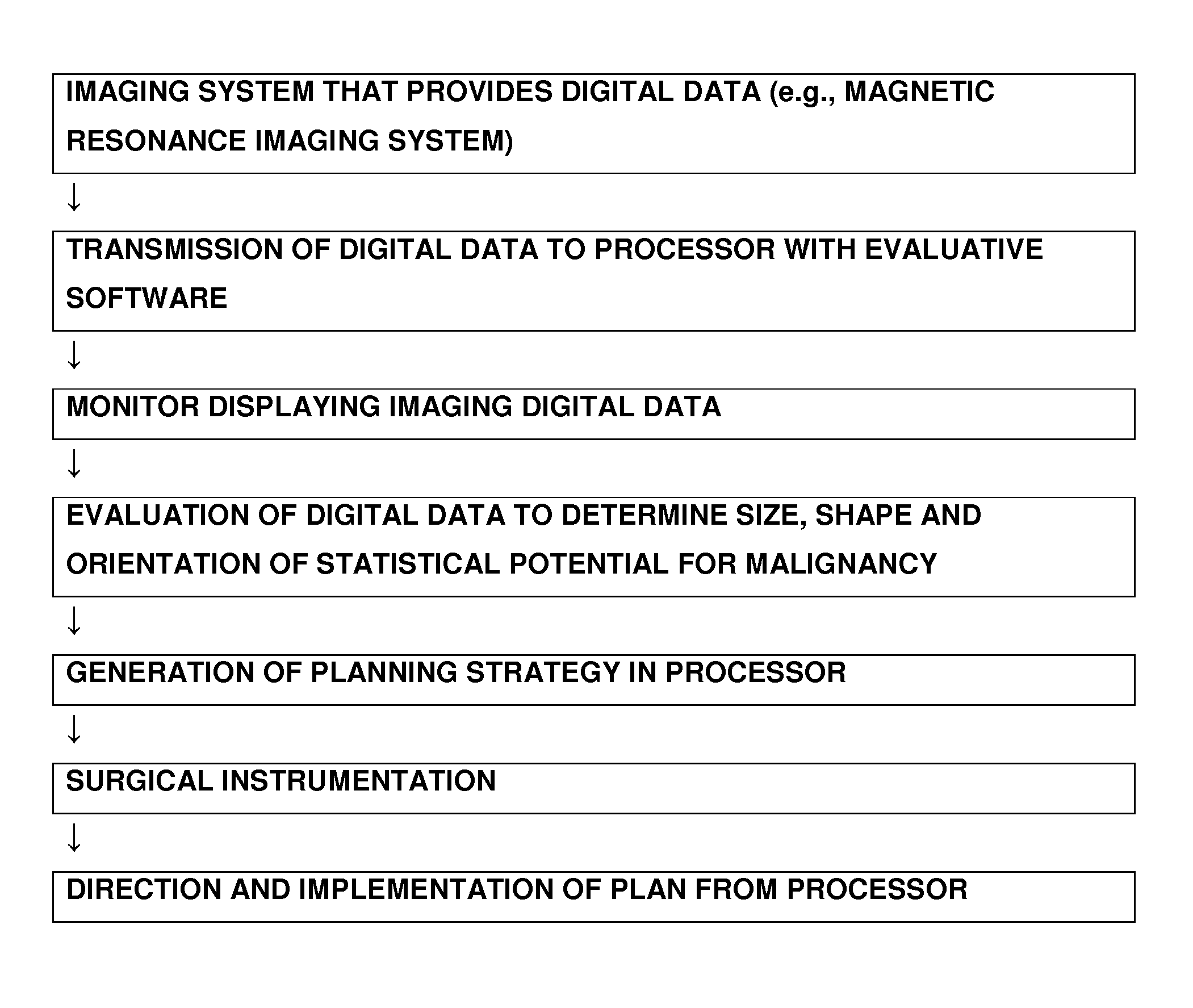
[0033]The technology described herein relates generally to apparatus, systems and methods for the treatment of cancer by removal of cancerous (malignant) tissue and cells, while attempting to minimize the removal of or damage to benign (non-cancerous) cells and tissue. The technology described herein is particularly useful for the treatment of prostate cancer where visualization of the tumors, cancerous tissue and differentiation from benign tissue has proven to be difficult by other means. The technology includes, by way of a non-limiting description, at least one imaging system (particularly an imaging system that directly provides digital image information or an analog imaging system having a processor that can convert analog imaging data into digital data) that provides data for differentiating between malignant and non-malignant tissues, especially within the prostate region of a patient. The system may also enable guided (automated, robotic, processor plan directed) delivery o...


PUM


Abstract

An imaging and diagnostic system and method to differentiate between malignant and non-malignant tissue of a prostate and surrounding region. The system acquires imaging data from the prostate and surrounding proximal region, and processes the data to differentiate areas of tissue malignancy from non-malignant tissue. A sectioning device or ablative device is provided. The ablative device is operable by automation for receiving the imaging output coordinates and defining the trajectory and quantity of energy or power to be delivered into the malignant tissue. A control system determines calculated energy or power to be deposited into the malignant tissue during ablation, to minimize destruction of the non-malignant tissue within the prostate and surrounding tissue. The system operates on generated ablative device output data.

Description

RELATED APPLICATION DATA[0001]This application claims priority from U.S. Provisional Patent Application No. 60 / 788,954 filed Apr. 4, 2006.BACKGROUND OF THE INVENTION[0002]1. Field of the Invention[0003]The invention relates to systems and method of planning treatments, execution of treatments and imaging. By using imaging techniques and data to differentiate between malignant and non-malignant prostate tissue, tissue removal such as by means of sectioning and ablation is directed to the malignant tissue, for preservation of the non-malignant tissue in the prostate and surrounding region, thereby minimizing the destructive effects of tissue removal.[0004]2. Background of the Art[0005]Prostate cancer is widely believed to be the most common cancer in men and the second most common cause of death due to cancer. There were approximately 230,000 reported cases of prostate cancer diagnosed in North America in 2005 and over 30,000 deaths. Furthermore, the true prevalence of the disease has...

Claims


Application Information

Patent Timeline
Patent Type & Authority Patents(United States)
IPC IPC(8): A61B5/05
CPCA61B18/20A61B2017/00274A61B2018/00547A61B2018/00636A61B2019/5236A61B2019/5238A61B2019/5276A61N7/02A61B2090/376A61B2090/374A61B2090/378
Inventor TRACHTENBERG, JOHNHAIDER, MASOOM A.WILSON, BRIAN CAMPBELL
Owner TRACHTENBERG JOHN
Why Eureka
Try Eureka