Surgical tool

a surgical tool and electrode technology, applied in the field of tissue cauterization, can solve the problems of affecting the healing effect of the tissue, the time-consuming and labor-intensive sealing of the blood vessel, and the inability to remove most organs intact, so as to facilitate tissue cauterization, reduce the bleeding capacity of the tissue, and eliminate the effect of the bleeding

Active Publication Date: 2013-11-05
AESCULAP AG
View PDF613 Cites 112 Cited by
  • Summary
  • Abstract
  • Description
  • Claims
  • Application Information

AI Technical Summary

Benefits of technology

[0012]The invention provides methods, systems, and apparatus which facilitate tissue cauterization in connection with such procedures as tissue resection and removal from patients undergoing a wide variety of procedures. The procedures may involve removal of an entire organ, e.g. hysterectomies, cholecystectomies, prostate removal, lung resection, and the like. Alternatively, the methods may involve removal of a portion of an organ or other tissue, such as tumor removal, often from highly vascularized organs, such as the liver, lung, or the like. The methods generally involve two steps, where the tissue is first necrosed or cauterized in whole or in part using radio frequency energy. In particular, the cauterization is effected at least along a desired resection plane within the tissue. The tissue is then resected along said plane(s). Advantageously, it has been found that resection within the necrosed or cauterized tissue substantially minimizes, and in some cases eliminates, the bleeding caused by the tissue resection. Preferably, the tissue cauterization is effected over a target volume of tissue, typically an entire organ or a portion thereof, e.g. the uterus, the lobe of a liver, a lung section, the prostate, or the like. By effecting substantially complete cauterization of a target tissue volume, the bleeding capacity of that tissue is reduced or eliminated, thus facilitating subsequent morcellization and tissue removal. Thus, organ and tissue removal is greatly facilitated with a substantial reduction in bleeding and the time needed for the surgical procedure.
[0014]High frequency (usually radio frequency) power is applied to the tissue mass through the electrically active regions(s) of the electrode structures, and the power is applied for a time and in an amount sufficient to cauterize or necrose tissue between said electrodes, preferably at least along a desired resection plane. Often, a volume of the tissue is resected by morcellating, e.g. grinding, comminuting, cutting into small pieces, or the like. Such morcellation is greatly facilitated by necrosis of the target tissue volume. By necrosed, it is meant that the cells of the tissue have been killed and that bleeding of the tissue, upon subsequent resection, has been substantially inhibited. The tissue are usually resected along a plane within the necrosed tissue mass, with minimal bleeding as described above.
[0015]The electrically active regions of the electrode structures have an area of at least 1 cm2, more usually at least 2 cm2, often having areas of 5 cm2 or larger, more often having areas of 10 cm2 or larger, still more often having areas of 50 cm2 or larger. The electrodes may have a wide variety of characteristics, may generally be rigid, flexible, elastic, malleable, conformable, or the like. Preferably, the electrodes are flexible to facilitate engagement of the electrode against, and conformance to, a tissue surface. In at least some instances, it is desirable to provide flexible, elastic electrodes which may be conformed about the outer periphery of a tissue or organ surface, where the elastic nature of the electrode assures firm engagement and electrode contact. In other instances, however, the electrodes may be specifically configured to have a desired geometry for engaging a particular tissue surface.
[0016]The high frequency energy applied to the organ or tissue is generally provided at radio frequency typically, but not limited to the range from 100 kHz to 10 MHz, usually from 200 kHz to 750 kHz. The power levels depend on the surface area and volume of tissue being treated, but generally fall within the range from 10 W to 500 W, usually from 25 W to 250 W, more usually from 50 W to 200 W. Power is usually applied at a level of from 1 W / cm2 to 500 W / cm2, more usually from 10 W / cm2 to 100 W / cm2. The power is applied for a time sufficient to raise the tissue temperature in the tissue mass being treated to above a threshold level required for cauterization or necrosis, usually being above at least 60° C., frequently being above 70° C., and often above 80° C., or higher. The application of energy should be limited, however, so that adjacent tissue is not significantly heated or otherwise damaged. The use of opposed, bipolar electrodes is particularly advantageous in this regard because it concentrates the energy flux between the electrodes and limits the effect on adjacent tissue which are not confined within the opposed electrodes. The resulting necrosed tissue may comprise substantially the entire organ being treated, or in other cases may comprise a more narrow region, e.g. a planar region.

Problems solved by technology

The liver is a highly vascularized organ and sealing of the blood vessels is quite time consuming.
In the latter case, however, because of the limited access through cannula and other small passages, sealing of blood vessels can be even more time consuming and problematic.
Most organs are too large to be removed intact through a cannula or other limited access passage, thus requiring that the tissue be morcellated, e.g. cut, ground, or otherwise broken into smaller pieces, prior to removal.
It will be appreciated that morcellation of vascularized tissue can be very problematic.

Method used


Image

  • Surgical tool
  • Surgical tool
  • Surgical tool

Examples


Embodiment Construction

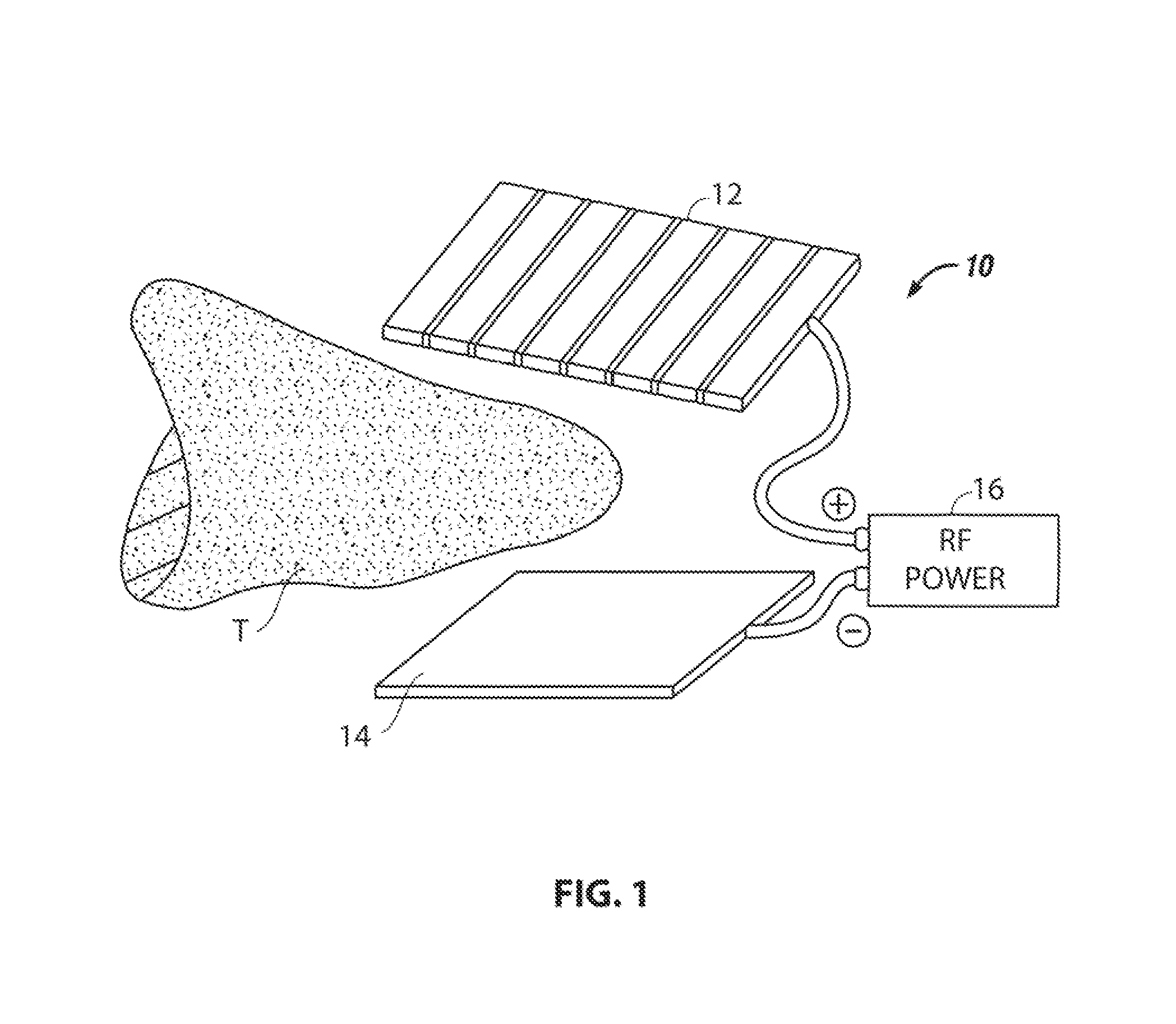
[0026]The methods, systems, and apparatus of the invention are useful for treating a variety of organs, portions of organs, or other solid tissue regions in a patient. The organ or other tissue mass have spaced-apart tissue surfaces, usually opposed tissue surfaces, which are accessible to electrode structures and which permit the application of radio frequency power between said surfaces. The tissue surfaces may be readily accessible or may require pre-treatment to gain access, e.g. blunt dissection, resection of small tissues or blood vessels using conventional surgical techniques, or the like. Organs which may be treated by the invention include the uterus, liver, prostate, kidney, bowel, pancreas, lung, breast, muscle, and the like.

[0027]The organs and other tissue are treated with bipolar radio frequency power directed at target tissue regions which are defined by spaced-apart placement of the electrode structures. The radio frequency power may be supplied by conventional gener...


PUM


Abstract

The invention is concerned with cauterizing and resecting tissue. A pair of electrodes are placed on opposed tissue surfaces, and radio frequency power is applied through the electrodes to cauterizing a tissue mass therebetween. After cauterization has been effected, the tissue may be resected along a plane within the cauterized region with minimum or no bleeding. The tissue mass may then be removed.

Description

CROSS REFERENCE TO RELATED APPLICATIONS[0001]This application is a Continuation in Part (CIP) of U.S. patent application Ser. No. 11 / 382,652, filed May 10, 2006 now U.S. Pat. No. 7,942,874, and claims priority to U.S. provisional patent application Ser. No. 60 / 746,256 filed May 2, 2006, the entirety of each of which is incorporated herein by this reference thereto.BACKGROUND OF THE INVENTION[0002]1. Field of the Invention[0003]The invention relates generally to tissue cauterization. More particularly, the invention relates to an improved electrode for tissue cauterization.[0004]2. Description of the Background Art[0005]Tissue and organ removal are required in a number of surgical procedures for a number of purposes. A major concern in all tissue removal procedures is hemostasis, i.e. cessation of bleeding. All blood vessels supplying an organ or a tissue segment to be removed have to be sealed, either by suturing or cauterization, to inhibit bleeding when the tissue is removed. For ...

Claims


Application Information

Patent Timeline
Patent Type & Authority Patents(United States)
IPC IPC(8): A61B18/14A61B17/29
CPCA61B18/1442A61B2018/00505A61B2018/00595A61B2018/0063A61B2018/00678A61B2019/464A61B2018/00791A61B2018/00827A61B2018/00875A61B2018/1412A61B2018/1455A61B2018/00702A61B2090/064A61B18/12A61B18/14A61B18/16
Inventor EDER, JOSEPHNEZHAT, CAMRANNORDELL, II, BENJAMIN THEODOREWALBERG, ERIKEDELSTEIN, PETER SETH
Owner AESCULAP AG
Why Eureka
Try Eureka