Method for processing radiological images to determine a 3D position of a needle

a radiological image and 3d technology, applied in the field of medical imaging, can solve the problems of non-negligible x-ray dose and time-consuming to determine the 3d imag

Active Publication Date: 2013-12-03
GENERAL ELECTRIC CO
View PDF26 Cites 12 Cited by
  • Summary
  • Abstract
  • Description
  • Claims
  • Application Information

AI Technical Summary

Problems solved by technology

One problem is that the determining of a 3D image takes time.
In addition, for images obtained by tomography, determining of the 3D image requires the acquisition of numerous 2D images resulting in a non-negligible X-ray dose.

Method used


Image

  • Method for processing radiological images to determine a 3D position of a needle
  • Method for processing radiological images to determine a 3D position of a needle
  • Method for processing radiological images to determine a 3D position of a needle

Examples


Embodiment Construction

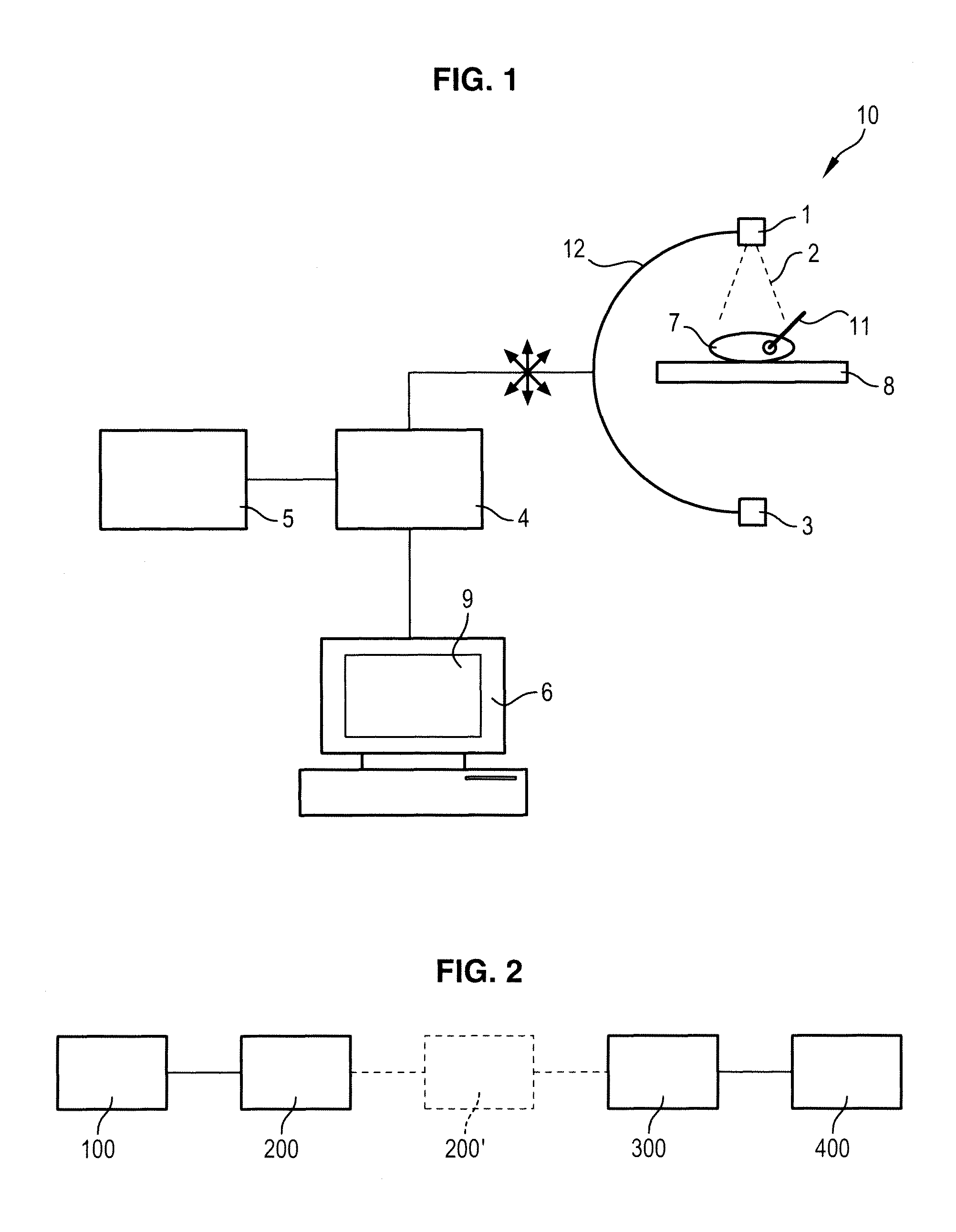
[0017]FIG. 1 illustrates a medical imaging system in accordance with one embodiment of the invention. In FIG. 1, the medical imaging system comprises a source 1 intended to emit a beam 2 of X-rays, a detector 3 arranged facing the source 1 and configured to detect the rays emitted by the source 1, a support 8 arranged between the source 1 and the detector 3, a processing unit 4, a storage unit 5 and an interface unit 6.

[0018]The X-ray source 1 and the detector 3 are connected via a C-arm 12. The arm 12 is more commonly called a vascular C-arm. The arm 12 can be orientated over three degrees of freedom as is illustrated by the arrows in FIG. 1.

[0019]It is the processing unit 4 which controls the position of the arm 12 i.e. the position of the X-ray source 1 in relation to the detector 3.

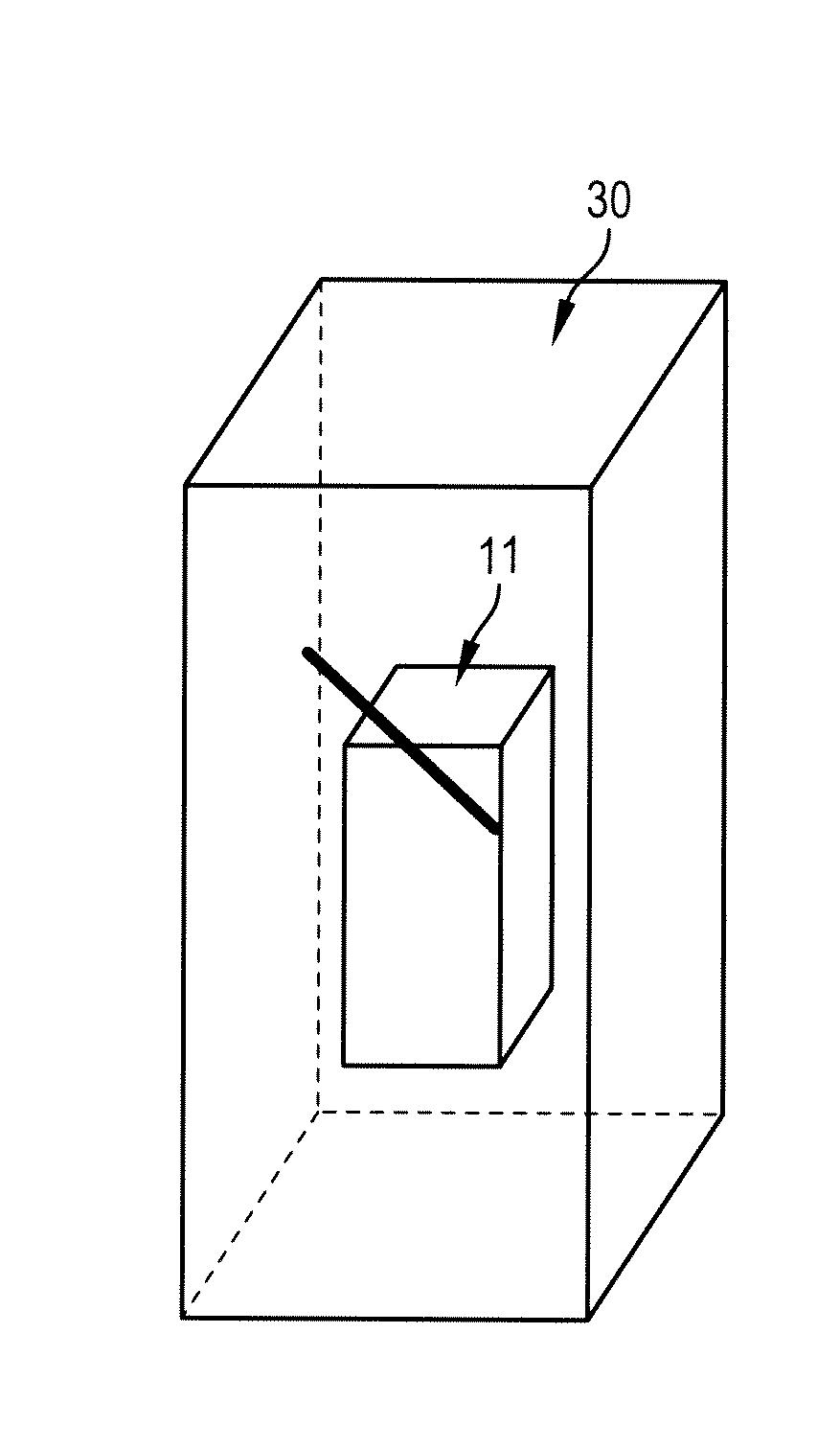
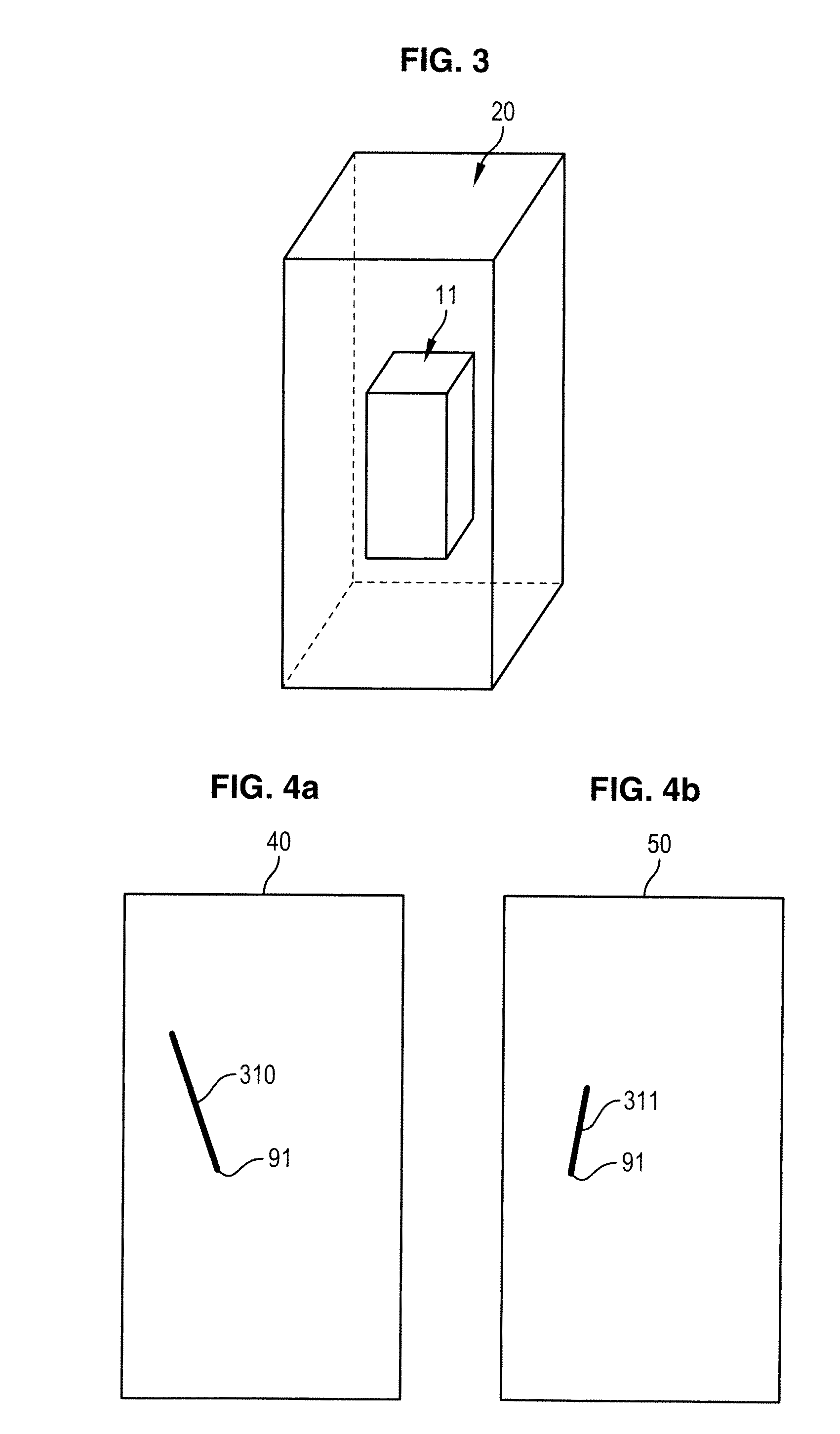
[0020]The support 8 is intended to receive a patient 7 in whom the surgeon is to perform surgery, such as vertebroplasty.

[0021]The processing unit 4 is configured to command emission of X-rays by the ...


PUM


Abstract

A method to process images for interventional imaging, wherein a 3D image of an object is visualized with a medical imaging system, the medical imaging system comprising an X-ray source and a detector, is provided. The method comprises acquiring a plurality of 2D-projected images of the object along a plurality of orientations of the imaging chain, wherein a rectilinear instrument has been inserted into the object. The method also comprises determining a 3D reconstruction of the instrument such that a plurality of 2D projections of the 3D image of the instrument, along the respective orientations in the 2D-projected images of the object were acquired, are closest to the acquired 2D-projected images of the object. The method further comprises superimposing the 3D reconstruction of the instrument over the 3D image of the object so as to obtain a 3D image comprising the object and the instrument.

Description

CROSS-REFERENCE TO RELATED APPLICATIONS[0001]This application claims priority under 35 U.S.C. §119(a) to prior-filed, co-pending French patent application serial number 1053955, filed on May 22, 2010, the disclosure of which is hereby incorporated by reference in its entirety.BACKGROUND OF THE INVENTION[0002]1. Field of the Invention[0003]Embodiments of the present invention relate to the field of medical imaging and more particularly relate to the processing of interventional radiology images. Embodiment of the present invention further particularly relate to a method and system for allowing the real-time display of the position of a rectilinear instrument, notably a needle, in a region of interest of a patient.[0004]2. Description of the Prior Art[0005]During a surgical operation, a surgeon must insert a needle in a region of interest of a patient, for example, into the patient's spine. To control the insertion of the needle, the surgeon must be able to visualize the region of int...

Claims


Application Information

Patent Timeline
Patent Type & Authority Patents(United States)
IPC IPC(8): G06K9/00
CPCG06T7/0044G06T7/0065G06T11/006G06T2207/10081G06T2207/10116G06T2207/30021G06T2211/436G06T7/55G06T7/74
Inventor GORGES, SEBASTIENTROUSSET, YVES
Owner GENERAL ELECTRIC CO
Why Eureka
Try Eureka