
Intraocular pressure reduction with intracameral bimatoprost implants

a bimatoprost and intraocular pressure technology, applied in the field of ocular conditions, can solve the problems of difficult glaucoma management, layer does not offer much resistance to aqueous humor outflow, etc., and achieve the effect of preventing progressive optic neuropathy and profound reduction of iop

Active Publication Date: 2014-03-18
ALLERGAN INC
View PDF264 Cites 15 Cited by
  • Summary
  • Abstract
  • Description
  • Claims
  • Application Information

AI Technical Summary

Benefits of technology

The patent text describes a new treatment for glaucoma that reduces the pressure in the eye. This treatment involves using a special implant that is placed in the eye and can help to lower the pressure gradually over a period of time. This implant can offer an alternative to other treatments like eye drops and surgery, which may be less effective or have potential risks. By using this new method, many patients with glaucoma can achieve significant reduction in pressure and potentially avoid the need for these other treatments.

Problems solved by technology

This layer does not offer much resistance to aqueous humor outflow because intercellular spaces are large.
Increases in the episcleral venous pressure such as seen with carotid-cavernous sinus fistulas, orbital varices, and Sturge-Weber Syndrome, can lead to difficult to manage glaucoma.

Method used


Image

  • Intraocular pressure reduction with intracameral bimatoprost implants
  • Intraocular pressure reduction with intracameral bimatoprost implants
  • Intraocular pressure reduction with intracameral bimatoprost implants

Examples


example 1

Intracameral Bimatoprost Implant with High Initial Release Rate

[0093]A bimatoprost implant comprising Bimatoprost 30%, R203S 45%, R202H 20%, PEG 3350 5% was manufactured with a total implant weight of 900 mg (drug load 270 ug). The in vitro release rates of this implant are shown in FIG. 8. This implant releases ˜70% over first 30 days. An implant with a 270 ug drug load would release 189 ug over first 30 days or 6.3 ug per day. The remainder of the implant (81ug) is released over the next 4 months (i.e. 675 ng per day).

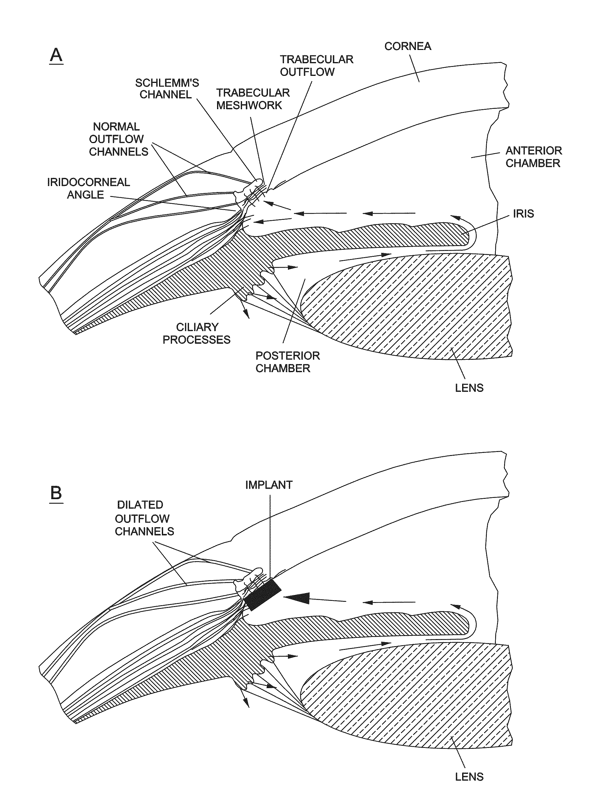
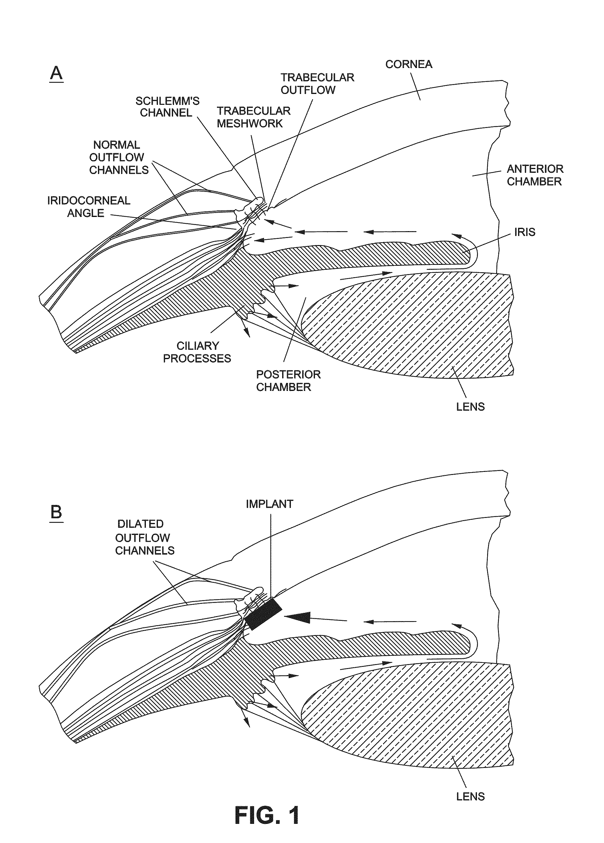
[0094]A normal beagle dog was given general anesthesia and a 3 mm wide keratome knife was used to enter the anterior chamber of the right eye. The intracameral bimatoprost implant was placed in the anterior chamber and it settled out in the inferior angle within 24 hours. As shown in FIG. 5, the IOP was reduced to approximately −60% from baseline and this was sustained for at least 5 months (See FIG. 5). As shown in FIG. 4,the episcleral vessels are dilated.

example 2

Intracameral Bimatoprost Implant with Slow Initial Release Rate

[0095]A bimatoprost implant comprising Bimatoprost 20%, R203S 45%, R202H 10%, RG752S 20%, PEG 3350 5% was manufactured with a total implant weight of 300 ug or 600 ug (drug loads of 60 or 120 ug, respectively). The in vitro release rates of this implant are shown in FIG. 9. The implant releases ˜15% of the drug load over the first month. An implant with a 60 ug drug load would release 9 ug over first 30 days or 300 ng per day, thereafter, it releases ˜50 ug over 60 days or ˜700 ng / day. Like Example 1, it was found that the episcleral vessels were dilated.

example 3

[0096]The following experiment was carried out by inserting the implants described below in six Beagle dogs:

[0097]Implant Formulations:

[0098]2 mm Bimatoprost implant in applicator (20% Bimatoprost, 45% R203s, 20% RG752s, 10% R202H, 5% PEG-3350)

[0099]2 mm, Placebo implant in applicator (56.25% R203s, 25% RG752s, 12.25% R202H, 6.25% PEG-3350)

[0100]Dog 1,2,3: API implant intracameral OD (one 2 mm implant), OS placebo implant

[0101]Dog 4,5,6: API implant intracameral OD (two 2 mm implants), OS placebo implant

[0102]

Implant WeightDrug DoseDog ID(mg)(20% load, ug)CYJ AUS0.31763.4CYJ AYE0.32665.2CYJ AUR0.31563.0CYJ AUG0.302126.60.331CYJ BAV0.298125.40.329CYJ BBY0.306126.60.327

[0103]Surgical Procedure: Implants were loaded in a customized applicator with a 25G UTW needle. Under general anesthesia, normal beagle dogs had the implant inserted in the anterior chamber through clear cornea and the wound was self-sealing. The applicator is described in Published United States Patent Application 200...


PUM

PropertyMeasurementUnit
timeaaaaaaaaaa
lengthsaaaaaaaaaa
lengthsaaaaaaaaaa

Abstract

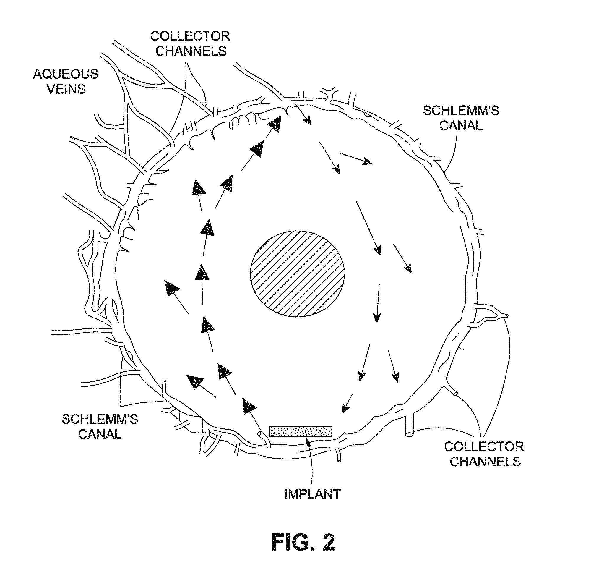
The present invention provides a method of treating an ocular condition in an eye of a patient, comprising the step of placing a biodegradable intraocular implant in an eye of the patient, the implant comprising a prostamide and a biodegradable polymer matrix that releases drug at a rate effective to sustain release of an amount of the prostamide from the implant to provide an amount of the prostamide effective to prevent or reduce a symptom of an ocular condition of the eye, wherein said ocular condition is elevated IOP and said implant is placed in an intracameral location to dilate the outflow channels of the eye emanating from Schlemm's Canal.

Description

CROSS REFERENCE TO RELATED APPLICATIONS[0001]This application is a continuation in part of U.S. patent application Ser. No. 10 / 837,260, filed on Apr. 30, 2004 in the names of Hughes et al, incorporated entirely by reference.BACKGROUND OF THE INVENTION[0002]1. Field of the Invention[0003]The present invention relates to a method of treating an ocular condition, comprising the step of placing a biodegradable intraocular implant in an eye of the patient, the implant comprising a prostamide and a biodegradable polymer matrix that releases drug at a rate effective to sustain release of an amount of the prostamide from the implant to provide an amount of the prostamide effective to prevent or reduce a symptom of the ocular condition, wherein said ocular condition is elevated IOP.[0004]2. Summary of the Related Art[0005]The anterior and posterior chambers of the eye are filled with aqueous humor, a fluid predominantly secreted by the ciliary body with an ionic composition similar to the bl...

Claims


Application Information

Patent Timeline
Patent Type & Authority Patents(United States)
IPC IPC(8): A61F2/14
CPCA61K9/204A61K31/557A61K47/34A61K9/0051A61K31/5575A61K31/165A61P27/00A61P27/02A61P27/06A61P9/12
Inventor HUGHES, PATRICK, M.
Owner ALLERGAN INC
Learn More