Sinus valved glaucoma shunt

Inactive Publication Date: 2005-11-22
CLARITY
View PDF7 Cites 95 Cited by
  • Summary
  • Abstract
  • Description
  • Claims
  • Application Information

AI Technical Summary

Benefits of technology

[0021]According to a further aspect of the present invention, a method is provided for preventing postoperative hypotony comprising implanting in an animal's eye in need thereof a shunt, said shunt comprising a tubing; a crossbeam affixed to the tubing; a bulb; and a slit valve, wherein the tubing's length allow for aqueous humor fluid flow from the anterior chamber of an animal's eye directly into the frontal sinus cavity.
[0022]The device is an implantable shunt for diverting or draining aqueous humor from the anterior chamber of an animal's eye to the frontal sinus via tubing, having a guide needle at one end thereof, for flushing the shunt preoperatively to ensure slit-valve function and a plug tip at an opposite end of the tubing. The guide needle is removable from the tubing and does not remain in the frontal sinus. Slits are formed at 90° on the tubing adjacent to the pl

Problems solved by technology

Despite its importance, the long-term control of canine primary glaucoma, whether medical or surgical, continues to elude the veterinary profession.
One of the disadvantages of the art is that implants in dogs fail to maintain normotensive intraocular pressures for more than 6 months postoperatively or do so only when combined with other forms of glaucoma therapy including medical and surgical management.
In other words, one of the problems with the art is that the implantable shunts do not remain secure to the site for the

Method used


Image

  • Sinus valved glaucoma shunt
  • Sinus valved glaucoma shunt
  • Sinus valved glaucoma shunt

Examples


Embodiment Construction

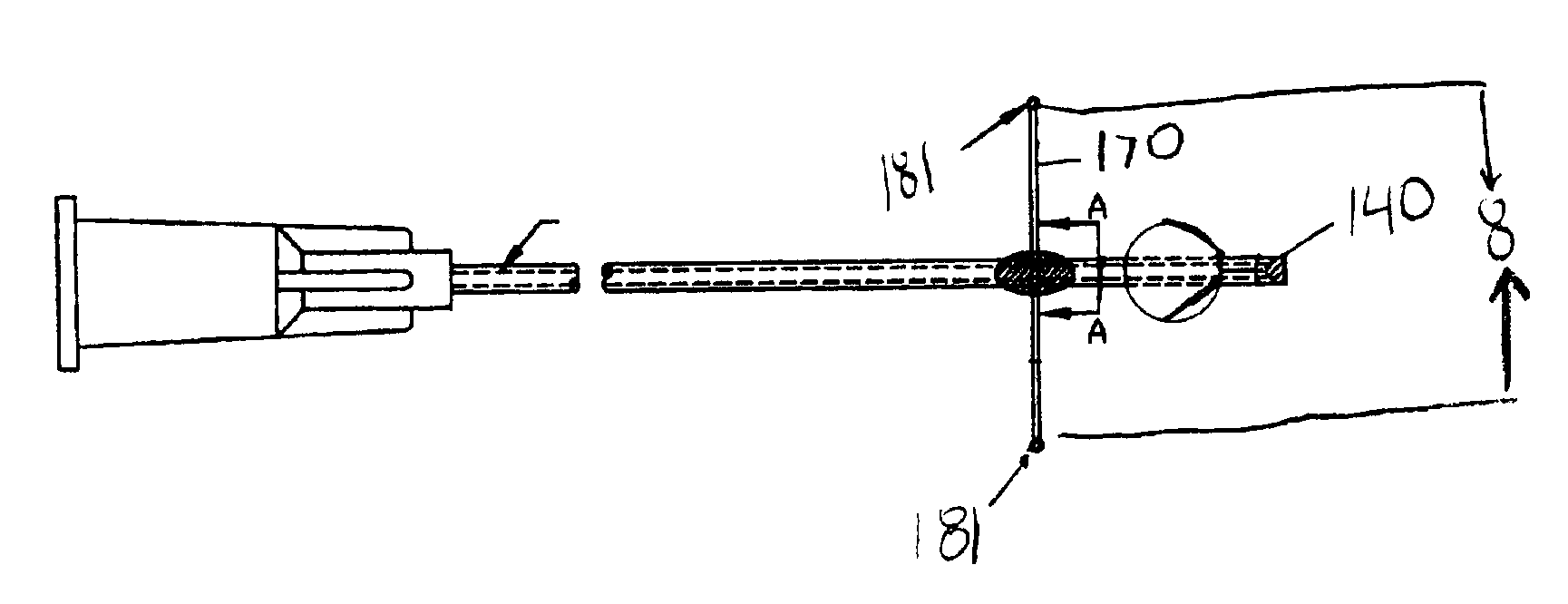
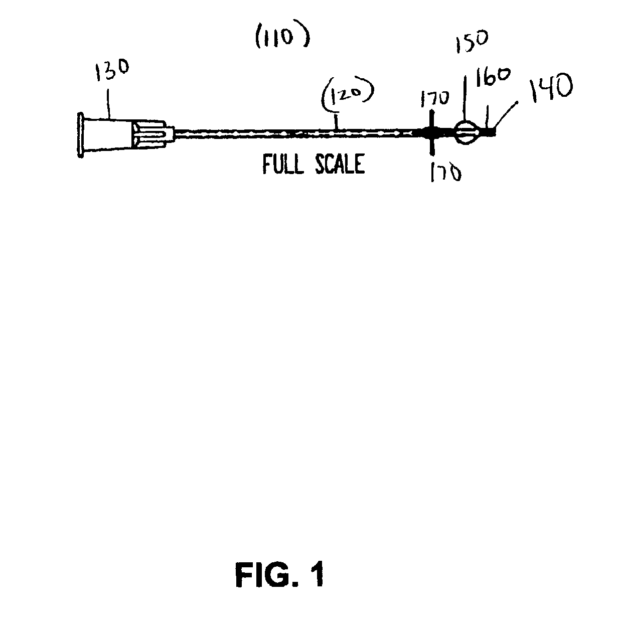
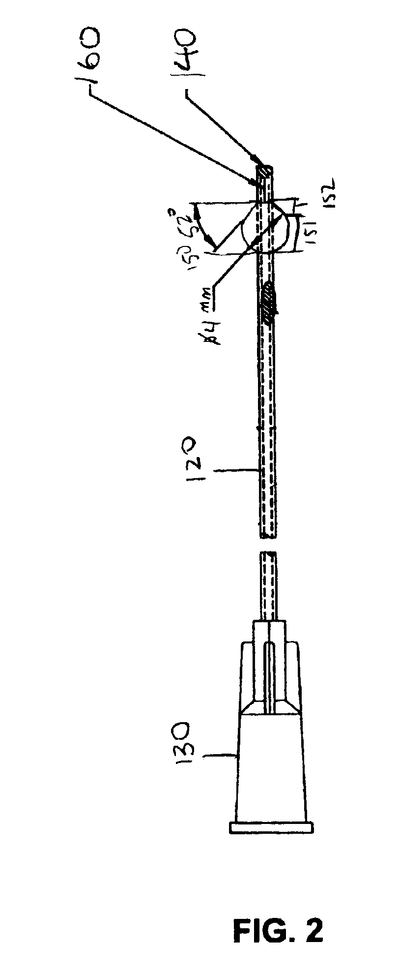
[0029]The present invention provides an a method for treating primary glaucoma and an anterior chamber shunt device to drain or divert aqueous humor in an animal's eye from the anterior chamber into the frontal sinus cavity, in which the shunt device comprises a first end, adapted to be fitted with a guide needle, to be received within the anterior chamber following removal of the guide needle, and a second end having a crossbeam, bulb, slits and a plug tip to be received within the frontal sinus cavity, wherein the device permits aqueous humor communication from the anterior chamber to the frontal sinus cavity through the slit valves. Fluid communication can be facilitated by intraocular pressure directing the aqueous humor into the slits, as described below.

[0030]The embodiments of the present invention can be used to treat animals with primary glaucoma, particularly to drain or divert aqueous humor extraocularly and, more particularly to prevent postoperative hypotony.

[0031]Refer...


PUM


Abstract

The invention relates to a device and method for treating animals, such as humans and dogs, with primary glaucoma by draining or diverting aqueous humor extraocularly comprising a shunt implant wherein the length and tubing of the shunt ensure fluid flow from the eye directly into the frontal sinus cavity via tubing. The device has crossbeam aids in anchoring the device; four slit valves to control fluid flow at required volume; and bulb that anchors in the frontal sinus cavity. The device is preferably made of medical grade radiopaque silicone rubber, and is flexible. Other improvements and a method for implanting the device are disclosed as well.

Description

FIELD OF THE INVENTION[0001]The present invention relates generally to the field of glaucoma treatment and glaucoma devices, in particular to a new and useful implantable glaucoma shunt for relieving internal pressure in an animal's eye. More particularly, the present invention provides for a device and method for draining or diverting aqueous humor. Even more particularly, the invention provides for an apparatus and method of preventing postoperative hypotony.BACKGROUND OF THE INVENTION[0002]Glaucoma is a relatively common ocular disorder in animals. For example, glaucoma has long been recognized as a leading cause of human and canine blindness. The incidence of canine glaucoma, for example, is 0.5%. Increased incidence however is noted in specific breeds. Despite its importance, the long-term control of canine primary glaucoma, whether medical or surgical, continues to elude the veterinary profession.[0003]The current state of the art for long term control of glaucoma in humans an...

Claims


Application Information

Patent Timeline
IPC IPC(8): A61M5/00A61BA61F9/007
CPCA61F9/00781
Inventor CULLEN, CHERYLMARTEN, LEWIS H.
Owner CLARITY
Why Eureka
Try Eureka