Compositions and methods for inhibition of MMP13:MMP-substrate interactions
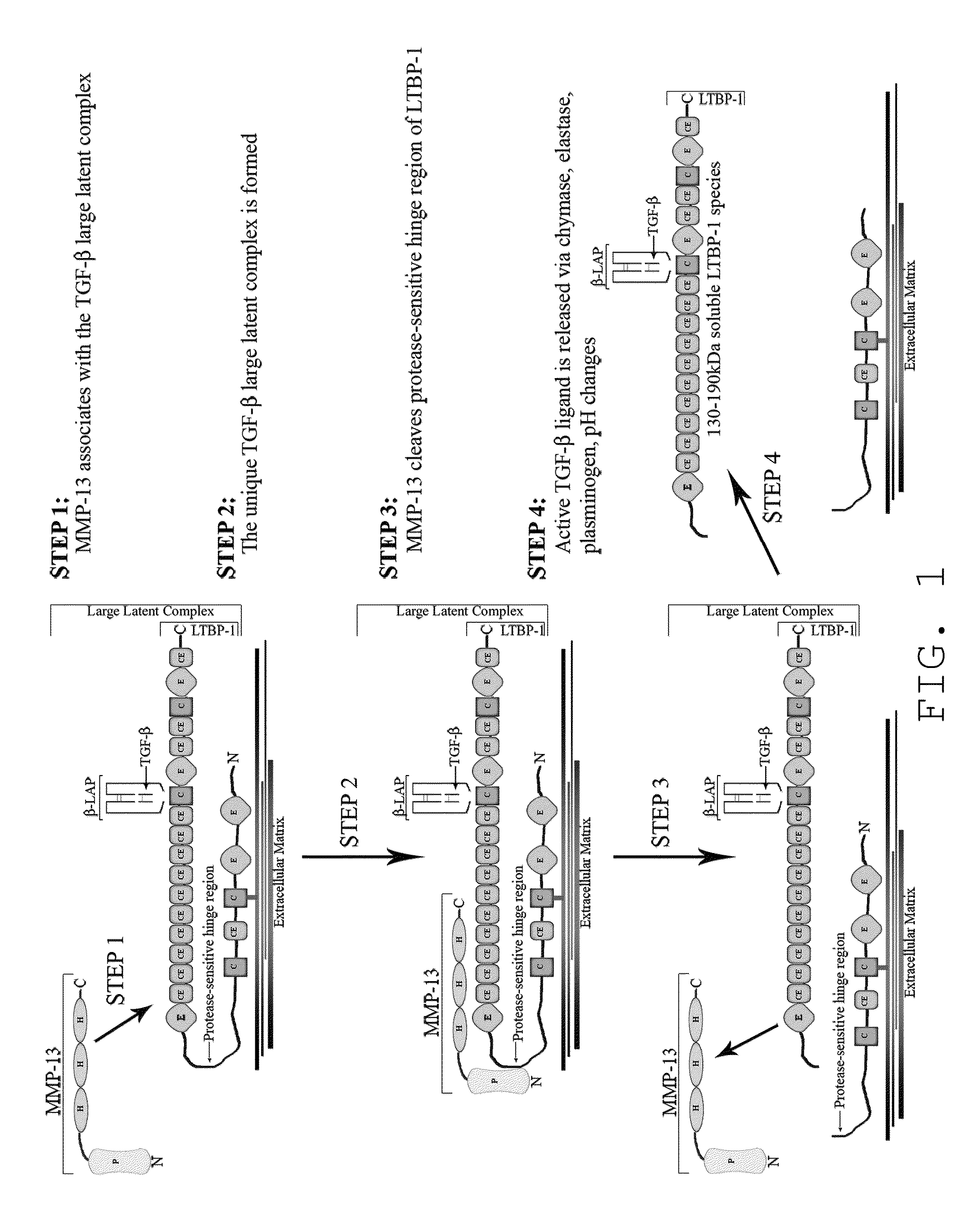
a technology of mmp13 and substrate, which is applied in the field of compositions and methods for inhibiting mmp13 and mmpsubstrate interactions, can solve problems such as severe side effects, and achieve the effects of preventing ltbp1, and inhibiting the activation of tg
- Summary
- Abstract
- Description
- Claims
- Application Information
AI Technical Summary
Problems solved by technology
Method used
Image
Examples
example 1
Materials and Methods
[0135]Cartilage Extract Preparation, Immunoprecipitation and Immunoblot Analysis
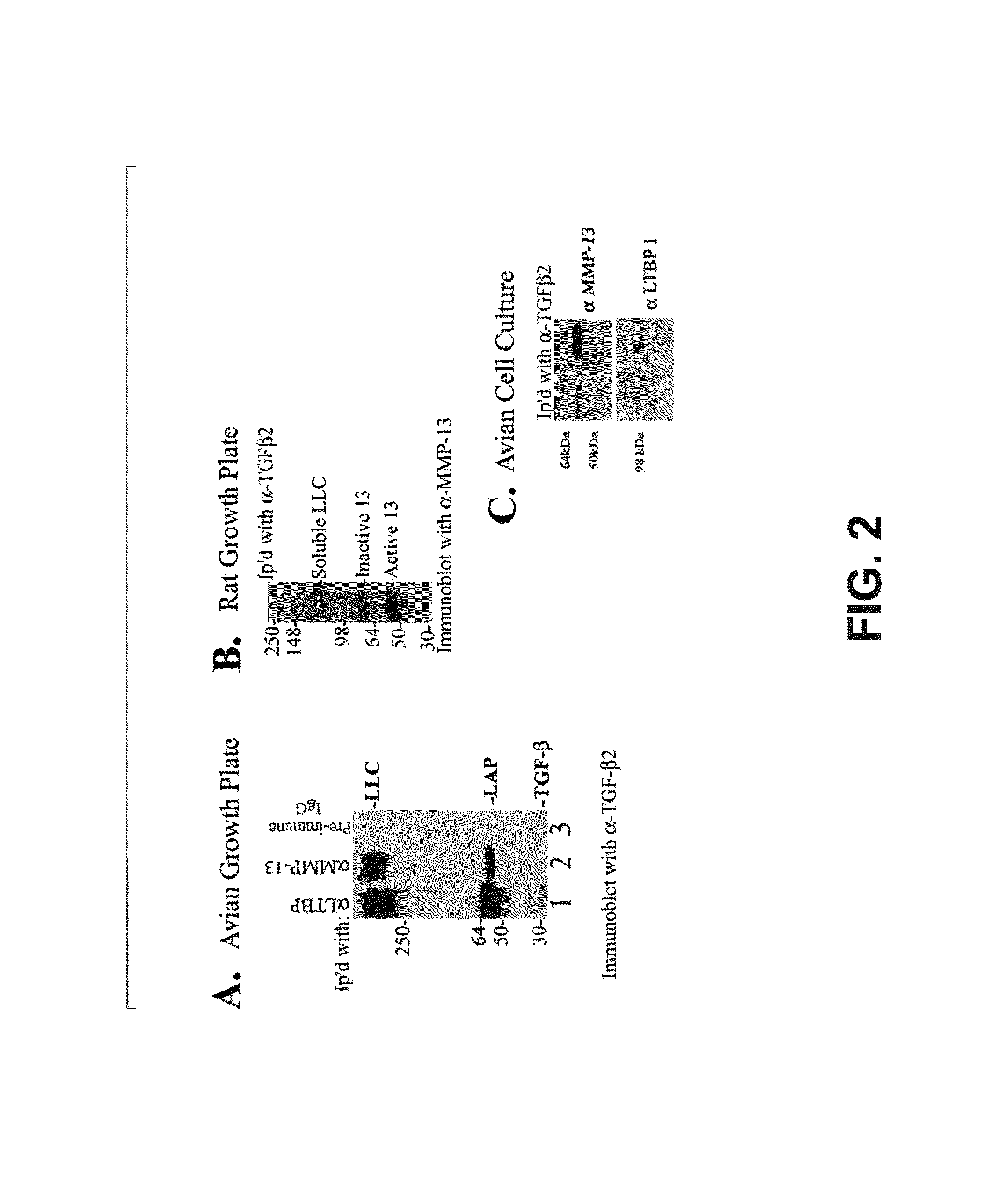
[0136]Tibia from normal Sprague-Dawley 14 day old newborn rat pups or day 19 chick embryos were dissected free of tissue and the tibial growth plates isolated by microdissection. The cartilage was minced and extracted overnight in 0.5% 3-([3-chlomadipropyl]dimethylammonio)-1-propane-sulfonate (CHAPS) buffer [10 mM Tris, 100 mM NaCl, 2 mM EDTA, pH 7.6] (Sigma, St. Louis, Mo., USA). Avian tissue was immunoprecipitated with rabbit anti human LTBP-1 (a kind gift of Dr. Kohei Miyazono), rabbit anti avian MMP-13 (D'Angelo, et al., 2000) or rabbit pre-immune serum (Pierce Biochemicals, Rockford, Ill., USA). Rat tissue was immunoprecipitated with rabbit anti human LTBP-1 or rabbit anti human TGFβ2 polyclonal antibodies (Santa Cruz Biotechnology, Inc., Santa Cruz, Calif., USA). Concurrently, TGFβ antibody was biotinylated using the EZ-Link Sulfo-NHS-LC-Biotinylation Kit according to protocol ...
example 2
Materials and Methods
[0158]Avian Chondrocyte Isolation and Serum-Free Cell Culture
[0159]Sterna were removed from day 17 chick embryos using microsurgical techniques as previously described (D'Angelo et al., J. Bone Miner. Res. 12:1368-1377, 1997). Cells were released by digestion of the extracellular matrix in 0.25% trypsin and 0.1% crude collagenase mixture (Sigma, St. Louis, Mo., USA) in Hanks' buffered saline solution for 3-4 hours at 37° C. Digestion was halted by suspension in high glucose Dulbecco's modified Eagle's medium (DMEM) containing 10% fetal bovine serum (FBS) (Invitrogen Life Technologies, Carlsbad, Calif., USA). The cell suspensions were then filtered through a 0.2 μm filter, counted, seeded onto Falcon 6-well, tissue culture treated plates and covered with 2 ml of high glucose DMEM containing 10% NuSerum and 50 μg / ml of penicillin / streptomycin, 2 mM of L-glutamine (Invitrogen Life Technologies, Carlsbad, Calif., USA). After 48 hours the cells were rinsed and proces...
example 3
[0177]To measure the ability of MMP13-19 peptide to bind endogenous large latent complex of TGFβ and interfere with the activation of this growth factor, hypertrophic chondrocytes were cultured in alginate beads in serum-free medium for 24 hours with varying concentrations of MMP13-19 peptide. Conditioned media was then subjected to an ELISA to measure total TGFβ produced and the percentage of endogenously activated TGFβ (FIG. 11). All three doses (10, 100, and 750 nM) of MMP13-19 peptide resulted in a statistically significant decrease in endogenously activated TGFβ although the total amount of TGFβ produced was not affected. These data indicate that the MMP13-19 peptide can be used as an inhibitor of TGFβ activation.
PUM
| Property | Measurement | Unit |
|---|---|---|
| voxel size | aaaaa | aaaaa |
| pKa | aaaaa | aaaaa |
| temperature | aaaaa | aaaaa |
Abstract
Description
Claims
Application Information
 - R&D
- Intellectual Property
- Life Sciences
- Materials
- Tech Scout
- Unparalleled Data Quality
- Higher Quality Content
- 60% Fewer Hallucinations
Browse by: Latest US Patents, China's latest patents, Technical Efficacy Thesaurus, Application Domain, Technology Topic, Popular Technical Reports.
© 2025 PatSnap. All rights reserved.Legal|Privacy policy|Modern Slavery Act Transparency Statement|Sitemap|About US| Contact US: help@patsnap.com


