Method and system for ultrasound based automated detection, quantification and tracking of pathologies

an automated detection and pathology technology, applied in the field of ultrasonography, can solve the problems of increasing the time and cost of providing care to patients, failing to paint an accurate picture of the underlying 3d structure of the anatomy, and other imaging methods such as ct and mr are relatively expensiv

Active Publication Date: 2014-07-15
GENERAL ELECTRIC CO
View PDF8 Cites 48 Cited by
  • Summary
  • Abstract
  • Description
  • Claims
  • Application Information

AI Technical Summary

Problems solved by technology

Unfortunately, this practice leads to additional time and cost in providing care to the patient.
However, modalities like X-ray entail use of 2D projections of the anatomy and fail to paint an accurate picture of the underlying 3D structure of the anatomy.
Also, other imaging methods like CT and MR are relatively expensive and are contraindicated for certain groups of patients (for example, patients with pacemakers cannot undergo MR scans).
In addition, use of 2D ultrasound leads to subjective diagnosis even among relatively skilled ultrasound practitioners.

Method used


Image

  • Method and system for ultrasound based automated detection, quantification and tracking of pathologies
  • Method and system for ultrasound based automated detection, quantification and tracking of pathologies
  • Method and system for ultrasound based automated detection, quantification and tracking of pathologies

Examples


Embodiment Construction

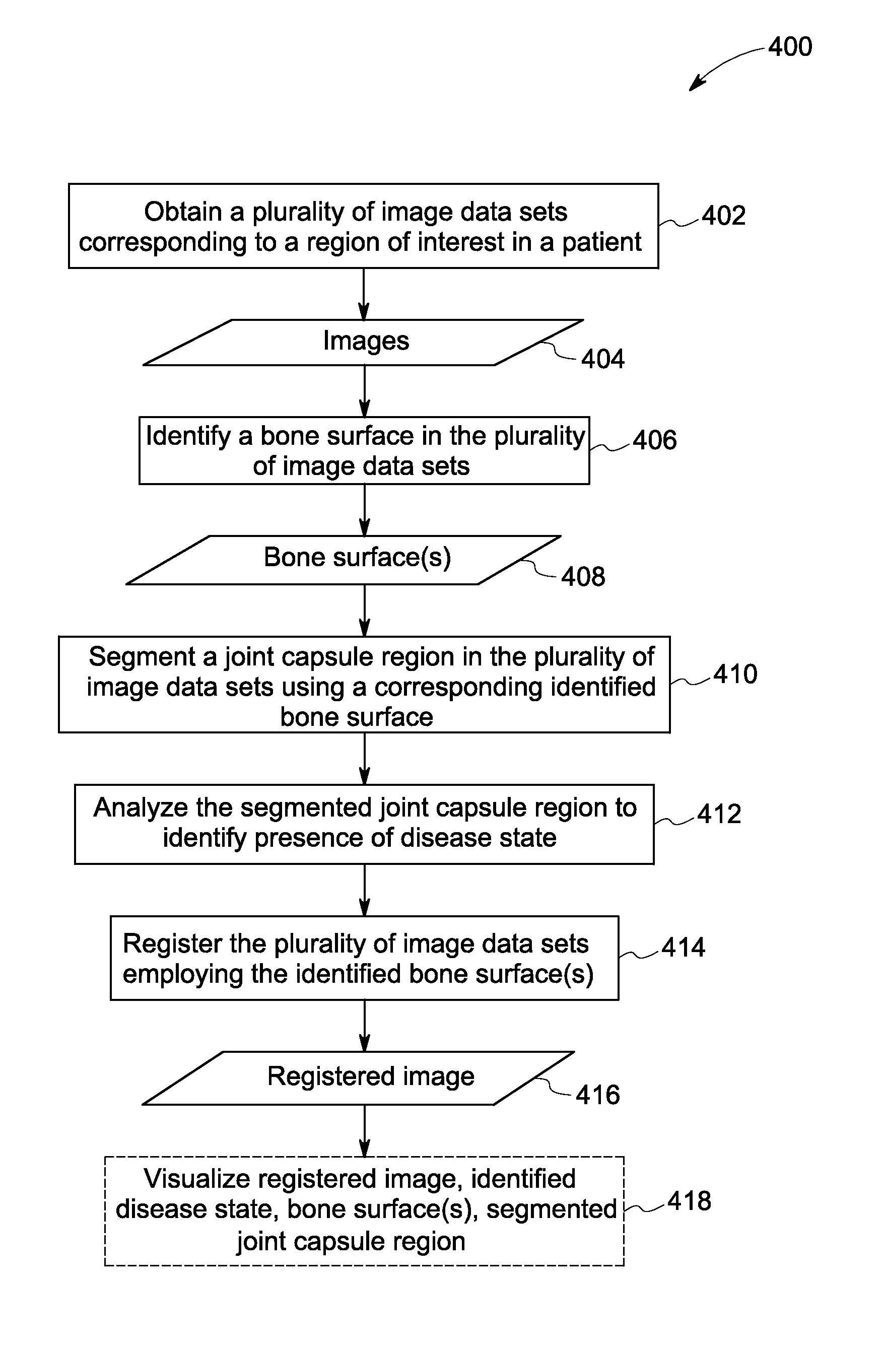
[0021]As will be described in detail hereinafter, various methods and systems for the automated ultrasound based detection, quantification and tracking of pathologies are presented. Employing the method and system described hereinafter facilitates early diagnosis, quantification (scoring) and enhances longitudinal tracking of the pathologies, while reducing operator dependence in the assessment of pathologies. Moreover, a method for the objective assessment of the pathologies is presented, thereby enhancing the efficiency of the diagnosis of the pathologies.

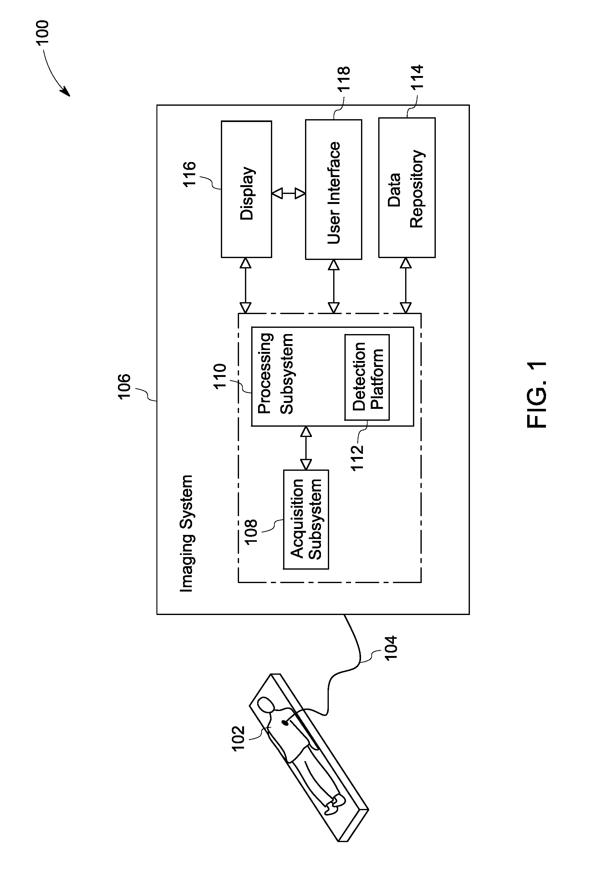
[0022]FIG. 1 is a block diagram of an exemplary system 100 for use in diagnostic imaging in accordance with aspects of the present technique. The system 100 is configured to facilitate automated detection, quantification and tracking of pathologies, such as musculoskeletal pathologies using ultrasound images corresponding to an anatomical region of interest in an object of interest. To that end, the system 100 may be configured t...


PUM


Abstract

An automated method for detecting a disease state is presented. The method includes identifying a bone surface in one or more image data sets, wherein the one or more data sets correspond to a region of interest in an object of interest. Furthermore, the method includes segmenting a joint capsule region corresponding to the one or more image data sets based on a corresponding identified bone surface. In addition, the method includes analyzing the segmented joint capsule region to identify the disease state. Systems and non-transitory computer readable medium configured to perform the automated method for detecting a disease state are also presented.

Description

CROSS REFERENCE TO RELATED APPLICATIONS[0001]This non-provisional application claims the benefit of priority under 35 U.S.C. §119(e) to U.S. Provisional Patent Application Ser. No. 61 / 531,165, filed Sep. 6, 2011, which is herein incorporated in its entirety by reference.BACKGROUND[0002]Embodiments of the present disclosure relate to ultrasound imaging, and more particularly to systems and methods for automated ultrasound based detection, quantification, and tracking of musculoskeletal pathologies over time.[0003]Arthritis is a common cause of disability especially in the older population and it is one of the most chronic diseases in the U.S. Generally, the current musculoskeletal practice relies heavily on highly trained radiologists to perform and analyze images of the anatomy of interest. Unfortunately, this practice leads to additional time and cost in providing care to the patient.[0004]Currently, imaging techniques like computed tomography (CT), magnetic resonance (MR) imaging,...

Claims


Application Information

Patent Timeline
Patent Type & Authority Patents(United States)
IPC IPC(8): A61B8/00
CPCG06T2207/30008A61B8/5246A61B8/483A61B8/0875A61B6/505A61B8/5223G06T2207/10132A61B6/5235A61B6/03G06T7/0012A61B8/469G06K9/3241G16H50/30G06V10/255
Inventor PATWARDHAN, KEDAR ANILMILLS, DAVID MARTIN
Owner GENERAL ELECTRIC CO
Why Eureka
Try Eureka