Eureka AIR delivers breakthrough ideas for toughest innovation challenges, trusted by R&D personnel around the world.

Transesophageal echocardiography capsule

a transesophageal echocardiography and capsule technology, applied in the field of ultrasound imaging via the esophagus, can solve the problems of further discomfort and discomfort for the patien

Active Publication Date: 2014-12-30
BIOSENSE WEBSTER (ISRAEL) LTD
View PDF14 Cites 3 Cited by
  • Summary
  • Abstract
  • Description
  • Claims
  • Application Information

AI Technical Summary

Problems solved by technology

Entry of the tube typically causes discomfort to the patient, even though the patient of necessity has been sedated.
Furthermore, an operator of the system typically needs to manipulate the system in the esophagus using the tube, generating further discomfort.

Method used


Image

  • Transesophageal echocardiography capsule
  • Transesophageal echocardiography capsule
  • Transesophageal echocardiography capsule

Examples


Embodiment Construction

Overview

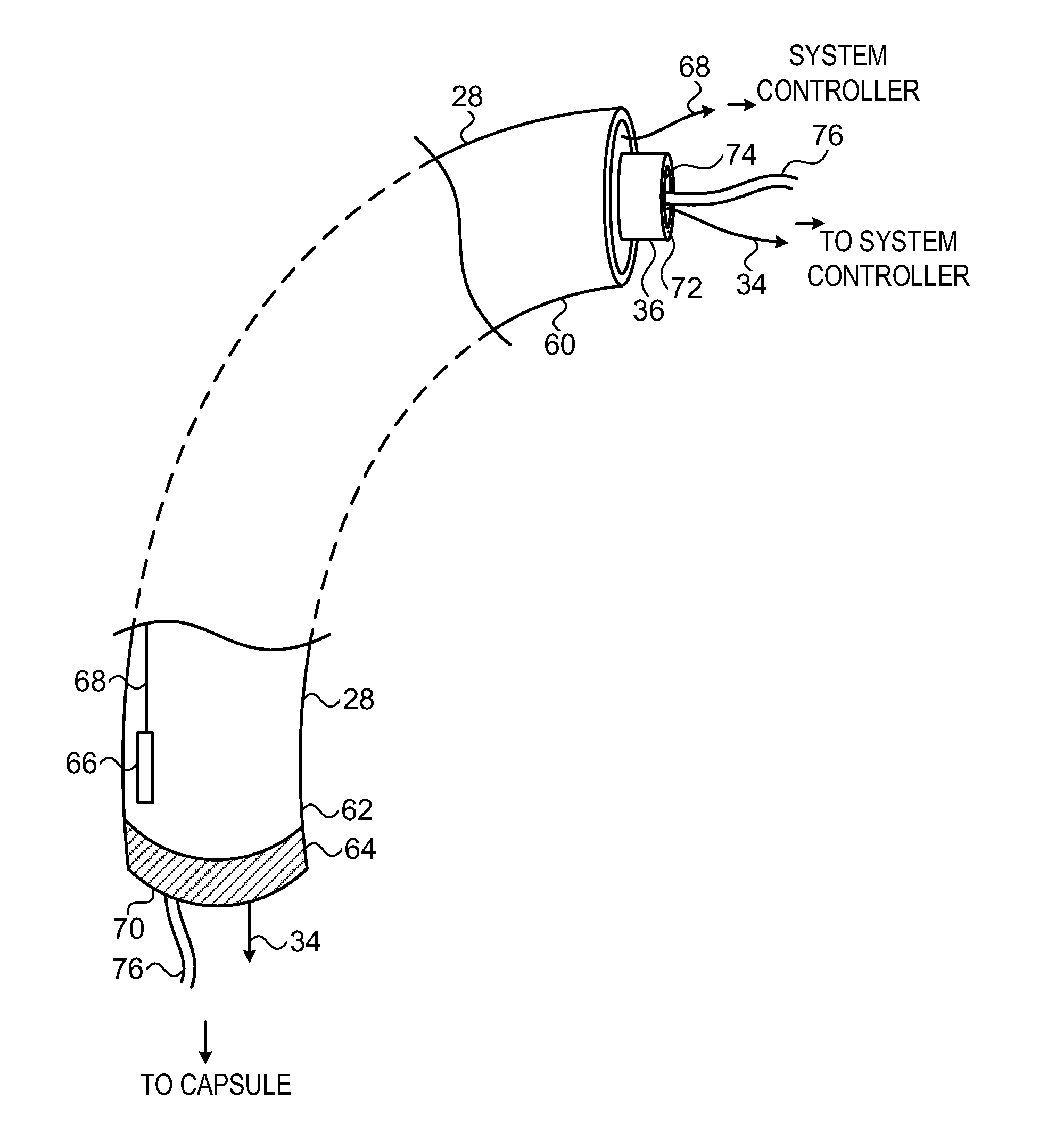
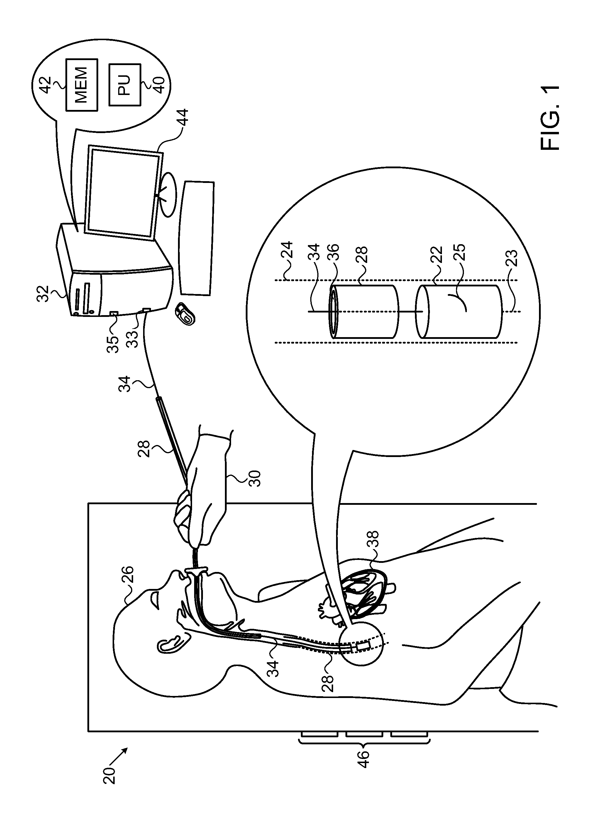
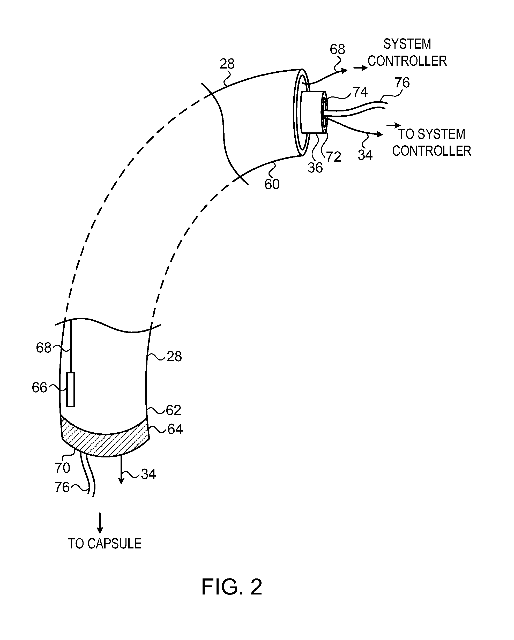
[0037]An embodiment of the present invention provides a transesophageal ultrasound imaging system, which may typically be used to provide ultrasound images of heart tissue of a patient. The system comprises an imaging capsule having an ultrasonic transducer, and the capsule is sized so as to be able to enter the esophagus of the patient. Typically, the transducer is mounted on one or more microelectronic mechanical system (MEMS) pistons, which allow the transducer to be translated and / or oriented while the capsule is fixed in the esophagus. The capsule itself may also include a MEMS rotation device allowing the whole capsule to be reoriented in the esophagus.

[0038]The system also comprises an applicator tube which is also sized to enter the patient's esophagus. The capsule and tube comprise locking and retaining mechanisms which enable the tube and capsule to be attached for positioning the capsule in the esophagus. Once the capsule is in a desired position in the esophagus,...


PUM


Abstract

An imaging system, including a capsule, configured to enter an esophagus of a patient, having an ultrasonic transducer configured to image tissue of the patient. The system further includes an applicator tube configured to enter the esophagus, the tube being attachable to the capsule for positioning the capsule within the esophagus, and being detachable from the capsule after positioning of the capsule so as to permit the tube to be withdrawn from the esophagus while the capsule remains in position in the esophagus.

Description

CROSS-REFERENCE TO RELATED APPLICATION[0001]This application claims the benefit of U.S. Provisional Patent Application 61 / 357,703, filed 23 Jun. 2010, which is incorporated herein by reference.FIELD OF THE INVENTION[0002]The present invention relates generally to imaging, and specifically to ultrasonic imaging via the esophagus of a patient.BACKGROUND OF THE INVENTION[0003]Transesophageal imaging systems are known in the art. Such systems typically require entry of a relatively thick tube into the esophagus of a patient being imaged. Entry of the tube typically causes discomfort to the patient, even though the patient of necessity has been sedated. Furthermore, an operator of the system typically needs to manipulate the system in the esophagus using the tube, generating further discomfort.[0004]Documents incorporated by reference in the present patent application are to be considered an integral part of the application except that to the extent any terms are defined in these incorpo...

Claims


Application Information

Patent Timeline
Patent Type & Authority Patents(United States)
IPC IPC(8): A61B1/04A61B8/00A61B1/00A61B8/12A61B1/273A61B5/05
CPCA61B1/00158A61B1/00101A61B1/00128A61B8/12A61B1/00154A61B1/041A61B1/00082A61B1/2733A61B8/4472
Inventor AMIT, MATIBAR-TAL, MEIRSCHWARTZ, YITZHACK
Owner BIOSENSE WEBSTER (ISRAEL) LTD
Why Eureka
Eureka Blog
Learn More