Method and system of automatic determination of geometric elements from a 3D medical image of a bone

a 3d medical image and geometric element technology, applied in the field of computer assisted surgery, can solve the problems of difficult to determine the correct and full diagnosis of the pathology, the repetitive impact of the proximal femoral neck, and the complex structure of the human body, so as to minimize the energy function

Active Publication Date: 2015-02-24
SMITH & NEPHEW ORTHOPAEDICS
View PDF19 Cites 20 Cited by
  • Summary
  • Abstract
  • Description
  • Claims
  • Application Information

AI Technical Summary

Benefits of technology

[0073]ii) minimizing the energy function by minimizing the distance between contiguous points.

Problems solved by technology

Articulations of the human body are often very complex systems and no precise generic model exists to capture all the variability from one articulation to another.
Structural abnormalities in the morphology of the hip can limit motion and result in repetitive impact of the proximal femoral neck against the acetabular labrum and its adjacent cartilage.
However, the correct and full diagnosis of this pathology is not easy to determine, especially when dealing with subtle deformities.
However, such processing of the 3D image remains a laborious manual task, comprising manual identification of the neck axis and manual fitting of a circle to the head of the bone in several 2D images, which cannot ensure accuracy and reproducibility, and can potentially mislead the diagnosis or the surgical indication.

Method used


Image

  • Method and system of automatic determination of geometric elements from a 3D medical image of a bone
  • Method and system of automatic determination of geometric elements from a 3D medical image of a bone
  • Method and system of automatic determination of geometric elements from a 3D medical image of a bone

Examples


Embodiment Construction

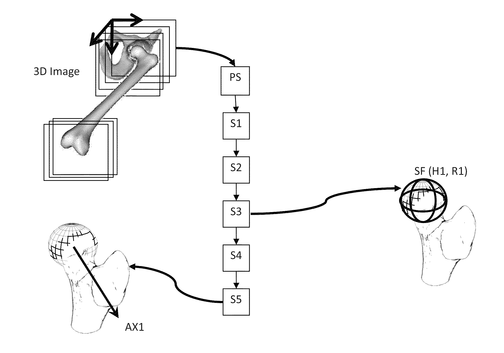
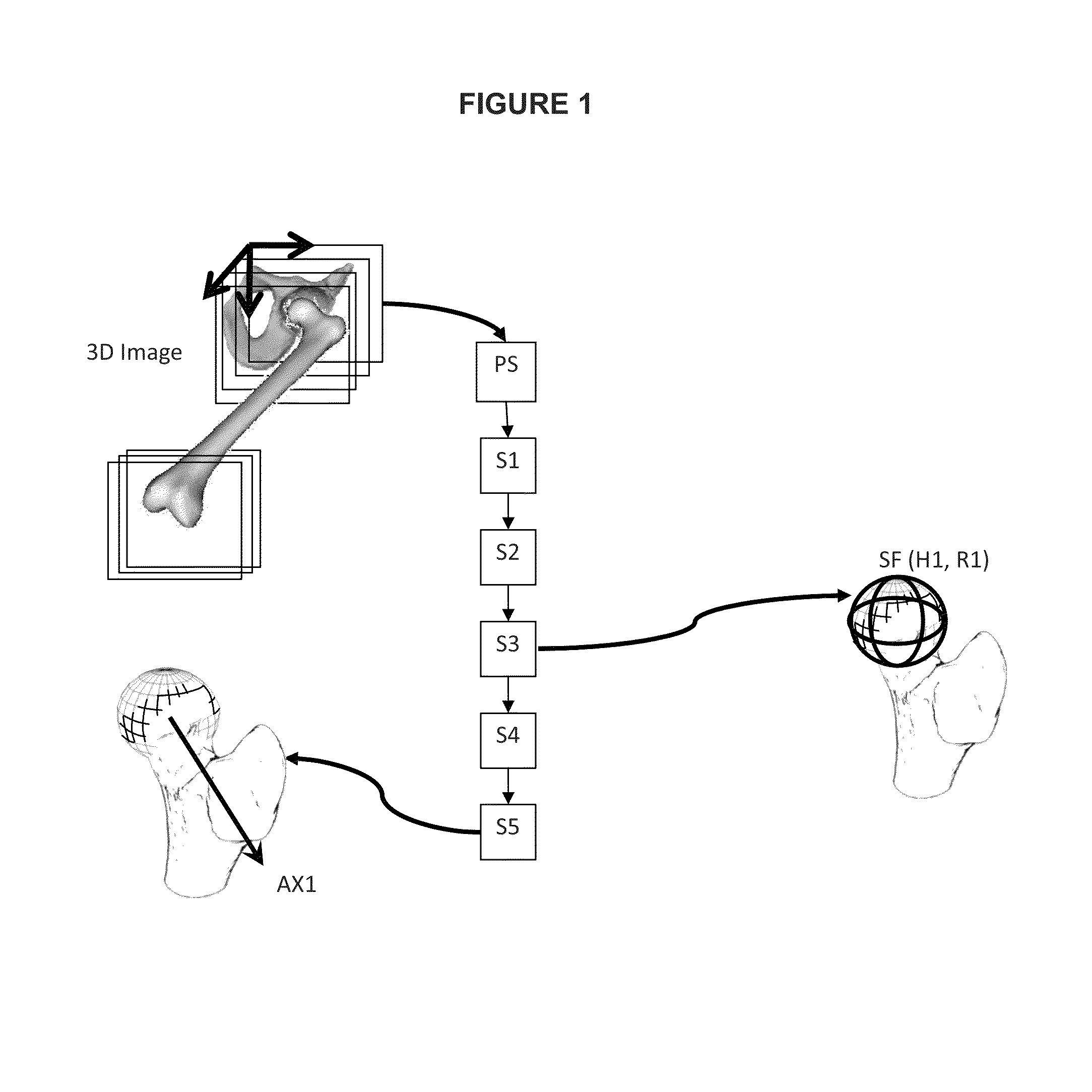
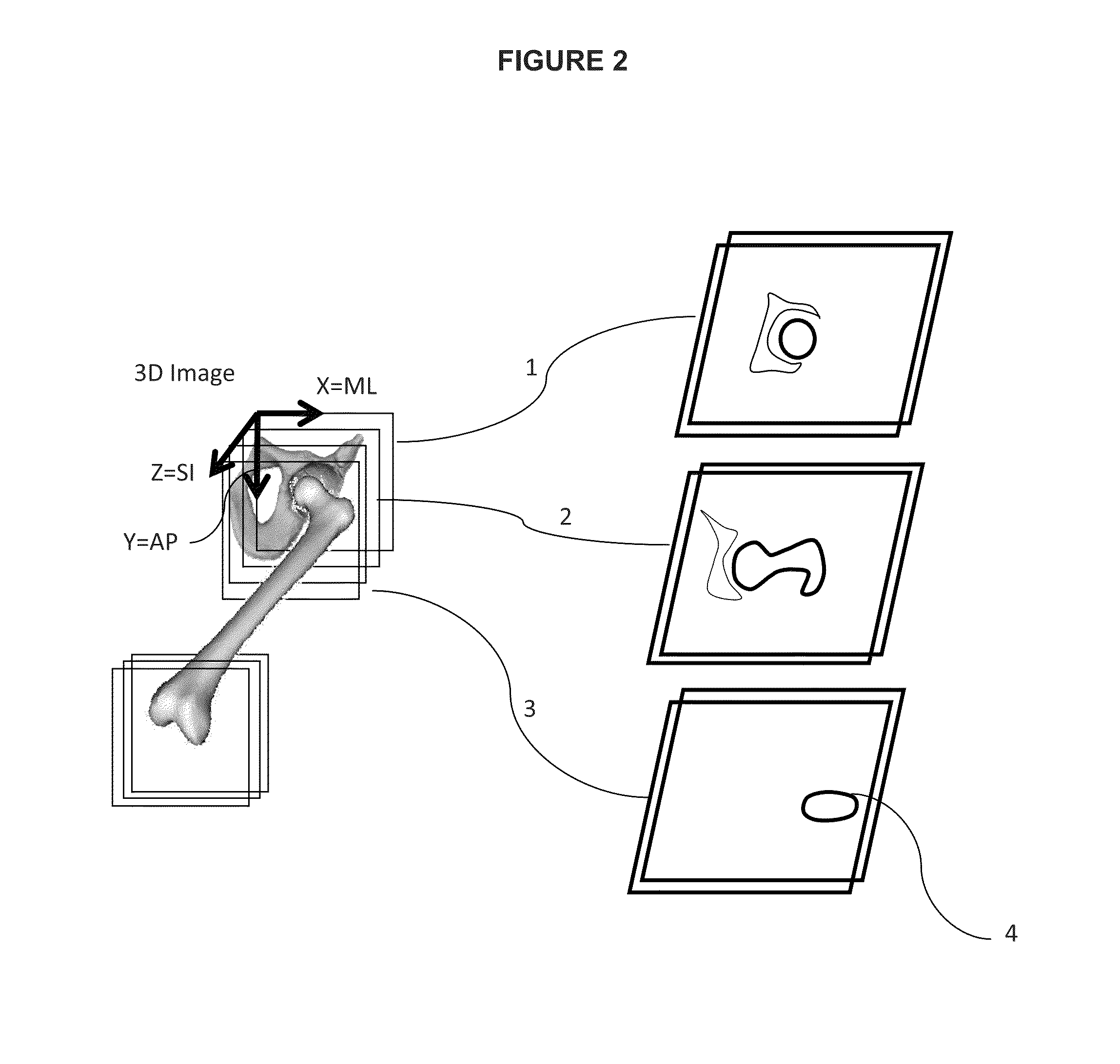
[0105]Hereafter, description of the invention will be made with reference to the articulation of the hip. However, the invention is not limited to this illustrative example and the person skilled in the art will easily transpose this description to any other articulation partially formed by a bone head, such as the shoulder.

[0106]Some critical anatomical elements are necessary to measure some specific anatomical characteristics of the proximal femur, such as the femoral neck version and inclination angles, and a newly defined 3D measure of alpha angle, which participates in the characterization of the proximal femur deformity in Femoro Acetabular Impingement (FAI) pathology.

[0107]The method is specifically addressing the femur but it can be extended to other bones of the human or animal body such as the humerus or other bones having a rotoid articulation. The general purpose of the invention is to determine automatically from the 3D image the major characteristic geometric elements ...


PUM


Abstract

The invention relates to an automated method for precise determination of the head center and radius and the neck axis of an articulated bone from acquired 3D medical image of an articulation, comprising the following steps: i) determining, from a 3D image of the bone, an approximate sphere (SFO) of the head of the bone that substantially fits the spherical portion of the head of the bone; ii) constructing from the 3D image and the approximate sphere (SFO), a 3D surface model (S) of the bone; iii) determining, from the 3D surface model (S) and from the approximate sphere (SFO), an approximate neck axis (AXO) of the neck of the bone; iv) determining, from the 3D surface model (S) and the approximate sphere (SFO), a precise sphere (SF); v) determining, from the 3D surface model (S), the precise sphere (SF) and the approximate neck axis (AXO), a precise neck axis (AX1).

Description

TECHNICAL FIELD[0001]The invention relates to the field of computer assisted surgery, and more particularly to a method and a system of determination of geometric elements from 3D medical image of a bone.BACKGROUND OF THE INVENTION[0002]Articulations of the human body are often very complex systems and no precise generic model exists to capture all the variability from one articulation to another. It is therefore necessary to use specific medical images or collection of digital patient data in order to get relevant information to develop techniques, devices and methods that will facilitate a treatment or a diagnosis. The present text focuses on the hip articulation between the acetabulum and the proximal femur although it can be easily extended to other articulations such as shoulder for example.[0003]Structural abnormalities in the morphology of the hip can limit motion and result in repetitive impact of the proximal femoral neck against the acetabular labrum and its adjacent carti...

Claims


Application Information

Patent Timeline
Patent Type & Authority Patents(United States)
IPC IPC(8): G06K9/00A61B19/00G06K9/62G06T7/00G06T7/60G06T17/20
CPCG06K9/00362A61B19/50G06K9/6205G06T7/0042G06T7/0083G06T7/0089G06T7/602G06T7/606G06T17/20A61B2019/505G06T2207/10072G06T2207/30008A61B34/10A61B2034/105G06T7/73G06T7/12G06T7/149G06T7/62G06T7/66G06V10/753G06V40/10
Inventor CHABANAS, LAURENCELAVALLEE, STEPHANENESME, MATTHIEUSCHERS, JONATHAN
Owner SMITH & NEPHEW ORTHOPAEDICS
Why Eureka
Try Eureka