Object recognition system for medical imaging

a technology of object recognition and medical imaging, applied in the field of medical imaging, can solve the problems of inability to generate segmented images of the prostate, inability to realize real-time imaging, and inability to segment the prostate, etc., to achieve stable and easy operation, minimize the energy function of the initial contour/boundary, and minimize the effect of the energy function

Active Publication Date: 2010-09-28
EIGEN HEALTH SERVICES LLC
View PDF90 Cites 4 Cited by
  • Summary
  • Abstract
  • Description
  • Claims
  • Application Information

AI Technical Summary

Benefits of technology

[0011]The predetermined models may provide an initial boundary that may be fitted to a boundary region of the image. That is, while the actual boundary may not be readily discernible the region in which the boundary lays may be identified. In one arrangement, the predetermined models are based on a plurality of stored boundaries that may be determined from actual patient data. For instance, a plurality of patients may provide ultrasound images and these ultrasound images may be segmented to identify boundaries within each image. These predetermined boundaries may provide an adequate boundary estimation to allow for subsequent automated boundary identification. That is, by providing the boundary estimate based on predetermined models, the present utility may be performed in an automated procedure.
[0013]In another aspect, an improved system and method (i.e. utility) for segmentation on prostate ultrasound images is introduced. The utility minimizes the energy function of an initial contour / boundary that is placed on the prostrate image in an automated selection and fitting process (e.g., initialization). The energy function of the contour is minimized based on the regional information of the image as well as the shape information of the contour, and each term in the energy functional represent a certain weighted “energy”. A level set frame work is applied, in which the contour is represented implicitly, while the necessary re-initialization step in the traditional level set method is totally eliminated. The final level set function which contains the edge information will be obtained by a finite difference (the steepest descent search) calculation scheme.
[0014]Generally, the system works as the following way: a predetermined boundary is fit to a prostrate image, in one arrangement a trained user selects a few points inside the prostate region of the image to allow for the automated fitting of the predetermined boundary; in another arrangement, the region inside the prostrate is identified in an automated process based, for example on the uniformity of the pixels inside the prostrate. The predetermined boundaries may include prostrate boundary models. Such model may be generated from actual prostrate image and stored for use in the present utility. After the initial boundary is selected a narrow band mask which includes the real boundary of the prostate will be generated. Then all the calculations of terms included in the finite difference update process will be carried out in this new domain (e.g., narrow band mask). After steps of iterations, the finite difference will converge due to the “balanced” regions by the different weighs in the cost function. The final edges can be estimated by smoothing the obtained contour (e.g.,. the converged contour). This utility is stable and easy for the chosen of initial condition, and use of penalized terms in the energy function of the contour limit the speed of expansion of the contour to ensure there no “split” or “blooding” before the convergence state is reached.
[0015]According to another aspect, a system and method for use in obtaining a boundary of a structure within ultrasound image is provided. The utility includes obtaining an ultrasound image of a patient where the ultrasound image includes a structure of interest. Additionally, the utility receives an input regarding demographic information associated with the patient. Based on the demographic information, an initial estimate of a boundary of the structure of interest is provided.

Problems solved by technology

Segmentation of the ultrasound prostate images is a very challenging task due to the relatively poor image qualities.
Generally, such a process of manual boundary identification and subsequent processing has made real time imaging (e.g., generating a segmented image while a TRUS remains positioned)of the prostrate impractical.

Method used


Image

  • Object recognition system for medical imaging
  • Object recognition system for medical imaging
  • Object recognition system for medical imaging

Examples


Embodiment Construction

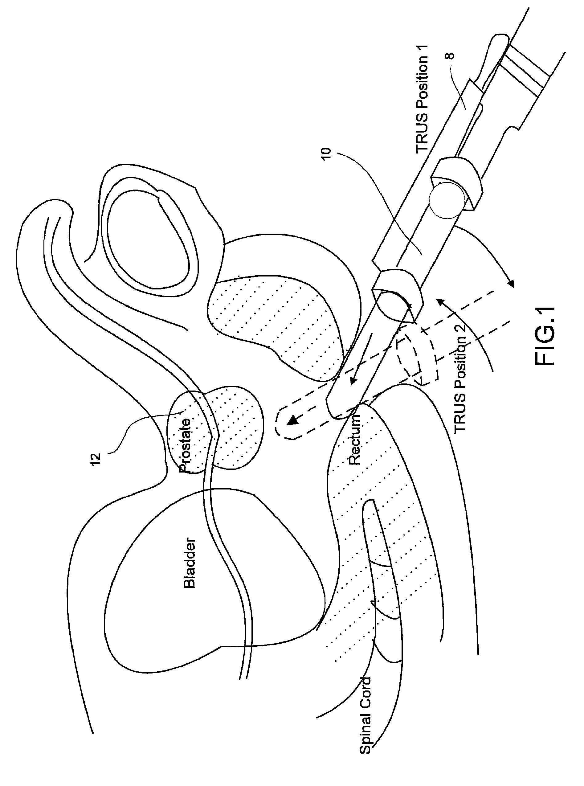
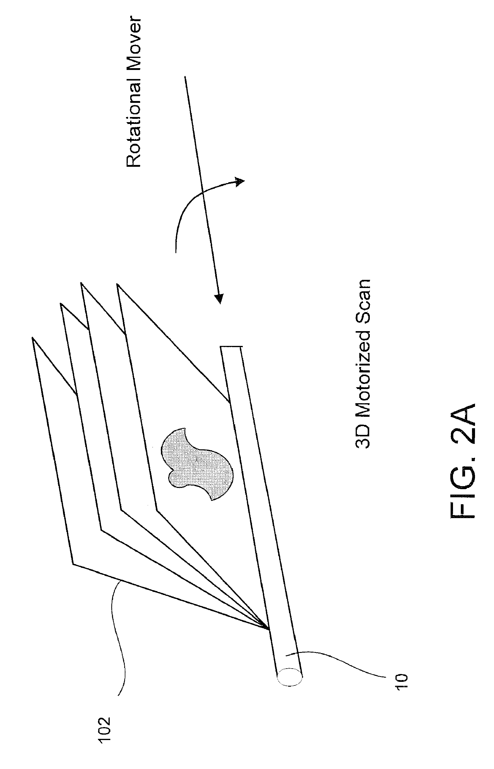
[0040]Reference will now be made to the accompanying drawings, which assist in illustrating the various pertinent features of the present disclosure. Although the present disclosure is described primarily in conjunction with transrectal ultrasound imaging for prostate imaging, it should be expressly understood that aspects of the present invention may be applicable to other medical imaging applications. In this regard, the following description is presented for purposes of illustration and description.

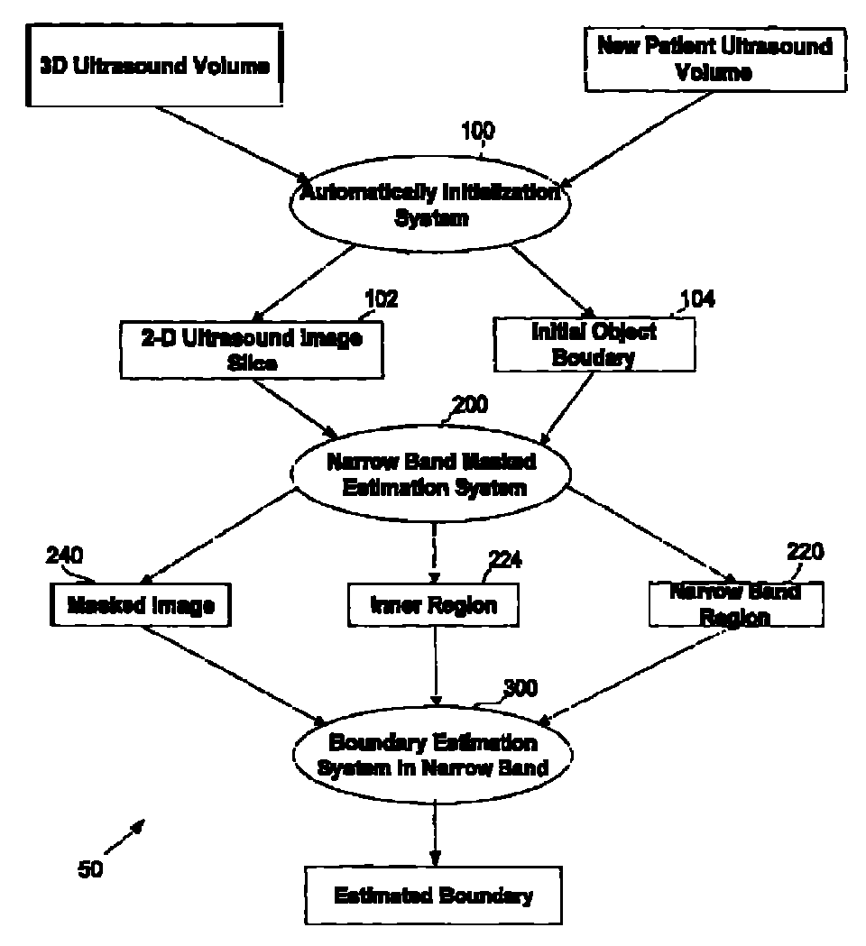
[0041]Disclosed herein are systems and methods that allow for obtaining ultrasound images and identifying structure of interest from those images. In this regard, a segmentation algorithm is utilized to analyze the images such that structures of interest may be delineated from background within the image. Specifically, in the application disclosed herein, a segmentation algorithm is provided that is utilized to delineate a prostrate gland such that volume and / or shape of the prostrate ...


PUM


Abstract

An improved system and method (i.e. utility) for segmentation of medical images is provided. The utility fits an estimated boundary on a structure of interest in an automated selection and fitting process. The estimated boundary may be a model boundary that is generated actual boundaries of like structures. In one arrangement, the boundaries may be selected based on the age and / or ethnicity of a patient. In further arrangements, narrow band processing is performed to estimate the actual boundary of the structure of interest.

Description

CROSS REFERENCE TO RELATED APPLICATION[0001]This application claims priority under 35 U.S.C. §119 to U.S. Provisional Application No. 60 / 863,505 entitled: “Object Recognition System” and having a filing date of Oct. 30, 2006, the entire contents of which are incorporated herein by reference.FIELD[0002]The present disclosure pertains to the field of medical imaging, and more particular to the segmentation of medical images to allow for accurate calculation of the volumes of structures of interest within the medical images.BACKGROUND[0003]Medical imaging, including X-ray, magnetic resonance (MR), computed tomography (CT), ultrasound, and various combinations of these techniques are utilized to provide images of internal patient structure for diagnostic purposes as well as for interventional procedures. One application of medical imaging (e.g., 3D imaging) is the measurement of volume and / or assessment of shape of internal structures of interest. These measurements typically require se...

Claims


Application Information

Patent Timeline
Patent Type & Authority Patents(United States)
IPC IPC(8): G06K9/00A61B8/00
CPCG06T7/0083G06T7/0089G06T2207/10072G06T2207/10136G06T2207/20161G06T2207/30081G06T2207/30096G06T7/12G06T7/149
Inventor SURI, JASIT SLI, LU
Owner EIGEN HEALTH SERVICES LLC
Why Eureka
Try Eureka