Interface devices for use with intracavity probes for high field strength magnetic resonance systems

a high field strength magnetic resonance and interface device technology, applied in the field of intracavity probes, can solve the problems of inability to use mr systems designed to operate, inherent single coil approach,

Inactive Publication Date: 2015-03-24
DXTX MEDICAL INC
View PDF71 Cites 6 Cited by
  • Summary
  • Abstract
  • Description
  • Claims
  • Application Information

AI Technical Summary

Benefits of technology

This patent describes an invention of an intracavity probe for magnetic resonance imaging (MRI) that has a broader frequency response than previous probes, without sacrificing signal-to-noise ratio. This means that the probe does not need to be tuned for each patient or coil. The invention also provides an interface device that interfaces the intracavity probe with an MR system to obtain high resolution images and spectroscopic results without the need for tuning. Additionally, the invention provides an interface device for interfacing an intracavity probe with an MR system that does not have its own preamplifier. This interface device includes a PIN diode and a preamplifier to enhance signal-to-noise ratio and pass MR signals to the probe input port of the MR system.

Problems solved by technology

The disadvantage inherent to the single coil approach is attributable to just one coil structure being used to acquire the MR signals over the entire region of interest.
Consequently, they are not suitable for use with MR systems designed to operate at higher field strengths, such as the 2.0 to 5.0 Tesla and particularly 3.0 Tesla MR systems that are capable of producing even higher quality images and spectrographic results.

Method used


Image

  • Interface devices for use with intracavity probes for high field strength magnetic resonance systems
  • Interface devices for use with intracavity probes for high field strength magnetic resonance systems
  • Interface devices for use with intracavity probes for high field strength magnetic resonance systems

Examples


Embodiment Construction

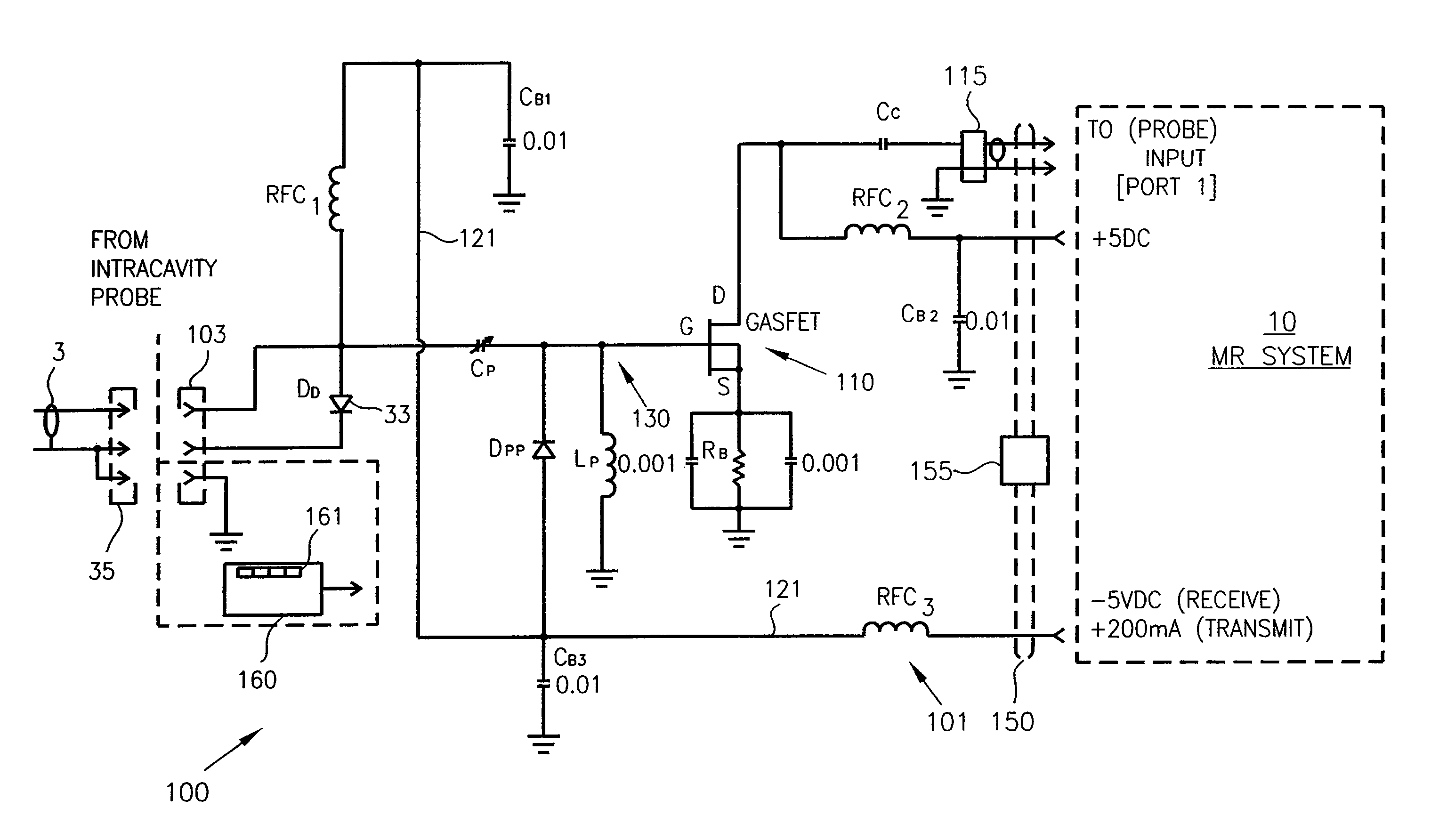
[0052]In all of its embodiments and related aspects, the present invention disclosed below is ideally used with magnetic resonance (MR) systems designed to operate at 3.0 Tesla field strengths, though it is also applicable to those operable from approximately 2.0 to 5.0 T. For purposes of illustration below, the invention will be described in the context of the 3.0 T systems produced by General Electric Medical Systems (GEMS).

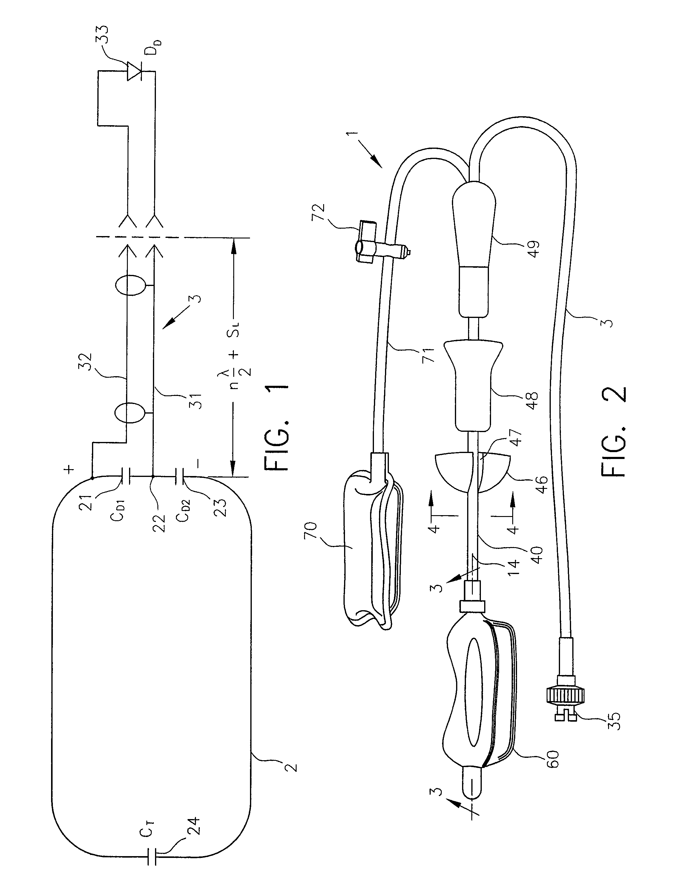
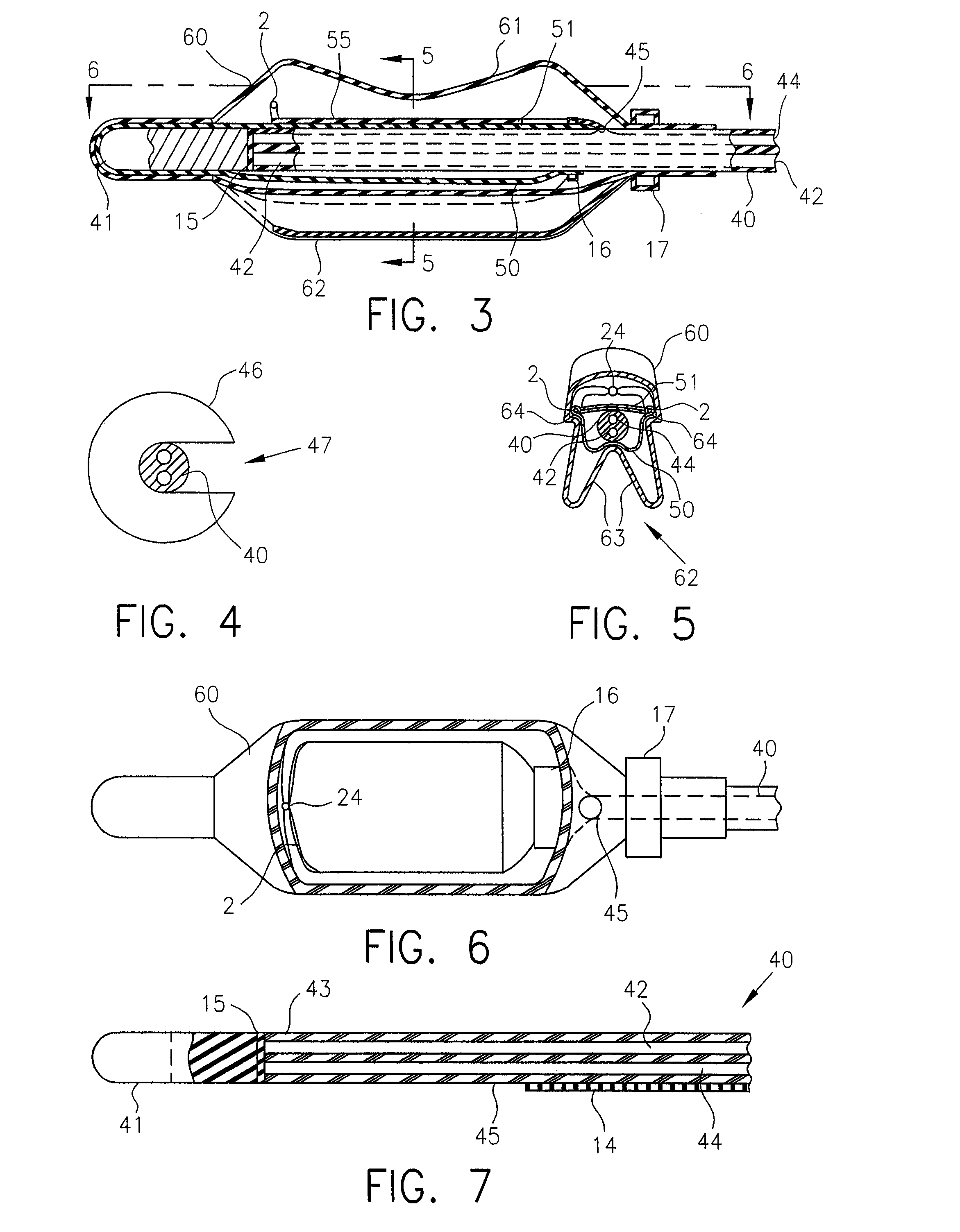
[0053]FIGS. 1-7 illustrate one aspect of a presently preferred embodiment of the invention, namely, an intracavity probe, generally designated 1. The probe is intended for use with an MR system to obtain images or spectra of a region of interest within a cavity of a patient. It is described herein in a specific implementation, i.e., as an endorectal probe designed to be inserted into the rectum to obtain images and / or spectra of the male prostate gland. Although presented herein as an endorectal probe, it should be understood that the invention is equally capab...


PUM


Abstract

An interface device enables an intracavity probe to be used with a magnetic resonance (MR) system for the purpose of obtaining images or spectra of a region of interest within a cavity of a patient. The probe includes a shaft, a balloon at one end thereof, and a coil loop within the balloon. The coil loop preferably includes two drive capacitors and a tuning capacitor, all of which in series. A junction node between the drive capacitors serves as a ground for electrically balancing the coil loop. Diametrically opposite the junction node, the tuning capacitor enables the coil loop to resonate at the operating frequency of the MR system. Across each drive capacitor is connected an output cable having an electrical length of SL+n(λ / 4). The output cables terminate in a plug that is used to connect the coil loop to the interface device.

Description

CROSS-REFERENCE TO RELATED APPLICATION[0001]This present application is a divisional of U.S. application Ser. No. 11 / 719,253 filed 14 May 2007 (now U.S. Pat. No. 7,885,704), which is a national stage entry of PCT International Application No. PCT / US2005 / 041912 filed 15 Nov. 2005, which is a continuation-in-part of PCT International Application PCT / US03 / 07774 filed 13 Mar. 2003 and which claims the benefit of U.S. Provisional Application 60 / 628,166 filed on 15 Nov. 2004, all of which incorporated by reference herein and assigned to the assignee of the invention disclosed below.FIELD OF THE INVENTION[0002]The present invention generally relates to systems and methods of obtaining images and spectra of intracavity structures using magnetic resonance (MR) systems. More particularly, the invention pertains to an intracavity probe capable of being inserted into various bodily openings, such as the rectum, vagina, mouth, etc., to obtain high resolution images of and spectroscopic results f...

Claims


Application Information

Patent Timeline
Patent Type & Authority Patents(United States)
IPC IPC(8): A61B5/055G01R33/28G01R33/34G01R33/36
CPCG01R33/285G01R33/3628A61B5/055G01R33/34084
Inventor MISIC, GEORGE J.
Owner DXTX MEDICAL INC
Why Eureka
Try Eureka