Three-dimensional localization, display, recording, and analysis of electrical activity in the cerebral cortex

a technology of electrical activity and cerebral cortex, applied in the field of medical devices concerning the brain, can solve the problems of large size, lack of proper objective diagnosis, and high cost of medical equipmen

Active Publication Date: 2015-11-10
DOIDGE MARK S +1
View PDF37 Cites 16 Cited by
  • Summary
  • Abstract
  • Description
  • Claims
  • Application Information

AI Technical Summary

Problems solved by technology

The cerebral cortex, largely due to its complexity, is also susceptible to a large number of disorders and diseases, many of which lack proper objective diagnoses, or require expensive medical equipment to properly diagnose.
Each of these technologies has its strengths and weaknesses.
MEG is also the most costly of the above technologies and the least accessible.
In general, these technologies are very expensive, typically $1,000,000-$10,000,000 USD per machine.
In addition to the cost of the machine itself, technical staff, maintenance fees, specialized environments, and chemical or radioactive agents may also be required.
These machines are physically large and require entire spaces to be devoted to their function, hence restricted to larger medical institutions and universities, and most definitely not portable.
It is primarily for the aforementioned reasons that these technologies are largely inaccessible to both members of the general population and much of the academic and private research community.
There exists, however, a related technology that can match the temporal resolution of MEG, is portable, but cannot localize in three dimensions events that take place within the cerebral cortex.
U.S. Pat. No. 5,307,807 to Valdes Sosa (1994) describes a method and system to localize EEG signals in three dimensions, using an inverse solution approximation source localization algorithm, however it does not reduce to practice any applications, nor is it performed in near real-time, significantly reducing the clinical relevance, and it relies on tomographs (2D slices) to convey three-dimensional data, which complicates the interpretation of contiguous 3D data.
The only other cortical imaging modality capable of this temporal resolution, MEG, is approximately 500 times more expensive than this setup and requires special magnetically shielded rooms;
In addition, in the absence of a rapid diagnosis, the delay leaves both doctor and patient alike wondering about the outcome;

Method used


Image

  • Three-dimensional localization, display, recording, and analysis of electrical activity in the cerebral cortex
  • Three-dimensional localization, display, recording, and analysis of electrical activity in the cerebral cortex
  • Three-dimensional localization, display, recording, and analysis of electrical activity in the cerebral cortex

Examples


Embodiment Construction

A Preferred Embodiment of the Present Invention—FIG. 1

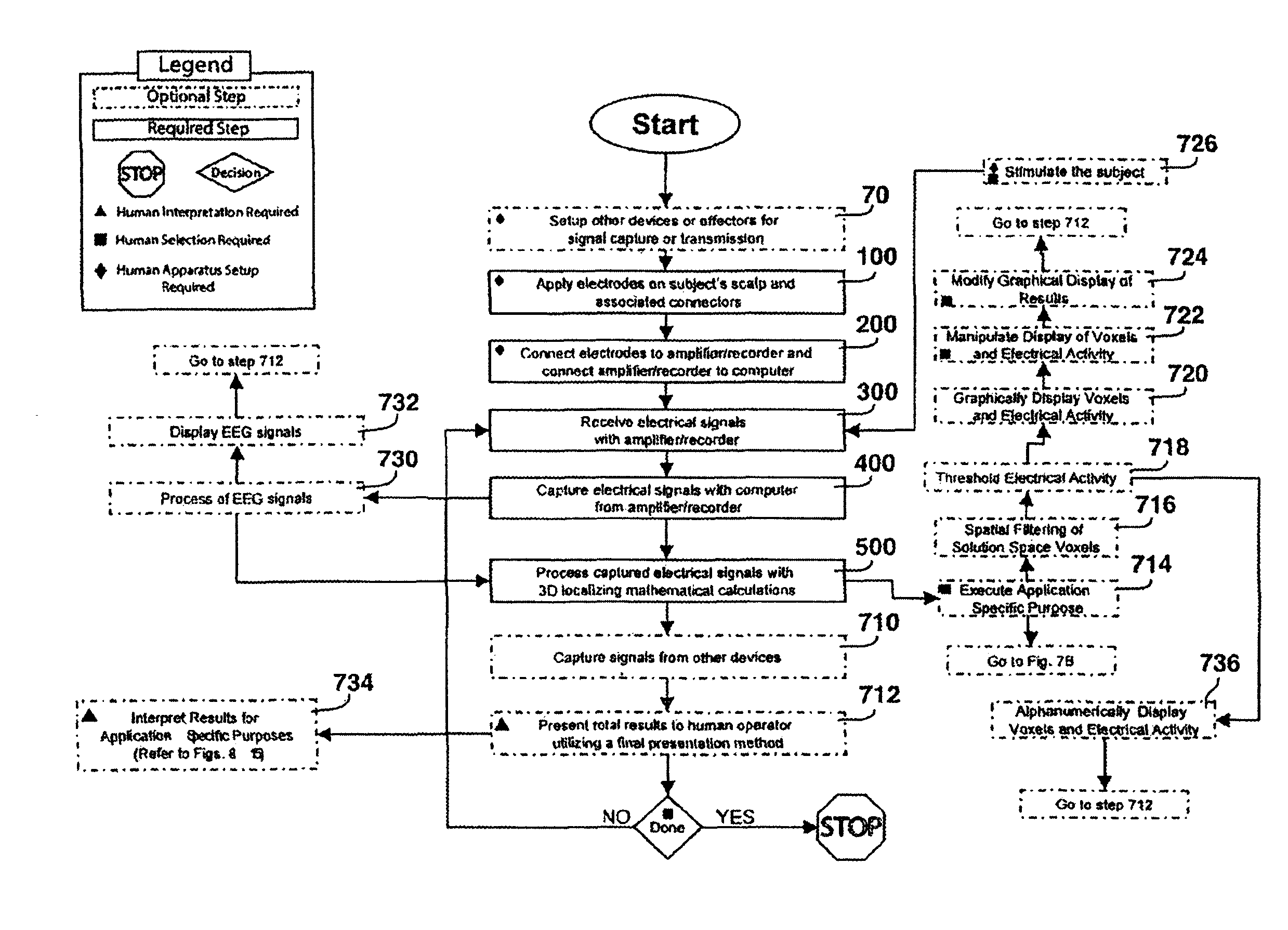
[0181]A preferred embodiment of the present invention is outlined in FIG. 1. It consists of several fundamental steps where:

[0182]Step 100 entails the application of electrodes on a subject's scalp. This requires a practical method to hold the electrodes in place on the scalp so as to make good electrical contact. Typically this is accomplished with manually attached electrodes with adhesives or caps / cap-like structures that fit over a subject's scalp that integrate or have adapters for the electrodes; a number of these are commercially available. A conductive medium is also generally required for the conductance of electrical signals between the scalp and electrodes; typically the conductive medium is the same as the adhesive used, although it can be separate. The positions of the electrodes may be known to assist in the localization calculations, or generalized electrode positions based on ratios or morphological features of th...


PUM


Abstract

The present invention describes a method and apparatus to localize the electrical signals measured from a subject's scalp surface, preferably in near-real time, and to generate dynamic three-dimensional information of the electrical activity occurring within the cerebral cortex of the brain. In the preferred embodiment, it can produce images that can be immediately inspected and analyzed by an operator in near-real time, resulting in a powerful new cortical imaging modality, which we denote as Dynamic Electrocortical Imaging (DECI). The present invention involves the use of a computer, an electroencephalographic (EEG) amplifier, EEG electrodes, and custom software. It can measure healthy and diseased cortical events and states in both conscious and unconscious subjects. This is useful, as it allows for the diagnosis, monitoring and treatment of cortical disorders, while also furthering the understanding of the human brain and lending use to additional non-medical applications such as in entertainment, education, lie-detection and industry. The invention in one embodiment is implemented using software in conjunction with readily available EEG hardware. Furthermore, this same method can be applied to pre-existing data and when doing so, EEG hardware is not required. Having a practical near-real time 3D imaging system brings a far more accessible technology to doctors, researchers, individuals, and private clinics to better diagnose, monitor, treat and understand many of the conditions and abnormalities of the brain.

Description

CROSS-REFERENCE TO RELATED APPLICATIONS[0001]This application is a Continuation of PCT application PCT / CA20061000776 filed May 16, 2006, which claims priority to U.S. provisional patent application 60 / 681,140, filed 2005 May 16. This application claims priority to each of these applications and incorporates the content of each by reference.FEDERALLY SPONSORED RESEARCH[0002]Not applicableSEQUENCE LISTING OR PROGRAM[0003]None attachedBACKGROUND OF THE INVENTION[0004]1. Field of Invention[0005]The present invention relates largely to medical devices concerning the brain, and is a method and apparatus to non-invasively localize, in three dimensions (3D), electrical activity in the cerebral cortex of the brain, display the localized electrical activity in the form of 3D “motion pictures”, and to perform statistical analysis and quantification of the localized electrical activity, all in near-real time.[0006]2. Prior Art[0007]The human cerebral cortex is the convoluted outer surface of th...

Claims


Application Information

Patent Timeline
Patent Type & Authority Patents(United States)
IPC IPC(8): A61B5/04A61B5/16A61B5/0476
CPCA61B5/0476A61B5/164A61B5/4023A61B5/369G06T17/10
Inventor DOIDGE, MARK S.MOCANU, JOSEPH D.
Owner DOIDGE MARK S
Why Eureka
Try Eureka