Endorectal prostate probe composed of a combined mini gamma camera and ultrasound sensor

a technology of ultrasound sensor and endorectal probe, which is applied in the field of hybrid imaging of organs and tumors, can solve the problems of high false negative diagnosis of many missed cancers, inaccurate assessment of today's imaging techniques, and negative biopsy results, and achieve the effect of improving 3d reconstruction

Active Publication Date: 2016-03-01
WEST VIRGINIA UNIVERSITY
View PDF31 Cites 1 Cited by
  • Summary
  • Abstract
  • Description
  • Claims
  • Application Information

AI Technical Summary

Benefits of technology

[0044]Another embodiment of this invention provides for the imaging system, as described herein, wherein the gamma imaging detectors are capable of being operated in a static mode in which each of the detectors are fixed in position with respect to a target organ of a patient lying on the bed, or in a dynamic mode in which each of the detectors are rotated with respect to the target organ of the patient lying on the bed to provide full angular projective sampling of the target organ for enhanced 3D reconstruction, and wherein the detectors can be rotated to a new viewing angle with respect to the target organ and then operated in the static mode to better view the target organ of the patient lying on the bed and to optimize gamma ray spatial resolution. Preferably, the rotating gantry of the imaging system enables 360 degree angular sampling in a 3D imaging mode with the probe and the gamma imaging detectors. Most preferably, the dual modality imaging system as described herein includes wherein the ultrasound sensor is placed in front of the gamma probe sensor within the housing of the probe. In a less preferred embodiment of this invention, the dual modality imaging system includes the probe, as described herein, wherein the ultrasound sensor is placed behind the gamma probe sensor and wherein the gamma probe sensor is placed in front of the ultrasound sensor within the housing of the probe.

Problems solved by technology

Biopsy results may be negative despite the presence of cancer due to sampling error.
The “blind” biopsy typically performed today under ultrasound (US) guidance results in high false negative diagnosis with many missed cancers.
Such accurate assessment today is not available with conventional imaging techniques [ultrasound (US), computed tomography (CT), magnetic resonance imaging and PET].
Standard PET scanners have spatial resolution inadequate to meet the clinical needs of prostate imaging—particularly when using specific, targeted imaging agents.
Conventional blind “biopsy” procedures under Tissue Differentiating Ultrasound are able to visualize only the structure and the margins of an organ, and thus do not provide differentiation between a cancerous tissue and healthy tissue.
Prostate cancer is the only human cancer that does not have a standard reliable method of imaging of the primary tumor.
Functionally blind biopsy typically performed today under transrectal ultrasound guidance results in high false negative diagnoses with many missed cancers.
Such accurate assessment is generally not available in the present state of the art, with the conventional imaging techniques available to urologists.
Thus, those persons skilled in the art appreciate that prostate cancer is difficult to visualize in its early stage using current imaging technology.
However, visible anatomic changes are not always present in early stages of the disease, making the use of current imaging modalities difficult in early detection of prostate cancer sites.
The key problem with conventional guiding systems during prostate biopsy is that they are based on symmetrical anatomical sampling of the prostate, and not on the location of the cancer.
The main challenge continues to be the inability to visualize the cancer in its early stages using current imaging technology.
U.S. Pat. No. 4,995,396 “Radioactive ray detecting endoscope” discloses an endoscope having both an ultrasonic imaging device and a radioactive ray (e.g., beta radiation) detecting device in the tip of the endoscope, but does not disclose use of a biopsy device.

Method used


Image

  • Endorectal prostate probe composed of a combined mini gamma camera and ultrasound sensor
  • Endorectal prostate probe composed of a combined mini gamma camera and ultrasound sensor
  • Endorectal prostate probe composed of a combined mini gamma camera and ultrasound sensor

Examples


Embodiment Construction

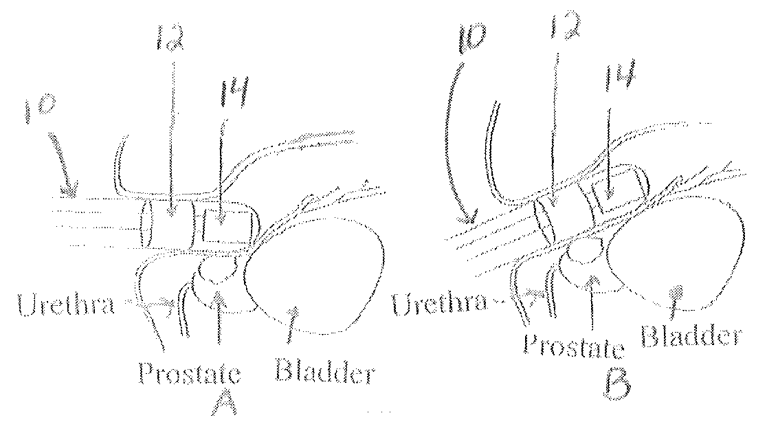
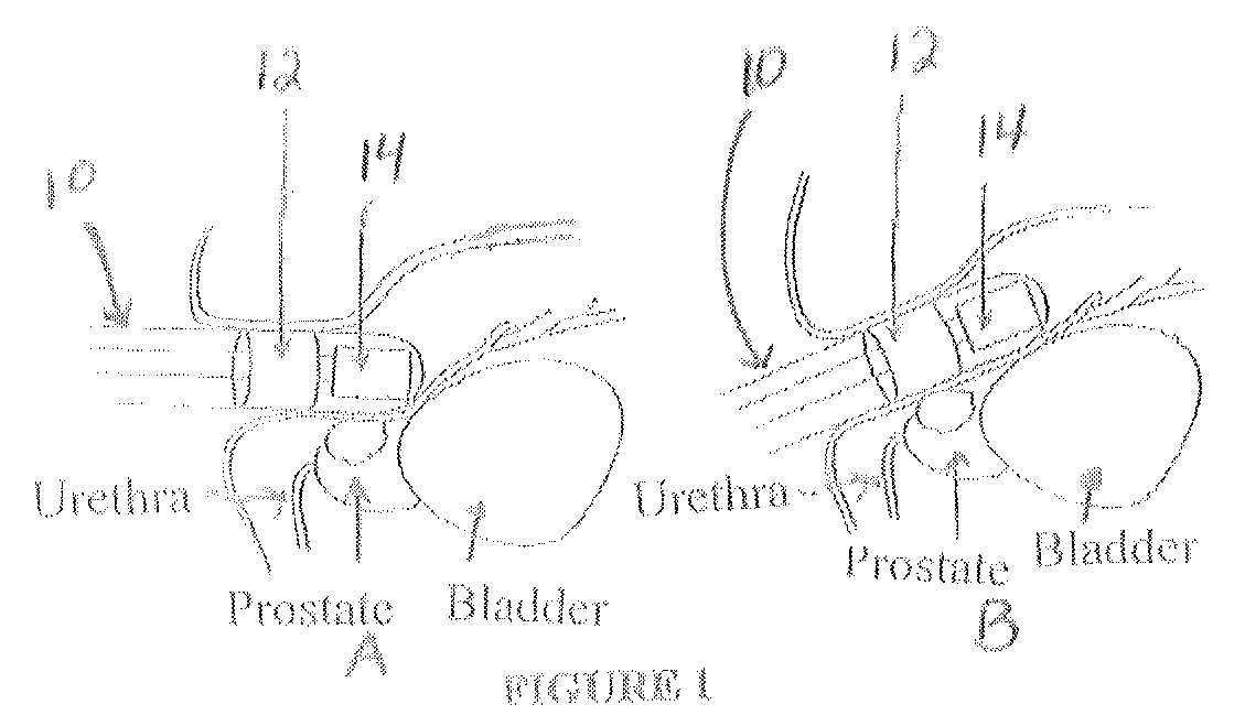
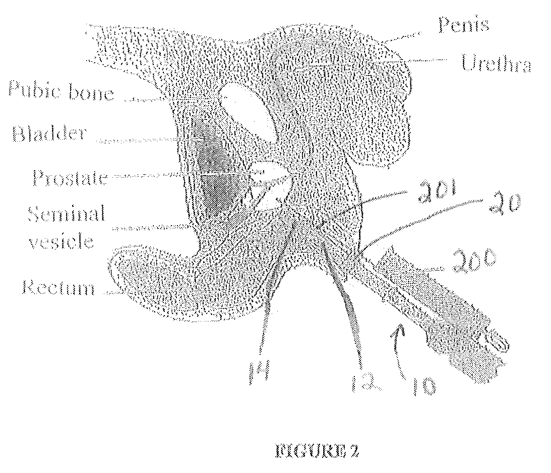
[0065]The gamma probe sensor and ultrasound dual modality probe and dual modality imaging system of this invention provide significant improvement over existing devices and methods to obtain evaluations of a target organ of a patient, to perform a biopsy of the target organ, and to perform localized surgery of the target organ. The target organ may be, for example, but not limited to the prostate gland of a male patient, a gynecological anatomical structure of a female patient (vagina, cervix, uterus, etc.), or the rectum of a patient, or other anatomical structure of a patient wherein an endoscopic probe is utilized.

[0066]In a preferred embodiment of this invention, the probe and imaging system and method of this invention is useful to guide prostate biopsy / surgery with high resolution combined dual-modality gamma probe / US (Ultrasound) probe imaging in one compact endorectal device. The 2D images from the two modalities are naturally fused because the corresponding images are obtai...


PUM


Abstract

A dual modality probe is disclosed having both a gamma probe sensor and an ultrasound sensor. A dual imaging system is provided having the probe and at least one external gamma imaging detector and a data acquisition computer system for collecting data simultaneously from the gamma probe sensor, the gamma imaging detector, and the ultrasound sensor of the probe. A method for evaluating a target organ of a patient utilizing the probe and imaging system, and performing a biopsy of the organ is disclosed.

Description

BACKGROUND OF THE INVENTION[0001]1. Field of the Invention[0002]The present invention relates to a device and method for performing hybrid imaging of organs and tumors. More particularly, an endorectal probe is provided for imaging of a prostate gland wherein the probe (device) has combined gamma camera and ultrasound imaging modalities.[0003]2. Description of the Background Art[0004]There are >200,000 new cases and nearly 30,000 deaths each year from prostate cancer (PCa) Prostate-specific antigen (PSA) testing has allowed early detection of impalpable PCa. Early detection has lowered the incidence of advanced disease with extracapsular extension and the subsequent early treatment appears to improve survival rate. After anomalous PSA results, the patient undergoes biopsy, and if the biopsy is positive, the patient undergoes surgery. The main objective of surgery is to remove the cancer at lowest functional cost, i.e. preserving continence and sexual function. The stage of the ca...

Claims


Application Information

Patent Timeline
Patent Type & Authority Patents(United States)
IPC IPC(8): A61B6/00A61B6/04A61B8/12A61B8/00
CPCA61B6/4417A61B6/0407A61B8/12A61B8/4245A61B8/4416A61B8/4444A61B5/4381A61B6/4429A61B6/481A61B6/5247A61B6/54A61B8/4254A61B8/5261A61B8/54A61B10/0241
Inventor MAJEWSKI, STANISLAWMARANO, GARYSTOLIN, ALEXANDERPROFFITT, JAMES
Owner WEST VIRGINIA UNIVERSITY
Why Eureka
Try Eureka