
Method and system for contrast inflow detection in 2D fluoroscopic images

a fluoroscopic image and contrast inflow technology, applied in image enhancement, image analysis, instruments, etc., can solve the problems of mislead detection and poorly worked in practice, and achieve the effect of efficient and robust detection

Active Publication Date: 2016-03-22
SIEMENS HEALTHCARE GMBH
View PDF12 Cites 1 Cited by
  • Summary
  • Abstract
  • Description
  • Claims
  • Application Information

AI Technical Summary

Benefits of technology

The present invention describes a way to automatically detect the arrival of a contrast agent in a sequence of fluoroscopic images. This is important because it can help medical professionals quickly and accurately identify the presence of contrast agent and determine when it reaches its maximum level. This technology is based on learning and can be used over a long period of time to improve its accuracy and reliability.

Problems solved by technology

However, this works poorly in practice since the moving diaphragm, which often appears in cardiac x-ray images, can easily mislead the contrast inflow detection.
A main challenge of contrast inflow detection is due to the variability of the data, especially when the images are acquired in low dose radiation with a lot of noise.

Method used

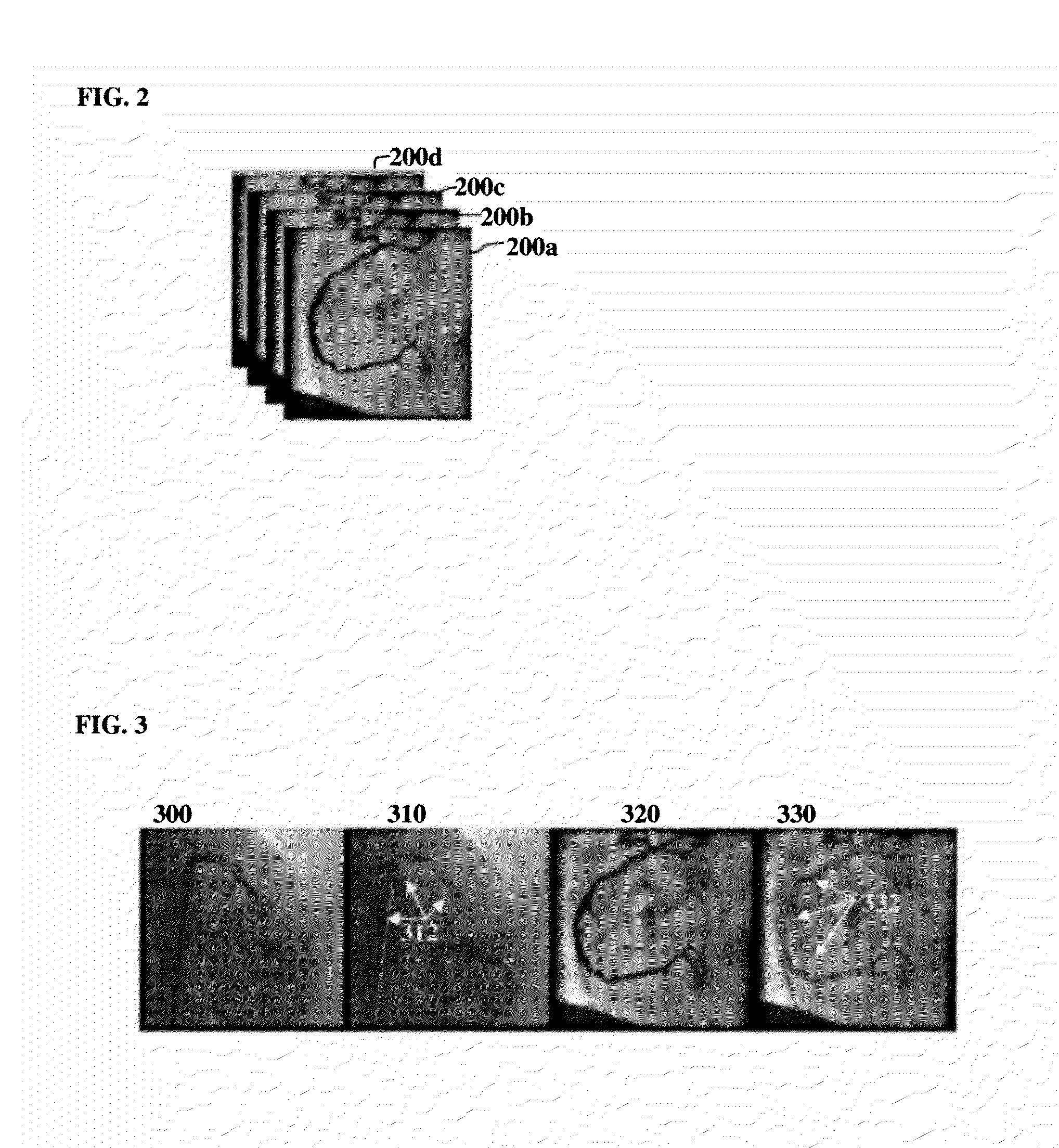

Image

  • Method and system for contrast inflow detection in 2D fluoroscopic images
  • Method and system for contrast inflow detection in 2D fluoroscopic images
  • Method and system for contrast inflow detection in 2D fluoroscopic images

Examples


Embodiment Construction

[0014]The present invention relates to a method and system for contrast inflow detection. Embodiments of the present invention are described herein to give a visual understanding of the contrast detection method. A digital image is often composed of digital representations of one or more objects (or shapes). The digital representation of an object is often described herein in terms of identifying and manipulating the object. Such manipulations are virtual manipulations accomplished in the memory or other circuitry / hardware of a computer system. Accordingly, is to be understood that embodiments of the present invention may be performed within a computer system using data stored within the computer system.

[0015]Although it is often easy for a physician to tell when the contrast agent is present in fluoroscopic images, an automatic contrast inflow detection method is desirable for many computer-aided intervention procedures. For example, in a stent enhancement application, the algorith...


PUM


Abstract

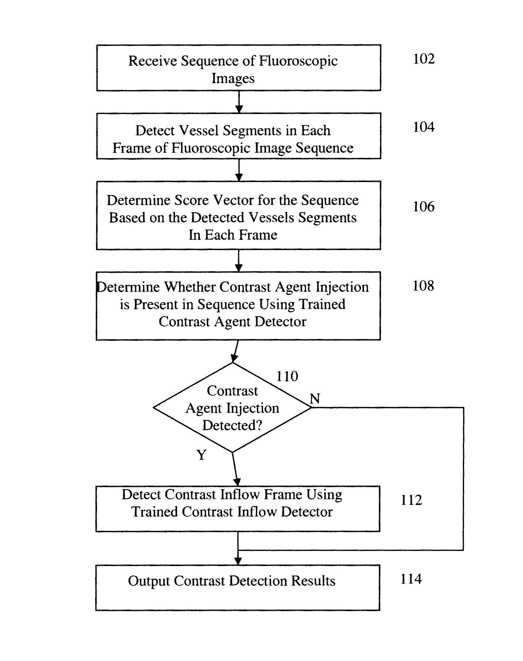
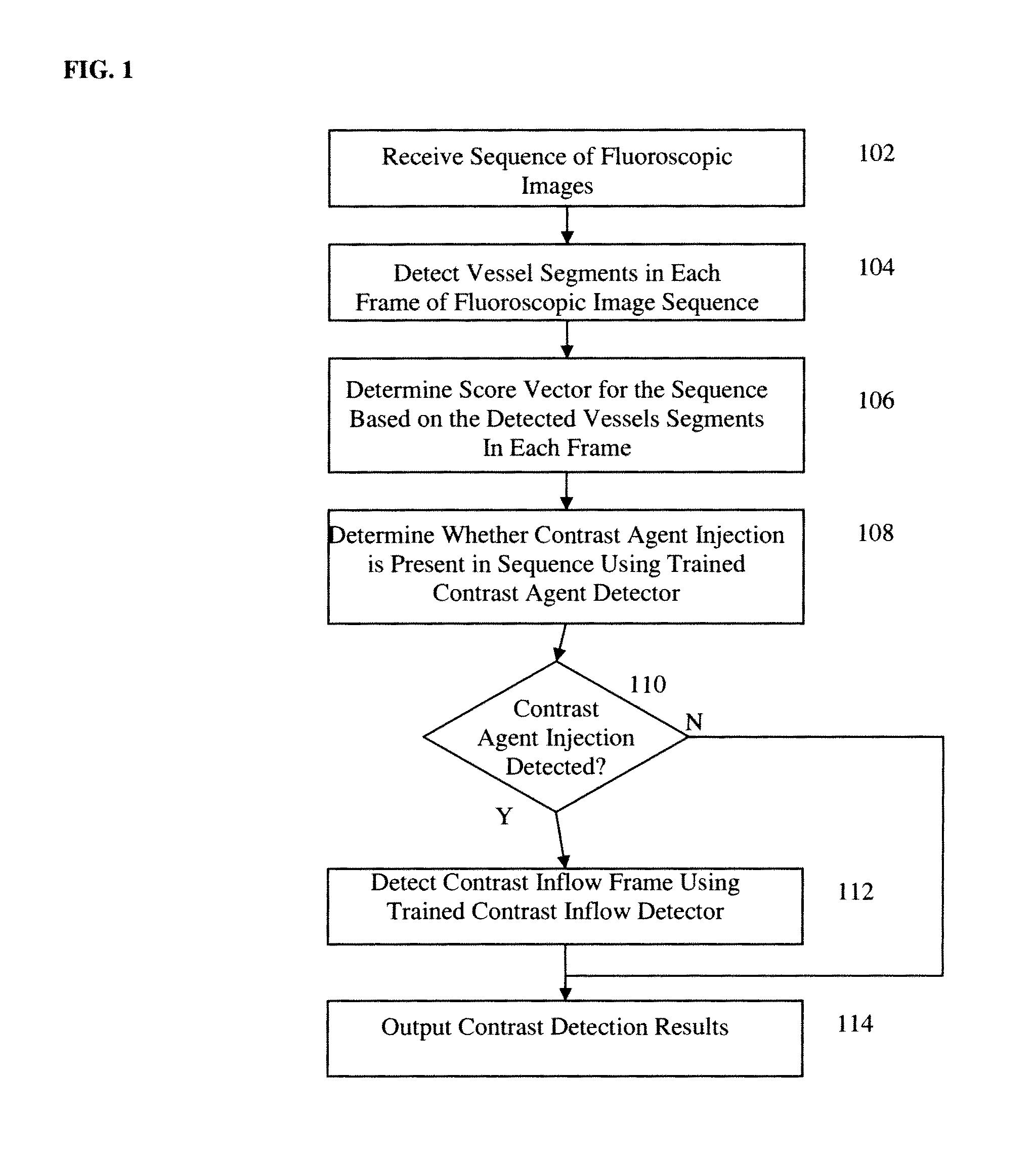
A method and system for contrast inflow detection in a sequence of fluoroscopic images is disclosed. Vessel segments are detected in each frame of a fluoroscopic image sequence. A score vector is determined for the fluoroscopic image sequence based on the detected vessel segments in each frame of the fluoroscopic image sequence. It is determined whether a contrast agent injection is present in the fluoroscopic image sequence based on the score vector. If it is determined that a contrast agent injection is present in the fluoroscopic image sequence, a contrast inflow frame, at which contrast agent inflow begins, is detected in the fluoroscopic image sequence based on the score vector.

Description

[0001]This application claims the benefit of U.S. Provisional Application No. 61 / 449,863, filed Mar. 7, 2011, the disclosure of which is herein incorporated by reference.BACKGROUND OF THE INVENTION[0002]The present invention relates to contrast inflow detection in fluoroscopic images, and more particularly, to detecting whether contrast medium is used in a sequence of fluoroscopic images and detecting at which frame in a sequence of fluoroscopic images the contrast inflow begins.[0003]Computer-aided and image-guided interventions have become more important in recent years for advanced cardiovascular treatments. Medical images, such as 2D fluoroscopic (x-ray) images are widely used for pre-operative planning, intra-operative guidance, post-operative assessment. During an intervention, an angiography is often acquired in multiple phases of the procedure. An angiography is typically acquired by injecting a radio-opaque contrast agent into the blood vessels of a patient and then acquiri...

Claims


Application Information

Patent Timeline
Patent Type & Authority Patents(United States)
IPC IPC(8): G06K9/00G06T7/00
CPCG06T7/0016G06T2207/10121G06T2207/30101
Inventor CHEN, TERRENCEFUNKA-LEA, GARETHCOMANICIU, DORIN
Owner SIEMENS HEALTHCARE GMBH
Learn More