
Image processing apparatus and image processing method

a technology of image processing and image processing, which is applied in the field of image processing apparatus and image processing method, can solve the problems of cumbersome finding out the change, decrease in the s/n ratio and the resolution of the slo image, and the accompanying diameter of the measuring beam, so as to achieve the effect of easy and accurate measurement of blood fluidity

Active Publication Date: 2016-04-12
CANON KK
View PDF16 Cites 3 Cited by
  • Summary
  • Abstract
  • Description
  • Claims
  • Application Information

AI Technical Summary

Benefits of technology

This patent describes an invention that can use multiple moving images to accurately measure the flow of blood at a specific location. This makes it easier and more accurate than existing methods.

Problems solved by technology

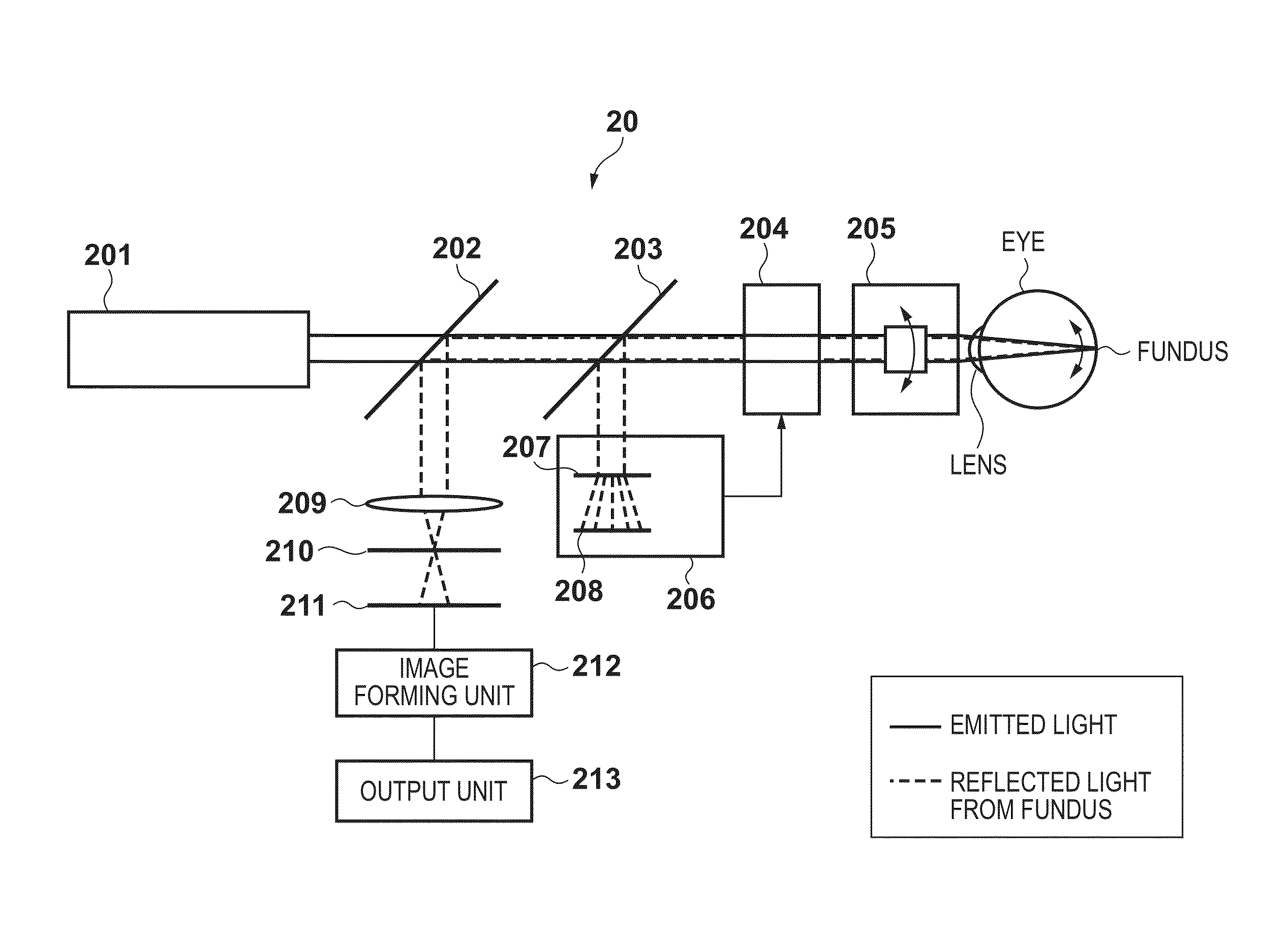
However, there has been a problem in acquiring a retinal SLO image in that increasing the diameter of the measuring beam is accompanied by a decrease in the S / N ratio and the resolution of the SLO image due to aberrations in the eye of the examination subject.
However, in the case of measuring blood fluidity using multiple SLO images that were captured in the same site in the eye area, there has been a problem in that it is cumbersome to find out the change over time.
Also, there has been a problem in that the reliability of measured values is low since statistical values for measured values are measured / calculated without giving consideration to the timing of the heartbeat.
However, the technique above does not give consideration to simply and accurately obtaining a measurement position that corresponds to a measurement position of an image that was captured in the past in order to easily measure and display the change over time in measured values in images captured at different timings, for example.

Method used


Image

  • Image processing apparatus and image processing method
  • Image processing apparatus and image processing method
  • Image processing apparatus and image processing method

Examples


Embodiment Construction

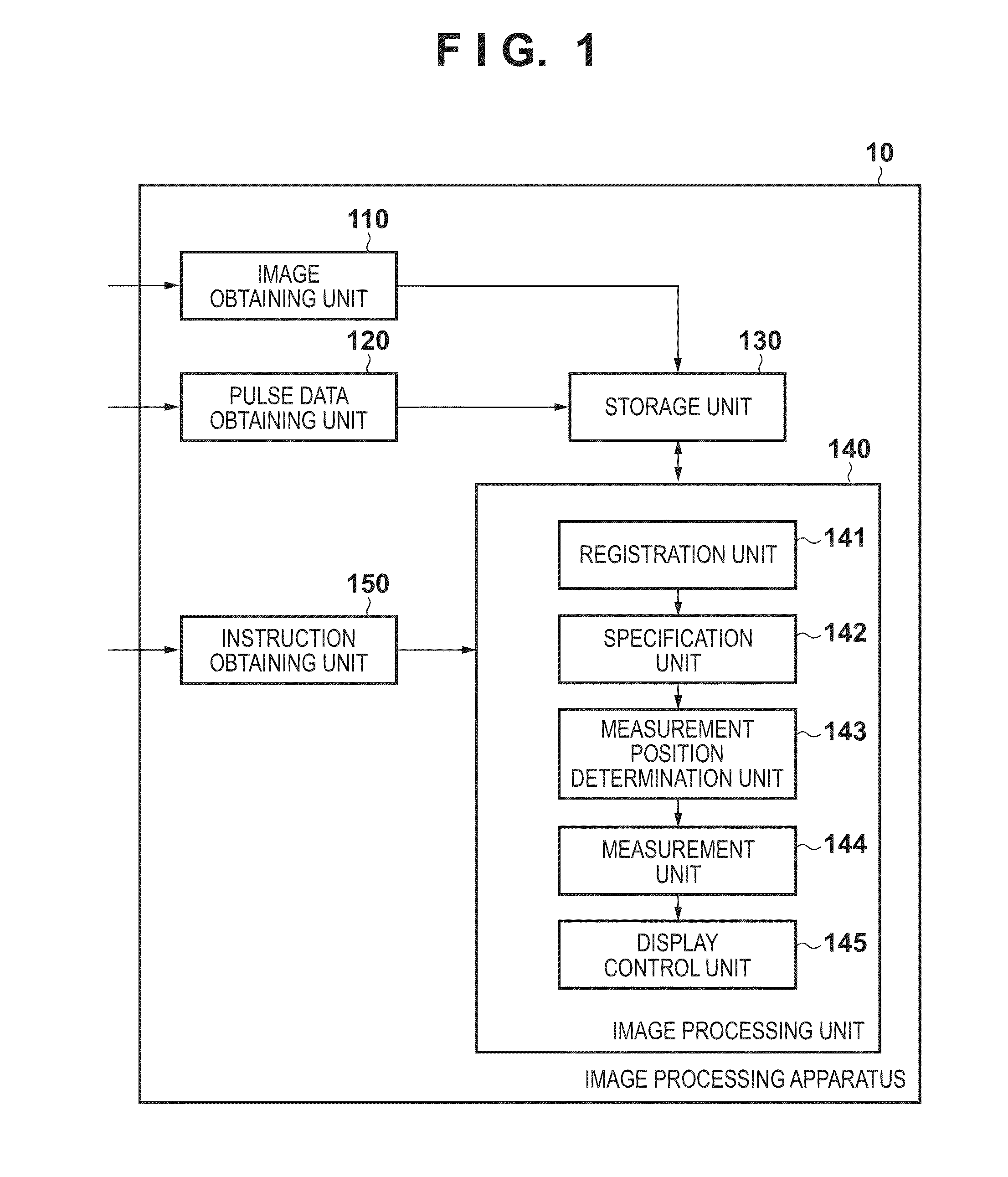
[0032]Preferred embodiments of the image processing apparatus and method according to the present invention will be described below in accordance with the accompanying drawings. Note that the present invention is not limited to the embodiments disclosed below.

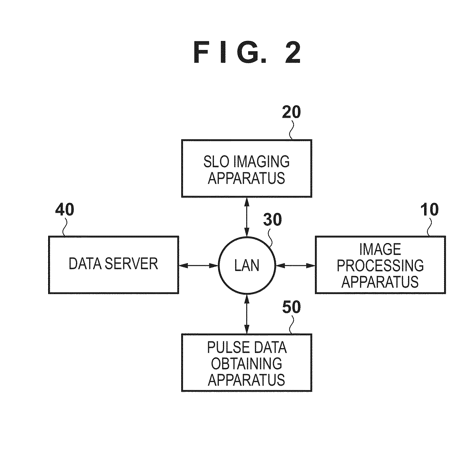
[0033]The image processing apparatus according to the present embodiment is configured to measure change in blood cell aggregate sizes in vascular branches (between bifurcations) using the respective SLO images captured at different dates / times and to display the change over time in the measured values from the different dates / times.

[0034]Specifically, the image processing apparatus registers multiple SLO moving images that have the same imaging site and were captured at different dates / times. Next, blood vessel extraction is performed on a reference image (described in detail later) and a composite image of blood vessel images is generated. A measurement target vascular branch is specified in the composite image based on the s...


PUM


Abstract

An image processing apparatus obtains a plurality of moving images of a predetermined site of an eye, specifies a vascular region by using at least one moving image of the plurality of moving images, and measures the sizes of blood cell aggregates in a specific vascular branch in the specified vascular region for each moving image of the plurality of moving images.

Description

BACKGROUND OF THE INVENTION[0001]1. Field of the Invention[0002]The present invention relates to an image processing apparatus and an image processing method used for ophthalmological consultations.[0003]2. Description of the Related Art[0004]Ophthalmic examinations are widely performed for the purpose of early diagnosis of lifestyle-related diseases and diseases that rank highly among causes of loss of eyesight. A scanning laser ophthalmoscope (SLO), which is an image processing apparatus that uses the principle of a confocal laser microscope, is an apparatus that performs Raster scanning of an eye fundus using a laser that is a measuring beam and obtains a planar image at a high resolution and a high speed based on the intensity of the return light. The apparatus that captures this planar image will be referred to as an SLO apparatus, and the planar image will be referred to as an SLO image below.[0005]In recent years, it has been possible to obtain a retinal SLO image with an imp...

Claims


Application Information

Patent Timeline
Patent Type & Authority Patents(United States)
IPC IPC(8): A61B3/14A61B3/00A61B3/12
CPCA61B3/1241A61B3/0025
Inventor IMAMURA, HIROSHI
Owner CANON KK
Learn More