
Magnetic resonance imaging white matter hyperintensities region recognizing method and system

a magnetic resonance imaging and hyperintensity region technology, applied in the field of image recognition technology, can solve the problems of difficult to clearly distinguish a wmh region and recognition becomes even more difficult, and achieve the effect of reducing the burden associated and improving the recognition efficiency

Active Publication Date: 2016-10-11
NAT CENT UNIV +1
View PDF8 Cites 1 Cited by
  • Summary
  • Abstract
  • Description
  • Claims
  • Application Information

AI Technical Summary

Benefits of technology

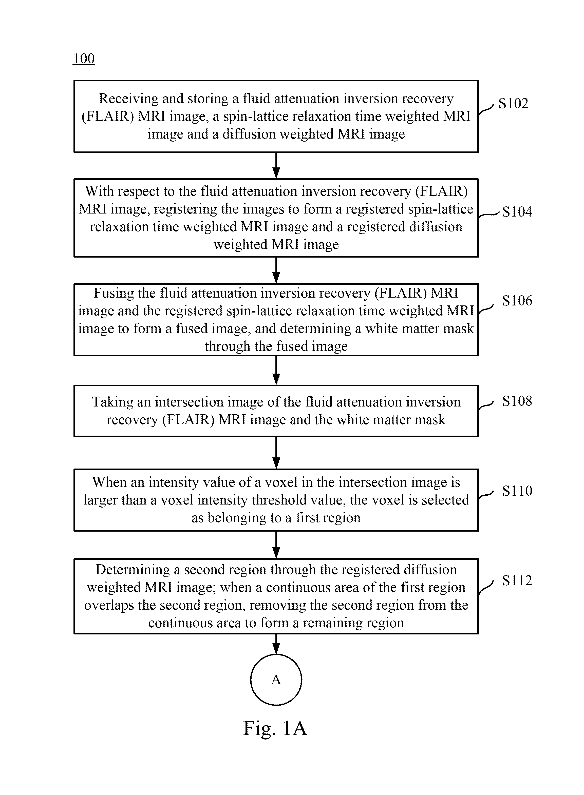
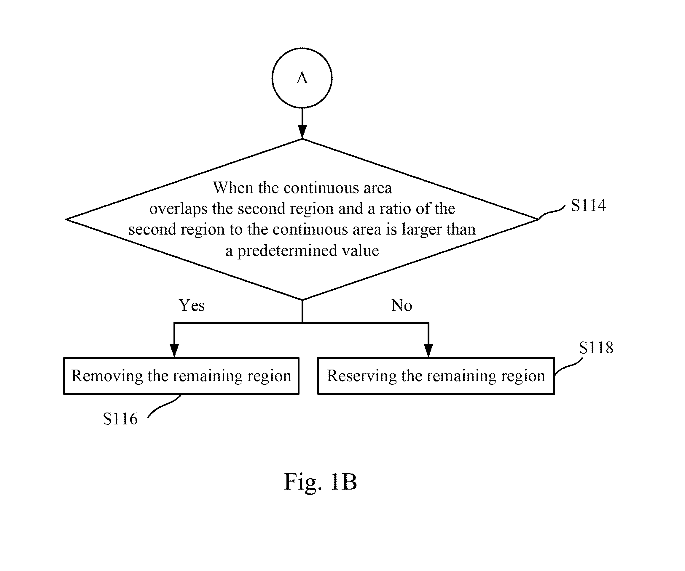
[0018]In sum, the present disclosure is directed to recognizing a WMH region and a cerebral infarct region in an MRI image through removing a cerebral infarct region with the assistance of a diffusion weighted MRI image and reserving a WMH region of a FLAIR MRI image by determining whether a remaining region is peri-infarct edema through the aforementioned embodiments. Moreover, the WMH region is automatically detected to eliminate the burden associated with manual recognition, build a consistent standard, and improve the recognizing efficiency.

Problems solved by technology

However, a cerebral infarct region has a voxel intensity that is similar to the voxel intensity of a general white matter region so that it is difficult to clearly distinguish a WMH region from a cerebral infarct region, easily leading to confusion if simply a FLAIR MRI image is utilized.
Recognition becomes even more difficult when the two regions overlap.

Method used


Image

  • Magnetic resonance imaging white matter hyperintensities region recognizing method and system
  • Magnetic resonance imaging white matter hyperintensities region recognizing method and system
  • Magnetic resonance imaging white matter hyperintensities region recognizing method and system

Examples


Embodiment Construction

[0030]Reference will now be made in detail to embodiments of the present disclosure, examples of which are described herein and illustrated in the accompanying drawings. While the disclosure will be described in conjunction with embodiments, it will be understood that they are not intended to limit the invention to these embodiments. On the contrary, the invention is intended to cover alternatives, modifications and equivalents, which may be included within the spirit and scope of the disclosure as defined by the appended claims.

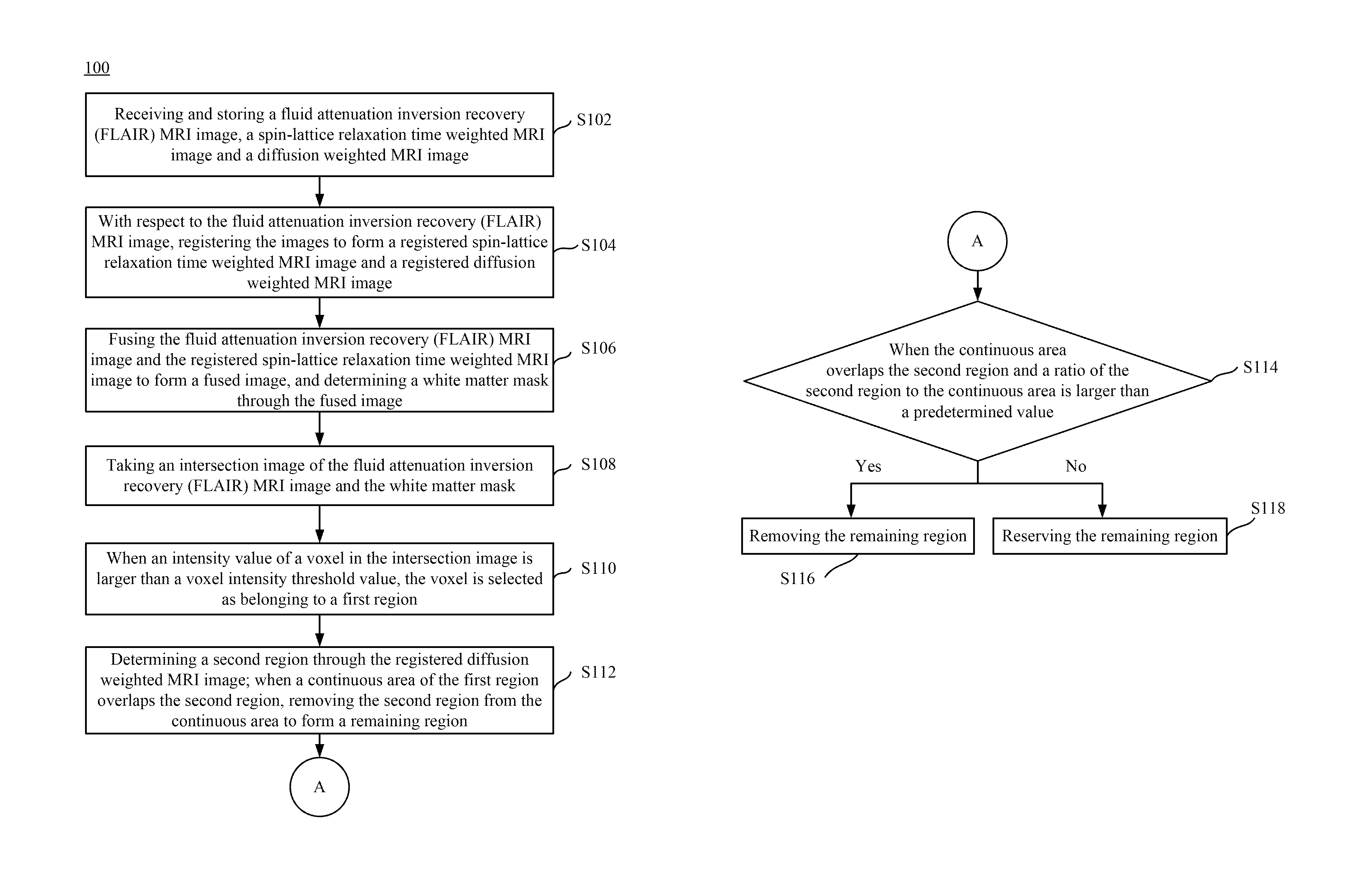
[0031]FIGS. 1A and 1B show flow charts of a magnetic resonance imaging (MRI) white matter hyperintensities region recognizing method according to an embodiment of the present disclosure, where point A is a connection point of the steps in FIGS. 1A and 1B. The MRI white matter hyperintensities region recognizing method 100 includes a plurality of steps S102-S118. However, those skilled in the art should understand that the sequence of the steps in the present...


PUM


Abstract

A magnetic resonance imaging white matter hyperintensities region recognizing method and system are disclosed herein. The white matter hyperintensities region recognizing method includes receiving and storing a FLAIR MRI image, a spin-lattice relaxation time weighted MRI image, and a diffusion weighted MRI image. Registration and fusion are preformed, and a white matter mask is determined. An intersection image of the FLAIR MRI image and the white matter mask is taken, a first region is determined after normalizing the intersection image, a cerebral infarct region is removed from the first image through the diffusion weighted MRI image, and then a determination is made as to whether to remove a remaining region in order to form a white matter hyperintensities region in the FLAIR MRI image.

Description

RELATED APPLICATIONS[0001]This application claims priority to Taiwanese Application Serial Number 104100102, filed Jan. 5, 2015, which is herein incorporated by reference.BACKGROUND[0002]1. Field of Invention[0003]The present invention relates to image recognizing technology. More particularly, the present invention relates to a magnetic resonance imaging white matter hyperintensities region recognizing method and system.[0004]2. Description of Related Art[0005]In the prior art, because a white matter hyperintensities (WMH) region is distinctive compared with a general white matter region in a fluid-attenuated inversion recovery (FLAIR) magnetic resonance imaging (MRI) image, such as FLAIR image is often used as a method to distinguish a WMH region from the general white matter region. However, a cerebral infarct region has a voxel intensity that is similar to the voxel intensity of a general white matter region so that it is difficult to clearly distinguish a WMH region from a cere...

Claims


Application Information

Patent Timeline
Patent Type & Authority Patents(United States)
IPC IPC(8): G06K9/00G01R33/56G06T7/00G01R33/563A61B5/00A61B5/055
CPCG06T7/0012G01R33/5602G01R33/5608G01R33/56341G06T7/0081A61B5/0042A61B5/055G06T2207/10088G06T2207/30016G06T7/11G06T7/174
Inventor PENG, SYU-JYUNCHEN, YU-WEITSAI, JANG-ZERNWANG, KUO-WEI
Owner NAT CENT UNIV
Learn More