Non-invasive deep muscle electromyography

a deep muscle electromyography and non-invasive technology, applied in the field of deep muscle electromyography, can solve the problems of inability to investigate the deep muscles located near major blood vessels, nerves, viscera, etc., and achieve the effect of not being able to investigate the superficial muscles, reducing the range of application, and reducing the number of procedures

Inactive Publication Date: 2017-06-27
UNIVERSITY OF CAPE TOWN +1
View PDF9 Cites 46 Cited by
  • Summary
  • Abstract
  • Description
  • Claims
  • Application Information

AI Technical Summary

Benefits of technology

[0011]It is an object of this invention to provide a method and apparatus for conducting electromyography of a deep muscle non-invasively.

Problems solved by technology

This category is accordingly limited in application to superficial muscles that are close to the skin's surface.
Not only is this technique invasive, but some deep muscles that are located close to major blood vessels, nerves, and viscera should not be investigated at all by such a technique for patient safety reasons.
Invasive electromyography carries with it a number of risks such as the potential for infliction of additional trauma on a patient as well as that of infection.
Such risks thus preclude its routine use in sports performance testing and training, particularly for elite athletes who may risk a performance-reducing or livelihood-destroying injury or infection as a result of such a procedure.
However this is only applicable to those specific muscles and not generalizable to all deep muscles.
Such inverse FEM models do not provide unique mathematical solutions unless there are a large number (>>100) of measurement points, and despite being introduced in 1996, to date there is no clinical evidence that this method is practically able to resolve deep from superficial muscle activity.

Method used


Image

  • Non-invasive deep muscle electromyography
  • Non-invasive deep muscle electromyography
  • Non-invasive deep muscle electromyography

Examples


Embodiment Construction

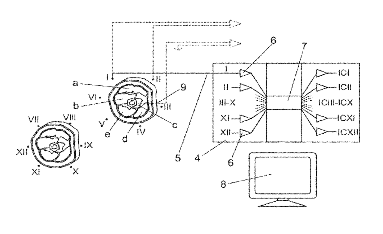
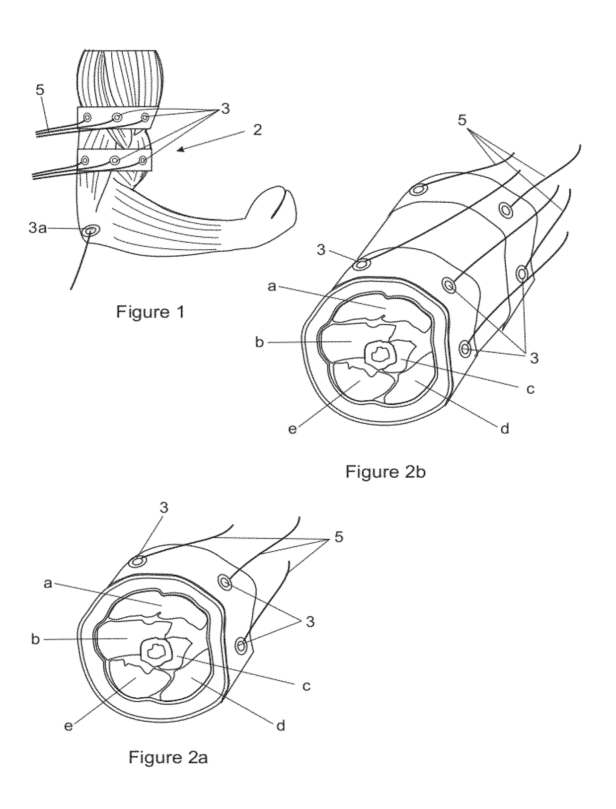
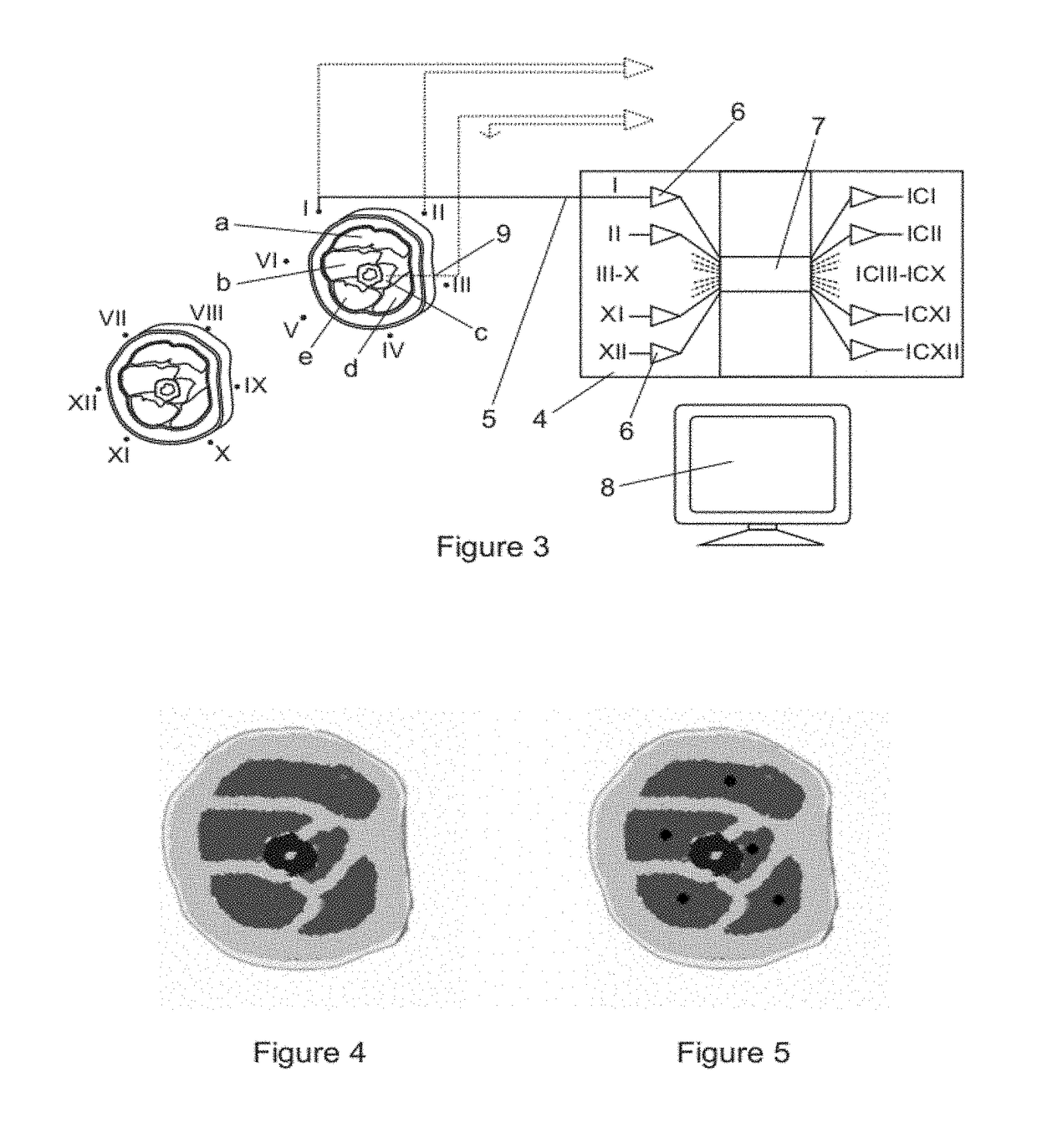
[0028]In one implementation of the invention the example of electromyography of the deep muscle brachialis (c) of the upper arm (2) as applied to an actual human subject in a laboratory setting, is given. In this implementation of the invention two arrays of suitable surface electromyography electrodes (3) are applied to the skin of a patient with the electrodes being arranged in two rings encircling the group of muscles in the appropriate part of the upper arm in which the relevant part of the brachialis is located.

[0029]In this particular instance, the two arrays of electrodes comprise a total of 12 electrodes, with 6 electrodes per ring that are arranged generally equally spaced around the periphery of the arm. The electrodes, that are indicated in FIG. 3 by the Roman numerals (I, II, III, IV, V, VI, VII. VIII, IX, X, XI, XII) sequentially in a clockwise direction around the two rings, are connected to a processing unit (4) by means of flexible conductors (5) in the usual way.

[00...


PUM


Abstract

A method and apparatus for conducting electromyography of a deep muscle non-invasively are provided. An array of suitable surface electromyography electrodes is arranged in one or more rings encircling a part of the human body in which a deep muscle being investigated is located. The potential of at least selected electrodes relative to another electrode selected from a common reference electrode (mono-polar) and other electrodes (bi-polar) in the array is recorded and the data is processed in respect of the recorded potentials of at least some of said selected electrodes in order to determine (optionally using approximations or algorithms, or both) the contribution being made by at least the deep muscle being investigated. Typically, this is done mathematically by resolving the electromyography signals into their constituent components using a suitable technique. Preferably, this is integrated with a static muscle imaging device that may use the same electrodes to obtain a static tomogram of the muscles encircled.

Description

CROSS-REFERENCE TO RELATED APPLICATIONS[0001]This application claims priority to PCT / IB2010 / 001876, filed Jul. 29, 2010, and published in English language as Publication No. WO 2011 / 012988 on Jul. 29, 2010, which claims priority to ZA Application No. 2009 / 05309, filed Jul. 30, 2009, incorporated herein by reference.FIELD OF THE INVENTION[0002]This invention relates to non-invasive deep muscle electromyography (EMG) to be used for recording the electrical activity of muscles in order to assess muscle function in a variety of different fields including sports, biomechanics research, physiotherapy and clinical neuro-muscular diagnostics.BACKGROUND TO THE INVENTION[0003]There are presently two categories of electromyography recordings. The first is a non-invasive surface electromyography (sEMG) in which adhesive bio-electrodes are placed on the surface of the skin directly above the muscle under investigation. This category is accordingly limited in application to superficial muscles th...

Claims


Application Information

Patent Timeline
Patent Type & Authority Patents(United States)
IPC IPC(8): A61B5/04A61B5/0492G06K9/62A61B5/0488A61B5/00G06K9/00A61B5/11A61B5/053A61B5/296
CPCA61B5/0492A61B5/04012A61B5/4519A61B5/7203A61B5/7264A61B2562/0209A61B2562/043G06K9/0057G06K9/624A61B5/0488A61B5/0536A61B5/1107A61B5/6802A61B5/6824A61B5/6831A61B5/316A61B5/296A61B5/389G06F2218/22
Inventor JOHN, LESTER RYAN
Owner UNIVERSITY OF CAPE TOWN
Why Eureka
Try Eureka