Methods of Distracting Tissue Layers of the Human Spine

a technology of human spine and tissue layer, applied in the field of apparatus and methods, can solve the problems of reducing bone density, weakening bones, and adding significant cost to the health care system, and achieve the effect of increasing the extent of the support structur

Active Publication Date: 2007-05-31
IZI MEDICAL PROD
View PDF81 Cites 207 Cited by
  • Summary
  • Abstract
  • Description
  • Claims
  • Application Information

AI Technical Summary

Benefits of technology

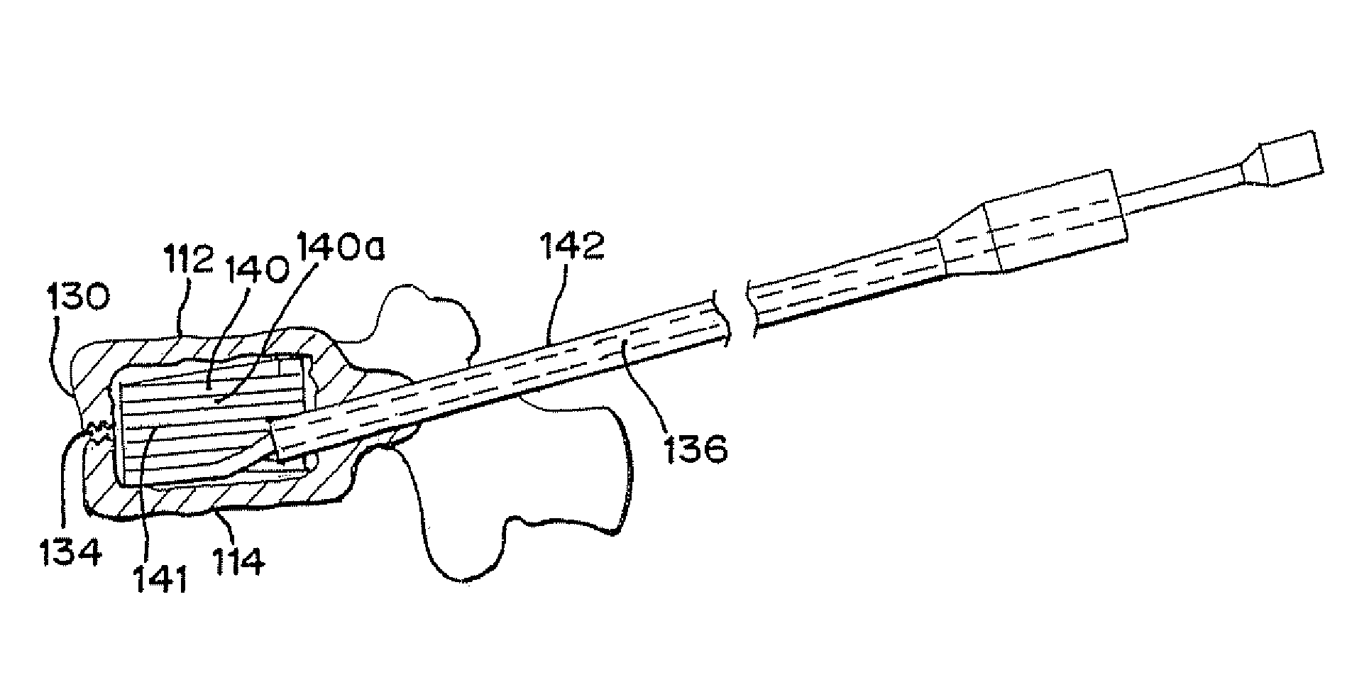
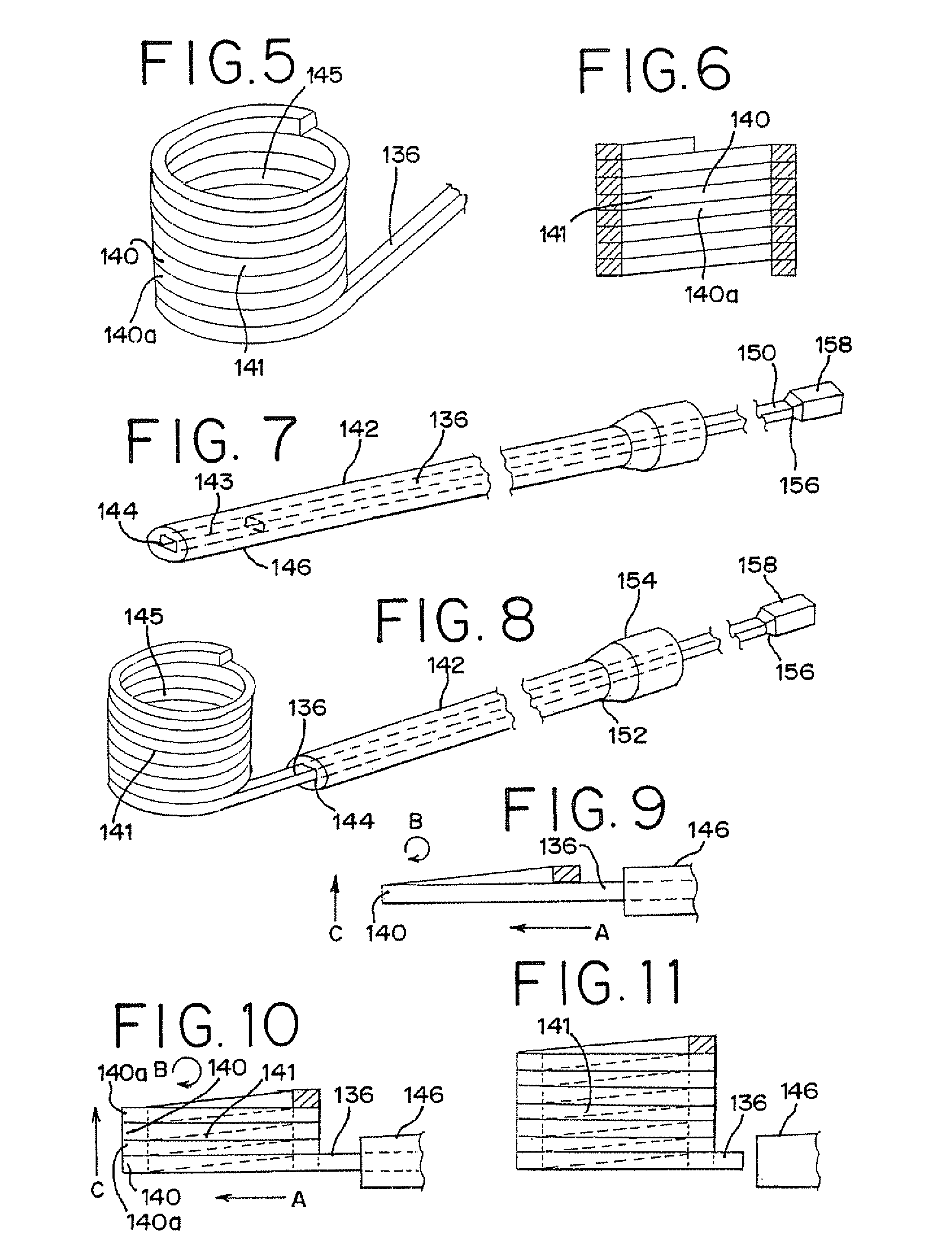
[0047] A further aspect of the present invention relates to methods for treating a vertebral body. In one embodiment, the method generally comprises inserting at least one elongated member into the body of a human vertebra, and allowing the elongated member to self-form into a generally helical shaped support structure that includes a plurality of stacked windings. The number of windings of the support structure increases as the elongated member is inserted, thereby increasing the extent of the support structure and separating and / or supporting the endplates of the vertebral body.

Problems solved by technology

Vertebral compression fractures affect a significant part of the population, and add significant cost to the health care system.
Osteoporosis reduces bone density, thereby weakening bones and predisposing them to fracture.
The osteoporosis-weakened vertebrae can collapse during normal activity and are also more vulnerable to injury from shock or other forces acting on the spine.
While the fractures may heal without intervention, the crushed bone may fail to heal adequately.
Spinal deformity may lead to breathing and gastrointestinal complications, and adverse loading of adjacent vertebrae.
Commonly, they cause loss of height and a humped back.
In severe cases, the body's center of mass is moved further away from the spine resulting in increased bending moment on the spine and increased loading of individual vertebrae.
When cancer cells spread to the spine, the cancer may cause destruction of part of the vertebra, weakening and predisposing the bone to fracture.
Osteoporosis and metastatic disease are common root causes leading to vertebral fractures, but trauma to healthy vertebrae can also cause fractures ranging from minor to severe.
Such trauma may result from a fall, a forceful jump, a car accident, or any event that compresses or otherwise stresses the spine past its breaking point.
However, they often cause a severe “band-like” pain that radiates from the spine around both sides of the body.
It is commonly believed that the source of acute pain in compression fractures is the result of instability at the fracture site, allowing motion that irritates nerves in and around the vertebrae.
Unfortunately, the typical patient is an elderly person.
As a class of patients, the elderly generally do not tolerate extended bed rest well.
However, such procedures dramatically increase the risk of bone filler extravasation from the vertebral body.
The difficulty of controlling or stopping bone filler flow into injury-sensitive areas increases as the required pressure increases.
Caution, however, must still be taken to prevent extravasation, with the greatest attention given to preventing posterior extravasation because it may cause spinal cord trauma.
If a foraminal leak results, the patient may require surgical decompression and / or suffer paralysis.
However, in practice it is difficult to consistently attain meaningful and predictable height restoration.
The inconsistent results may be due, in part, to the manner in which the balloon expands in a compressible media, such as the cancellous tissue within the vertebrae, and the structural orientation of the trabecular bone within the vertebra, although there may be additional factors as well.
The weight of the person generates a compressive load on the disks, but this load is not uniform during typical bending movements.
On the local (or cellular) level, decreased disk height results in increased pressure in the nucleus, which can lead to a decrease in cell matrix synthesis and an increase in cell necrosis and apoptosis.
In addition, increases in intra-diskal pressure create an unfavorable environment for fluid transfer into the disk, which can cause a further decrease in disk height.
Decreased disk height may also result in significant changes in the overall mechanical stability of the spine.
With decreasing height of the disk, the facet joints bear increasing loads and may undergo hypertrophy and degeneration, and may even act as a source of pain over time.
Increased stiffness of the spinal column and decreased range of motion resulting from loss of disk height can lead to further instability of the spine, as well as back pain.
This decrease may lead to spinal nerve impingement, with associated radiating pain and dysfunction.
The disks that must bear additional loading are susceptible to accelerated degeneration and compromise, which may eventually propagate along the destabilized spinal column.
However, a sudden decrease in disk volume caused by surgical removal of the disk or disk nucleus can heighten the local and global problems noted above.
Genetic and biochemical causes can result in changes in the extracellular matrix pattern of the disk and a decrease in biosynthesis of extracellular matrix components by the cells of the disk.
With a loss of water content, the nucleus becomes desiccated resulting in a decrease in internal disk hydraulic pressure that ultimately results in a loss of disk height.
This loss of disk height can cause non-tensile loading and buckling of the annulus.
The loss of disk height also causes the annular lamellae to delaminate, resulting in annular fissures and rupture of the annulus.
During a total diskectomy, a substantial amount (and usually all) of the volume of the nucleus pulposus is removed and immediate loss of disk height and volume can result.
Even with a partial diskectomy, loss of disk height can ensue.
A challenge of inserting a disk implant posteriorly is that a device large enough to contact the endplates and slightly expand the intervertebral space between the endplates must be inserted through a limited space.
This challenge is often further heightened by the presence of posterior osteophytes, which may cause converging or “fish mouthing” of the posterior endplates that results in very limited access to the disk.
A further challenge in degenerative disk spaces is the tendency of the disk space to assume a lenticular shape, which requires a relatively larger implant that often is not easily introduced without causing trauma to the nerve roots.
The size of rigid devices that may safely be introduced into the disk space is thereby limited.
Cages inserted from the posterior approach, however, are limited in size by the interval between the nerve roots.
It is commonly believed that instability can either be a source of back or neck pain or cause potential irritation or damage to adjacent nerves.
Although disk removal is commonly combined with fusion in the neck, this is not generally true in the low back (lumbar spine).
A major obstacle to the successful treatment of spine pain by fusion is the difficulty in accurately identifying the source of a patient's pain.
Vertebral bodies may be compromised due to disease, defect, or injury.
These prior art devices and the procedures associated therewith have difficulty in maintaining the proper structural scaffolding while a castable material, such as bone cement, is hardened in the cavity left by the removed vertebral body.
The maintaining of proper structural scaffolding has been especially difficult in a minimally invasive posterior surgical approaches.

Method used


Image

  • Methods of Distracting Tissue Layers of the Human Spine
  • Methods of Distracting Tissue Layers of the Human Spine
  • Methods of Distracting Tissue Layers of the Human Spine

Examples


Embodiment Construction

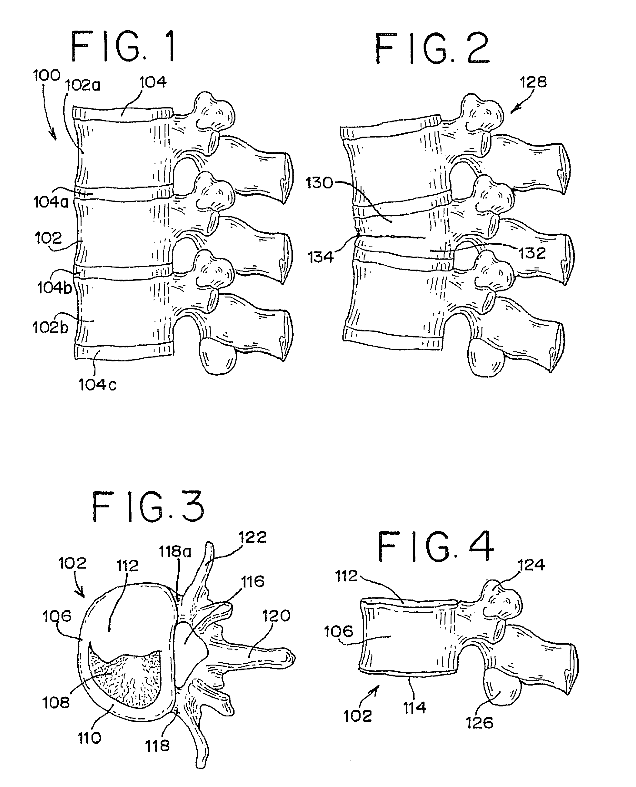
[0132]FIG. 1 illustrates a section of a healthy vertebral (spinal) column, generally designated as 100, without injury. The vertebral column 100 includes adjacent vertebrae 102, 102a and 102b and intervertebral disks 104, 104a, 104b and 104c separating adjacent vertebrae.

[0133]FIGS. 3 and 4 illustrate in more detail a normal vertebra and its attributes. The vertebra, generally designated as 102, includes a vertebral body 106 that is roughly cylindrically and comprised of inner cancellous bone 108 surrounded by the cortical rim 110, which is comprised of a thin layer of cortical compact bone. The cortical rim 110 can be weakened by osteoporosis and may be fractured due to excessive movement and / or loading. The body 106 of the vertebra is capped at the top by a superior endplate 112 and at the bottom by an inferior endplate 114, made of a cartilaginous layer. To the posterior (or rear) of the vertebral body 106 is the vertebral foramen 116, which contains the spinal cord (not shown)....


PUM

PropertyMeasurementUnit
internal diameteraaaaaaaaaa
diameteraaaaaaaaaa
sizesaaaaaaaaaa

Abstract

The present invention relates to methods of separating, supporting or both separating and supporting layers of tissue in the human spine. Such methods generally comprise inserting at least one member between layers of tissue in the human spine and changing the configuration of the member to define a support structure between the tissue layers.

Description

[0001] This application claims the benefit of U.S. Provisional Application No. 60 / 708,691, filed Aug. 16, 2005; U.S. Provisional Application No. 60 / 738,432, filed Nov. 21, 2005; and U.S. Provisional Application No. 60 / 784,185, filed Mar. 21, 2006, all of which are hereby incorporated herein by reference.FIELD OF THE INVENTION [0002] The present invention generally relates to apparatus and methods employed in minimally invasive surgical procedures and more particularly to such apparatus and methods in orthopedic procedures for separating and / or supporting tissue layers. BACKGROUND OF THE INVENTION [0003] A variety of physical conditions involve two tissue surfaces that, for diagnosis or treatment of the condition, need to be separated or distracted from one another and then supported in a spaced-apart relationship. Such separation or distraction may be to gain exposure to selected tissue structures, to apply a therapeutic pressure to selected tissues, to return or reposition tissue s...

Claims


Application Information

Patent Timeline
Patent Type & Authority Applications(United States)
IPC IPC(8): A61F2/44
CPCA61B17/70A61F2210/0085A61B2017/00867A61B2017/0256A61F2/44A61F2/4455A61F2/4611A61F2002/2835A61F2002/30092A61F2002/30153A61F2002/30235A61F2002/30263A61F2002/30289A61F2002/30462A61F2002/3052A61F2002/30594A61F2002/30601A61F2002/30677A61F2002/30772A61F2002/4415A61F2002/448A61F2002/4627A61F2002/4635A61F2002/4683A61F2002/4688A61F2210/0014A61F2220/0025A61F2220/0075A61F2230/0019A61F2230/0069A61F2230/0084A61F2230/0091A61F2310/00293A61F2/442A61B17/8852A61F2250/0067A61B17/7097A61F2/441
Inventor SCHALLER, LAURENT
Owner IZI MEDICAL PROD
Why Eureka
Try Eureka