MR imaging using apt contrast enhancement and sampling at multiple echo times

a contrast enhancement and multiple echo technology, applied in the field of magnetic resonance imaging, can solve the problems of insufficient clinical information, large artifacts, and insufficient contrast between different tissues, and achieve the effect of improving mr imaging technique, high contrast-to-noise mr imaging, and efficient and precise intrinsic b0 determination

Active Publication Date: 2017-09-19
KONINK PHILIPS ELECTRONICS NV
View PDF5 Cites 2 Cited by
  • Summary
  • Abstract
  • Description
  • Claims
  • Application Information

AI Technical Summary

Benefits of technology

[0011]From the foregoing it is readily appreciated that there is a need for an improved MR imaging technique. It is consequently an object of the invention to provide a MR imaging method and a MR device which enable high-quality and high contrast-to-noise MR imaging using APT / CEST with efficient and precise intrinsic B0 determination and possible robust elimination of adverse effects due to fat signal contributions.
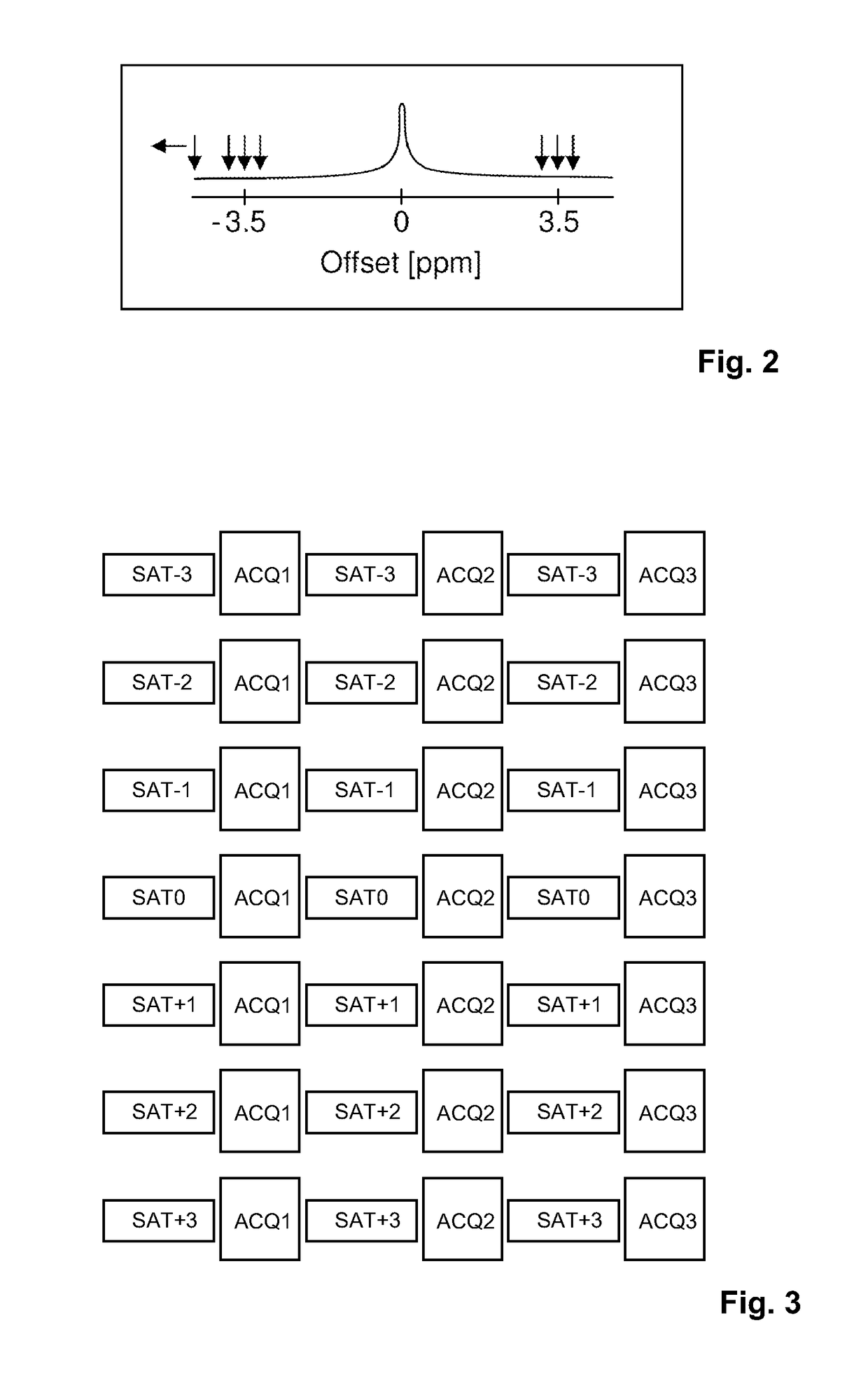
[0014]According to the invention, the combinations of saturation frequency offset values and echo time shift values is kept limited. In effect the plane spanned by the offset values and shift values is sparsely sampled by the interation of the imaging sequence at the selected combinations of offset values and shift values. As the number of interations at respective combinations of offset-values and shift-values is limited, the total acquisition time of the APT / CEST acquisition can be limited. In the optimal case, according to the invention, the extra time for B0 field mapping is fully avoided, while the overall acquisition time for a conventional APT / CEST acquisition (requiring B0 information in addition) is not significantly increased. As an example, an efficient 2D APT sampling scheme uses 7 different saturation frequency offsets (e.g. −4, −3.5, −3, +3, +3.5, +4.5 ppm and one image without or with far detuned saturation). The steps of saturation and signal acquisition are repeated, wherein the saturation frequency offset and / or the echo time shift in the imaging sequence are varied. This can be achieved, for example, by varying the timing of the RF refocusing pulse(s), thereby shifting the refocusing of the nuclear magnetization, and / or by varying the timing of the signal acquisition window and associated magnetic field gradients. An essential feature of the invention is that different and selected combinations of saturation frequency offset and echo time shift are applied in different repetitions. A subset of specific combinations are selected which can be efficiently used to obtain APT / CEST images with intrinsic magnetic field homogeneity correction. Finally, MR images is reconstructed from the acquired MR signals, which may be quantitative APT / CEST images or APT / CEST weighted images.
[0016]Since MR signals are acquired as spin echo signals at different echo time shifts, the spatial variation of B0 within the portion of the body can be determined from the acquired MR signals by means of a multi-point Dixon technique. According to the per se known Dixon technique, the spectral difference between fat and water spins is made use of for the purpose of separating MR signals emanating from water containing tissue and MR signals emanating from fat tissue. In spin echo Dixon imaging, multiple acquisitions of k-space are repeated with different echo time shifts. The simplest Dixon technique, a two-point Dixon technique, acquires two complete k-space data sets, wherein the fat magnetization in the second acquisition shows a phase difference (e.g. 180°=out phase) relative to the water magnetization, and a different phase difference (e.g. 0°=in phase) in the first acquisition. In the case of out phase and in phase images, separate and distinct water and fat images can be obtained by simple addition or subtraction of the complex MR signal data. In general, a B0 field map, a water image and a fat image are obtained by means of a Dixon technique, which may include an iterative reconstruction approach. Hence, also the spatial variation of B0 within the portion of the body can be determined from the MR signals acquired in accordance with the invention by means of the single- or multi-point spin echo Dixon technique. The method of the invention thus permits the application of Dixon methods for both B0 mapping as well as water / fat separation simultaneously in the context of spin echo MRI. The method of the invention integrates spin echo Dixon methods into APT / CEST MR imaging in an efficient manner.
[0017]The reconstruction of the MR image according to the invention may include deriving the spatial distribution of amide protons within the portion of the body from an asymmetry analysis or other z-spectral analysis technique based on the amplitude of the acquired MR signals as a function of the saturation frequency offset, wherein the z-spectral analysis involves a saturation frequency offset correction based on the spatial variation of B0 determined by means of the applied Dixon method. The approach of the invention thus enables correcting for B0 inhomogeneity in APT / CEST MR imaging by integration of spin echo Dixon methods.

Problems solved by technology

In some medical applications, the difference in MR signal intensity from standard MR protocols, i.e. the contrast between different tissues, might not be sufficient to obtain satisfactory clinical information.
This variation results in large artifacts in the finally reconstructed APT / CEST MR images.
This results in an extended overall imaging time.
Moreover, the separately performed measurement of to obtain the B0 inhomogeneity information is potentially inaccurate or inconsistent, for example because of patient motion, shimming or frequency drift of the used MR device between the field mapping and the actual APT / CEST acquisition.
Thus, the known technique may be severely limited for clinical applications with respect to scan time efficiency and precision.
Another issue in APT and CEST MR imaging is that a robust elimination of signal contributions from fat spins, e.g. by fat saturation RF pulses, is often difficult in the presence of B0 inhomogeneity.
However, residual fat signal contributions result in a strongly biased asymmetry of the amplitude of the acquired MR signals as a function of the saturation frequency offset near the chemical shift of fat protons at −3.4 ppm relative to the MR frequency of water protons.

Method used


Image

  • MR imaging using apt contrast enhancement and sampling at multiple echo times
  • MR imaging using apt contrast enhancement and sampling at multiple echo times
  • MR imaging using apt contrast enhancement and sampling at multiple echo times

Examples


Embodiment Construction

[0027]With reference to FIG. 1, a MR device 1 is shown. The device comprises superconducting or resistive main magnet coils 2 such that a substantially uniform, temporally constant main magnetic field B0 is created along a z-axis through an examination volume. The device further comprises a set of (1st, 2nd, and—where applicable—3rd order) shimming coils 2′, wherein the current flow through the individual shimming coils of the set 2′ is controllable for the purpose of minimizing B0 deviations within the examination volume.

[0028]A magnetic resonance generation and manipulation system applies a series of RF pulses and switched magnetic field gradients to invert or excite nuclear magnetic spins, induce magnetic resonance, refocus magnetic resonance, manipulate magnetic resonance, spatially and otherwise encode the magnetic resonance, saturate spins, and the like to perform MR imaging.

[0029]More specifically, a gradient pulse amplifier 3 applies current pulses to selected ones of whole-...


PUM


Abstract

The invention relates to a method of CEST or APT MR imaging of at least a portion of a body (10) placed in a main magnetic field B0 within the examination volume of a MR device. The method of the invention comprises the following steps: •a) subjecting the portion of the body (10) to a saturation RF pulse at a saturation frequency offset; •b) subjecting the portion of the body (10) to an imaging sequence comprising at least one excitation / refocusing RF pulse and switched magnetic field gradients, whereby MR signals are acquired from the portion of the body (10) as spin echo signals; •c) repeating steps a) and b) two or more times, wherein the saturation frequency offset and / or a echo time shift in the imaging sequence are varied, such that a different combination of saturation frequency offset and echo time shift is applied in two or more of the repetitions; •d) reconstructing a MR image and / or B0 field homogeneity corrected APT / CEST images from the acquired MR signals. Moreover, the invention relates to a MR device (1) for carrying out the method of the invention and to a computer program to be run on a MR device.

Description

CROSS-REFERENCE TO PRIOR APPLICATIONS[0001]This application is the U.S. National Phase application under 35 U.S.C. §371 of International Application No. PCT / IB2013 / 052244, filed on Mar. 21, 2013, which claims the benefit of U.S. Provisional Patent Application No. 61 / 619,601, filed on Apr. 3, 2012 and European Patent Application No. 12162970.3, filed on Apr. 3, 2012. These applications are hereby incorporated by reference herein.FIELD OF THE INVENTION[0002]The invention relates to the field of magnetic resonance (MR) imaging. It concerns a method of MR imaging of at least a portion of a body placed in a main magnetic field B0 within the examination volume of a MR device. The invention also relates to a MR device and to a computer program for a MR device.[0003]Image-forming MR methods which utilize the interaction between magnetic fields and nuclear spins in order to form two-dimensional or three-dimensional images are widely used nowadays, notably in the field of medical diagnostics,...

Claims


Application Information

Patent Timeline
Patent Type & Authority Patents(United States)
IPC IPC(8): G01R33/48G01R33/56G01R33/561A61B5/055G01R33/24
CPCG01R33/4828G01R33/5605A61B5/055G01R33/243G01R33/5617
Inventor EGGERS, HOLGERKEUPP, JOCHEN
Owner KONINK PHILIPS ELECTRONICS NV
Why Eureka
Try Eureka