
Silver nanocluster probe and target polynucleotide detection method using same, and silver nanocluster probe design method

a technology of silver nanocluster probes and target polynucleotides, which is applied in the direction of organic chemistry, biochemistry apparatus and processes, sugar derivatives, etc., can solve the problems of reproducibility of microfluidic chips, inconvenient measurement of impedance values, and difficulty in analysis, so as to achieve rapid and convenient detection, specificity and sensitivity, the effect of rapid and convenient detection

Active Publication Date: 2017-10-03
SEOULIN BIO SCI
View PDF10 Cites 0 Cited by
  • Summary
  • Abstract
  • Description
  • Claims
  • Application Information

AI Technical Summary

Benefits of technology

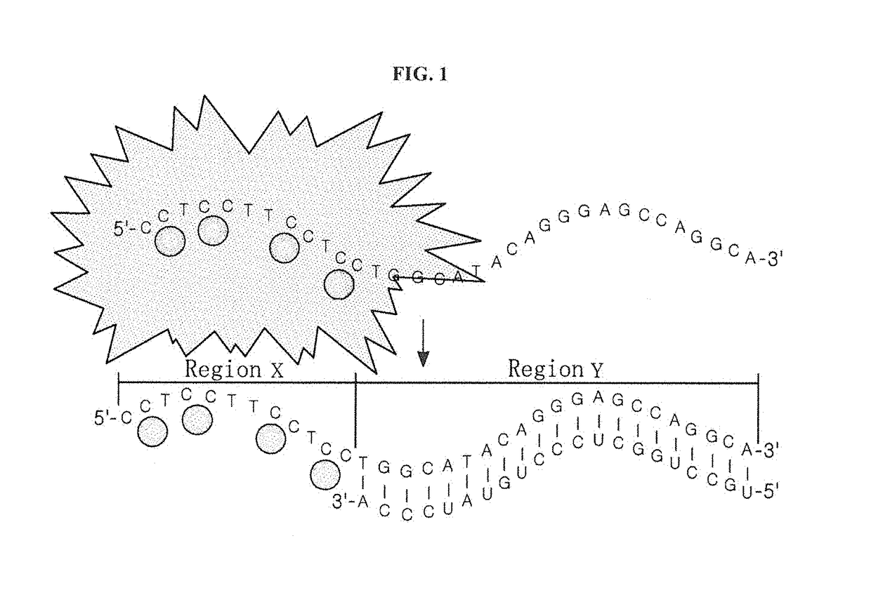
[0047]The silver nanocluster probe according to the present invention comprises a silver nanoparticle binding region and a specific nucleotide sequence region that specifically binds to a target polynucleotide. The silver nanocluster probe according to the present invention is constructed such that it will emit detectable light when silver nanoparticles bind to the silver nanoparticle binding region to form a silver nanocluster, but the light emission from the silver nanocluster probe will decrease or decay when the target polynucleotide binds to the specific nucleotide sequence region.
[0048]According to the present invention, either the presence of a target polynucleotide in a sample or a mutation in the target polynucleotide can be detected in a rapid and convenient manner by determining whether light emission decreases or decays when the target polynucleotide binds to the specific nucleotide sequence region of the silver nanocluster probe that emits detectable light. Specifically, when the silver nanocluster probe according to the present invention is used, either the presence of a target polynucleotide in a sample or a mutation in the target polynucleotide can be rapidly and conveniently detected within 1 hour without a separate labeling process while satisfying both the specificity and sensitivity of detection.
[0049]In addition, the present invention provides various silver nanocluster probes emitting light at various wavelengths, and these silver nanocluster probes can be provided as sensors corresponding to various target polynucleotides. Thus, the present invention can provide a biosensor capable of detecting either the presence of a target polynucleotide or a mutation in the target polynucleotide based on whether the emission of light at the wavelength of interest decreases or decays.

Problems solved by technology

However, this microfluidics chip is problematic in terms of the reproducibility of detection due to a buffer that is received in the reaction chamber.
It also has a problem in that a sweeping process for applying an alternating current at each frequency is required, making it inconvenient to measure an impedance value, resulting in difficulty in analysis.
In addition, a fluorophore such as Cy3 or Cy5, which is used in the microarray chip as described above, is inconvenient in that it should be previously labeled onto oligonucleotide primers.
In addition, it has problems such as poor optical stability and insufficient light intensity.
However, the prior art technologies as described above relate to either amplifying a signal for electron microscopic observation using silver nanoparticles as an electron microscopic dye or labeling an oligonucleotide with a cluster of silver nanoparticles in place of the fluorophore Cy3 or Cy5, and are far from a new base technology that rapidly and conveniently detects either the presence of a target polynucleotide in a sample or a mutation in the polynucleotide while satisfying both the specificity and sensitivity of detection.

Method used


Image

  • Silver nanocluster probe and target polynucleotide detection method using same, and silver nanocluster probe design method
  • Silver nanocluster probe and target polynucleotide detection method using same, and silver nanocluster probe design method
  • Silver nanocluster probe and target polynucleotide detection method using same, and silver nanocluster probe design method

Examples


example 1

ion of a Silver Nanocluster Probe and Analysis of its Emission

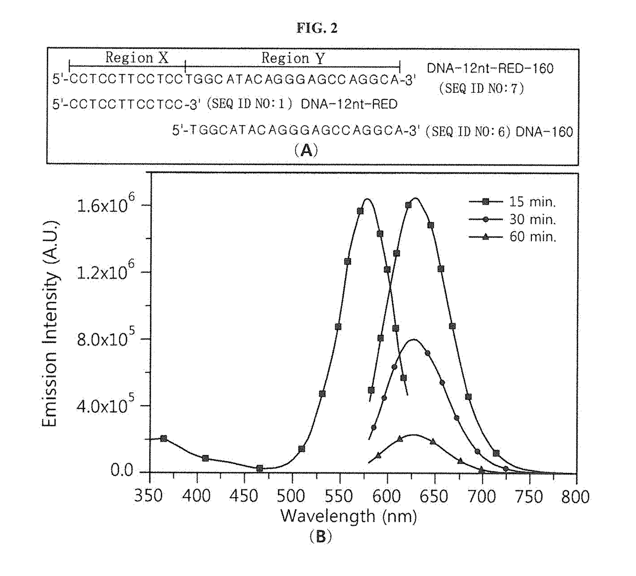
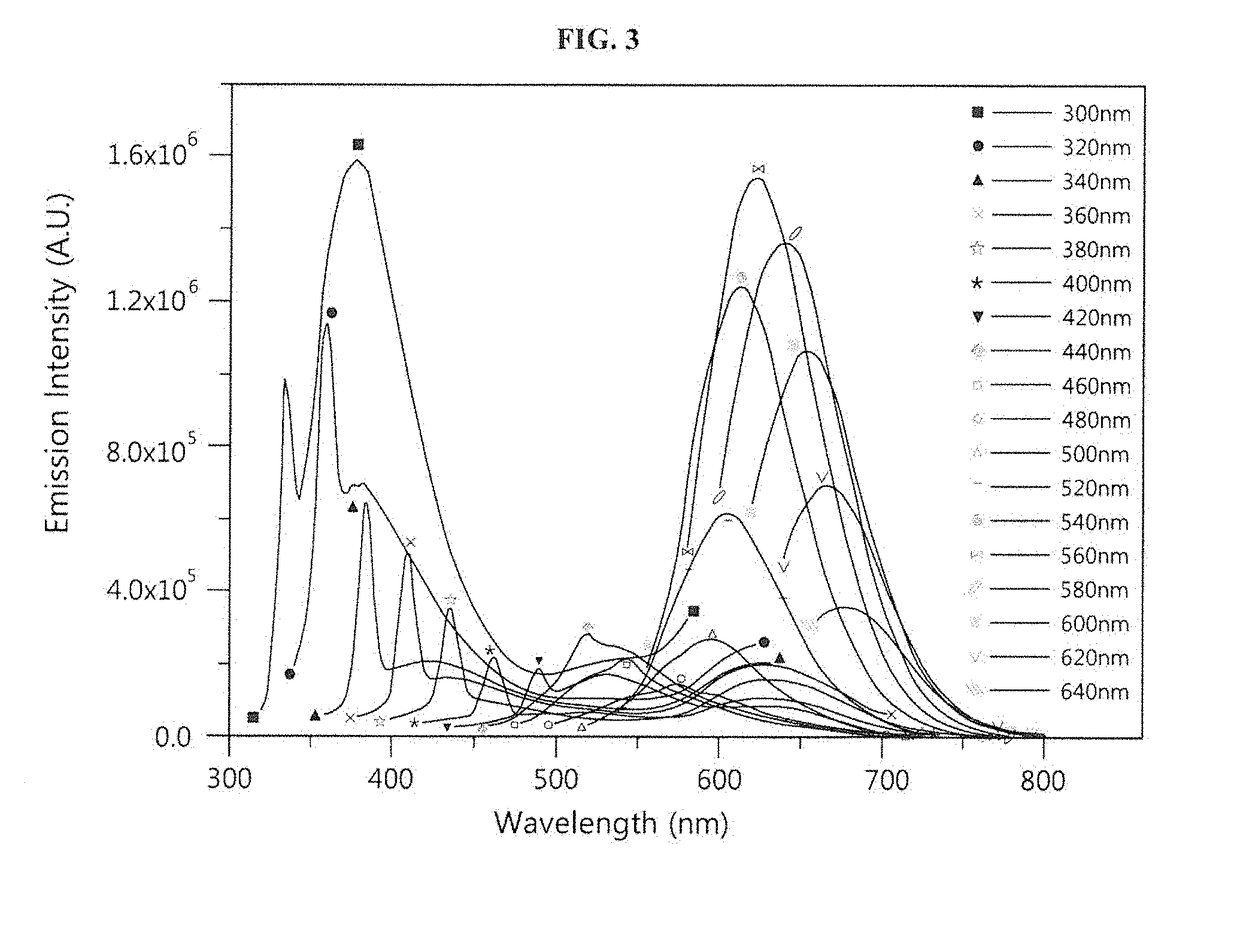
[0063]A nucleotide sequence (SEQ ID NO: 6; hereinafter abbreviated as “DNA-160”) complementary to miRNA 160 (SEQ ID NO: 12; hereinafter abbreviated as “miR160”) was added to the 3′ end of an oligonucleotide (SEQ ID NO: 1; hereinafter abbreviated as “DNA-12nt-RED”) of a silver nanoparticle binding region (region X), thereby constructing a silver nanocluster probe (SEQ ID NO: 7; hereinafter referred to as “DNA-12nt-RED-160 probe”) according to an embodiment of the present invention (see FIG. 2(A)). In this Example, the oligonucleotide of SEQ ID NO: 1 for region X, which emits red light when silver nanoparticles bind thereto, was linked to the specific nucleotide sequence region (region Y) that specifically binds to a target polynucleotide, but the present invention is not limited thereto. Various oligonucleotides capable of emitting light at various wavelengths can be used for region X. For example, the oligonucleotide of r...

example 2

on of the Ability of the Silver Nanocluster Probe to Detect a Target Polynucleotide by its Specific Binding

[0072]A mixture (final volume: 50 μL) of the silver nanocluster probe (1.5 μM; DNA-12nt-RED-160 probe) prepared in Example 1 and miR160 (target polynucleotide; SEQ ID NO: 12) in the concentration from 0 μM to 1.5 μM was incubated at 25° C. for 15 minutes. In the same manner as described in Example 1, AgNO3 and NaBH4 were added to the incubated mixture, followed by incubation at 25° C. for 1 hour. Then, 450 μL of distilled water was added thereto, and emission spectra were measured and recorded at 620 nm (after excitation at 560 nm) by a fluorimeter (Horiba Jobin Yvon, Fluoromax-4) in a 1 mm quartz cuvette (see FIG. 7(A)). As can be seen from the results in FIG. 7(A), the red emission intensity of the silver nanocluster probe (DNA-12nt-RED-160 probe) decreased depending on the concentration of the target polynucleotide miR160. In other words, it was shown that the amount of DNA-...

example 3

ion of the Specificity of the Silver Nanocluster Probe of the Present Invention

[0076]Solutions were prepared by adding various concentrations (0.2 μM to 15 μM) of miR160 (target polynucleotide; SEQ ID NO: 12) to 1.5 μM of the silver nanocluster probe (DNA-12nt-RED-160 probe; SEQ ID NO: 7) prepared in Example 1 of the present invention. In addition, as a control for testing the specificity of the silver nanocluster probe (DNA-12nt-RED-160 probe) of Example 1 for miR160, solutions were prepared by adding 0.5 μM of RNA-miR163 (SEQ ID NO: 13), 0.5 μM of RNA-miR166 (SEQ ID NO: 14), 0.5 μM of RNA-miR172 (SEQ ID NO: 15) or 0.5 μM of RNA-RY-1 (SEQ ID NO: 16) to 1.5 μM of the silver nanocluster probe (DNA-12nt-RED-160 probe).

[0077]The target polynucleotide miR160 and the control RNAs used to test the specificity of the silver nanocluster probe of the present invention have the following sequences (see FIG. 9(A)):

[0078]

RNA-miR160:(SEQ ID NO: 12)5′-UGCCUGGCUCCCUGUAUGCCA-3′;RNA-miR163:(SEQ ID N...


PUM

PropertyMeasurementUnit
wavelengthaaaaaaaaaa
excitation wavelengthaaaaaaaaaa

Abstract

The present invention provides a silver nanocluster probe which comprises a silver nanoparticle binding region and a specific nucleotide sequence region that specifically binds to a target polynucleotide, wherein the silver nanocluster probe is configured such that it will emit detectable light when silver nanoparticles bind to the silver nanoparticle binding region to form a silver nanocluster, but light emission from the silver nanocluster probe will decrease or decay when the target polynucleotide binds to the specific nucleotide sequence region. According to the present invention, either the presence of a target polynucleotide in a sample or a mutation in the target polynucleotide can be detected in a rapid and convenient manner by determining whether light emission decreases or decays when the target polynucleotide binds to the specific nucleotide sequence region of the silver nanocluster probe that emits detectable light.

Description

CROSS-REFERENCE TO RELATED APPLICATIONS[0001]his application is a §371 of PCT / KR2012 / 006659 filed on Aug. 22, 2012, which claims the benefit of Korean Patent Application No. 10-2012-0091554 filed on Aug. 22, 2012, the contents of each of which are incorporated herein by reference.TECHNICAL FIELD[0002]The present invention relates to a silver nanocluster probe and a method for designing the same, and more particularly, to a silver nanocluster probe and a method for designing the same, in which the silver nanocluster probe comprises a silver nanoparticle binding region and a specific nucleotide sequence region that specifically binds to a target polynucleotide, wherein the silver nanocluster probe is configured such that it will emit detectable light when silver nanoparticles bind to the silver nanoparticle binding region to form a silver nanocluster, but light emission from the silver nanocluster probe will decrease or decay when the target polynucleotide binds to the specific nucleo...

Claims


Application Information

Patent Timeline
Patent Type & Authority Patents(United States)
IPC IPC(8): C12Q1/68C07H21/00
CPCC12Q1/6816C12Q1/6876C12Q2525/161C12Q2563/149
Inventor YANG, SEONG WOOKVOSCH, TOM
Owner SEOULIN BIO SCI
Learn More