
Method for measuring protozoan oocyst and detecting reagent

a protozoan oocyst and reagent technology, applied in the field of protozoan oocyst and reagent measurement, can solve the problems of inability to completely destroy the oocyst in water by usual water purification treatment, inability to completely recover the oocyst in environmental samples, and inability to infect humans. , to achieve the effect of high accuracy

Active Publication Date: 2016-03-29
KOBE UNIV +1
View PDF9 Cites 0 Cited by
  • Summary
  • Abstract
  • Description
  • Claims
  • Application Information

AI Technical Summary

Benefits of technology

This method enables rapid, sensitive, and cost-effective detection of protozoan oocysts with high recovery rates and simplified staining processes, reducing the complexity and time required for analysis.

Problems solved by technology

Since the oocyst is stable and maintains activity for a long period of time, contamination of stream water and groundwater with oocysts discharged into for some reasons into drinking water may invite a situation that the oocyst infects human.
Oocyst of Cryptosporidium has an extremely strong resistance against disinfection by chlorine and treatment with ozone and thus it is impossible to completely annihilate the oocysts in water by usual water-purifying treatment.
However, in the case that oocysts are separated from an environmental sample using a centrifugal means, specific gravity of Cryptosporidium is close to specific gravities of water and other contaminants and hence oocysts of Cryptosporidium present in the environmental sample cannot be completely recovered by conducting common low-speed centrifugal separation alone.
Moreover, since a high-concentration salt is precipitated by the neutralization, the staining is not homogeneously effected and it is difficult to discriminate fluorescent oocysts from background in some cases.
Also, in the operation for dissociation, all the oocysts are not always dissociated and some oocysts may remain on the magnetic beads, so that the recovery ratio is also problematic.
Thus, the conventional method with the micron-size magnetic beads has defects that the steps are tedious and complex, a lot of skills are required for fluorescent staining, and also the recovery ratio of oocysts is insufficient.
Particularly, in the assay of tap water, since it is necessary to find one or two oocysts in a large amount of water, it is difficult even for those who attend skill-training to decide whether a substance emitting fluorescence belongs to Cryptosporidium or not and there are even confused cases induced by reports of mistaken detection, so that various problems remain on the method for detecting Cryptosporidium and thus water quality criteria therefor has not yet been defined.
The reasons for requiring considerable skill in this assay method are that it takes a lot of time to concentrate Cryptosporidium from an analyte and that it requires the skill to discriminate Cryptosporidium oocysts under a fluorescent microscope.
As another staining / microscopic inspection step, anti-acid staining has been also developed but non-specific reaction with substances other than oocysts occurs remarkably in this method and thus there is a problem that the judgment of oocyst is difficult for a person who is not considerably skilled specialist.
However, this method employs a thermal cycler for amplifying DNA and a primer having a specific base sequence and hence is by no means an inexpensive and convenient method.
However, since specific gravities of oocysts and sporozoites of Cryptosporidium are near to 1, there is a large loss part impossible to recover by general low-speed centrifugation and hence there is a problem that Cryptosporidium present in a sample cannot be sufficiently detected.

Method used


Image

  • Method for measuring protozoan oocyst and detecting reagent
  • Method for measuring protozoan oocyst and detecting reagent
  • Method for measuring protozoan oocyst and detecting reagent

Examples


example 1

Detection of Cryptosporidium Oocyst Using UCST-Type Thermo-Responsive Magnetic Nanoparticles

(i) Preparation of Standard Solution of Cryptosporidium Oocyst

[0073]A 500 μl portion of a commercially available standard reagent of Cryptosporidium parvum oocyst (Waterborne™, Inc., 1.25×10⁶ / ml) was placed in a 1.5 ml tube, followed by centrifugation (10000 rpm) at room temperature for 5 minutes. After removal of the supernatant under suction, 0.5 ml of TBST buffer (20 mM Tris-HCl, 150 mM NaCl, 0.05% by mass Tween 20, pH 7.5) was added to the tube and the whole was subjected to vortex stirring. Furthermore, a commercially available immunofluorescent labeling reagent FITC-Anti-Cryptosporidium IgM (mAb) (Waterborne™, Inc., A400FLR-20X) was added in an amount which is 1 / 2000 of the liquid volume, followed by a reaction at room temperature for 10 minutes. Thereafter, 2 μl (KPL 0.5 ug / μl) of a commercially available biotinylated antimouse anti-IgM secondary antibody (IgG), followed by a reaction ...

example 2

Comparison of Separation Efficiency Between UCST-Type Thermo-Responsive Magnetic Nanoparticles and Conventional Micron-Size Magnetic Beads

(i) Preparation of Sample

[0087]After 1.68×10, 1.68×10², 1.68×10³ or 1.68×10⁴ oocysts of a commercially available standard reagent of Cryptosporidium parvum oocysts (Waterborne™, Inc., 1.25×10⁶ / ml) in 500 μl of TBST buffer was added to a tube, a Cryptosporidium oocyst / antiCryptosporidium oocyst fluorescent antibody / antifluorescent antibody biotinylated antibody / avidin complex was formed according to Example 1. Thereafter, in accordance with Example 1, the above complex was reacted with biotinylated UCST-type thermo-responsive magnetic nanoparticles to separate Cryptosporidium oocysts. A finally recovered substance was suspended into 50 μl of a TBST buffer solution and the number of Cryptosporidium oocysts recovered was investigated on each sample.

(ii) Detection of Cryptosporidium Oocysts

[0088]In all the four samples, the equal number of oocysts to ...

example 3

Separation of Cryptosporidium Oocysts From a Mixed Liquid with Yeast Cells with UCST-Type Thermo-Responsive Magnetic Nanoparticles

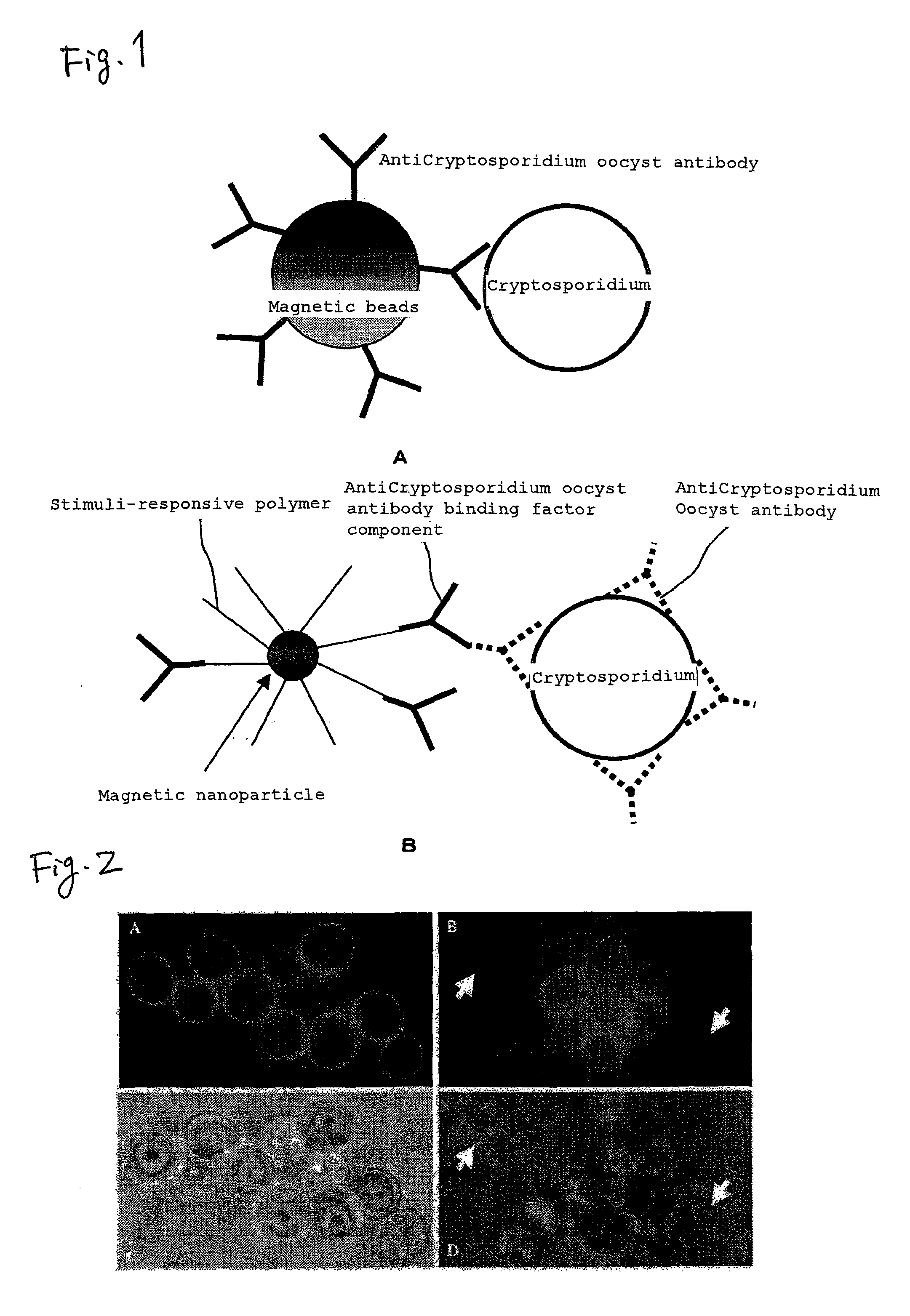
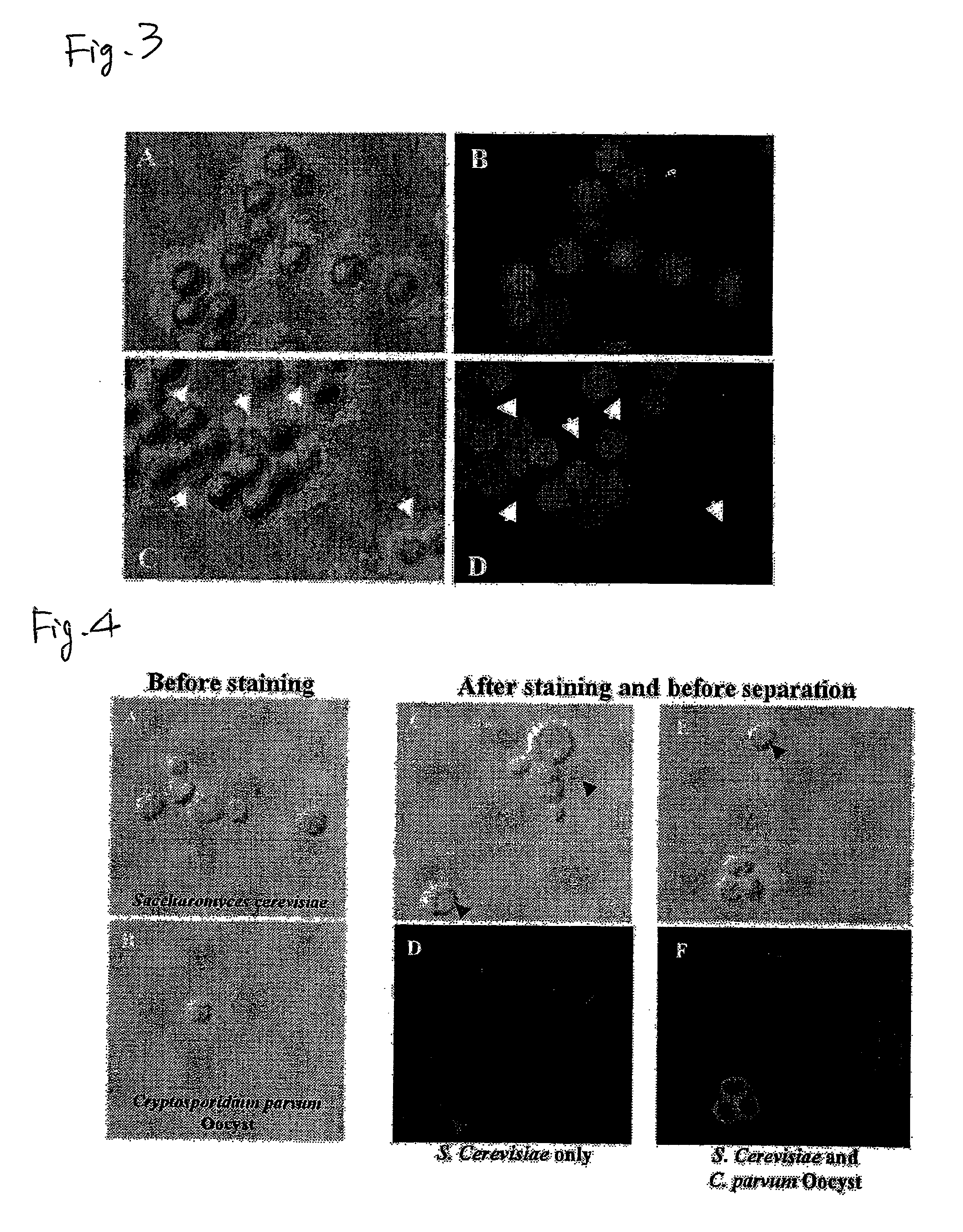
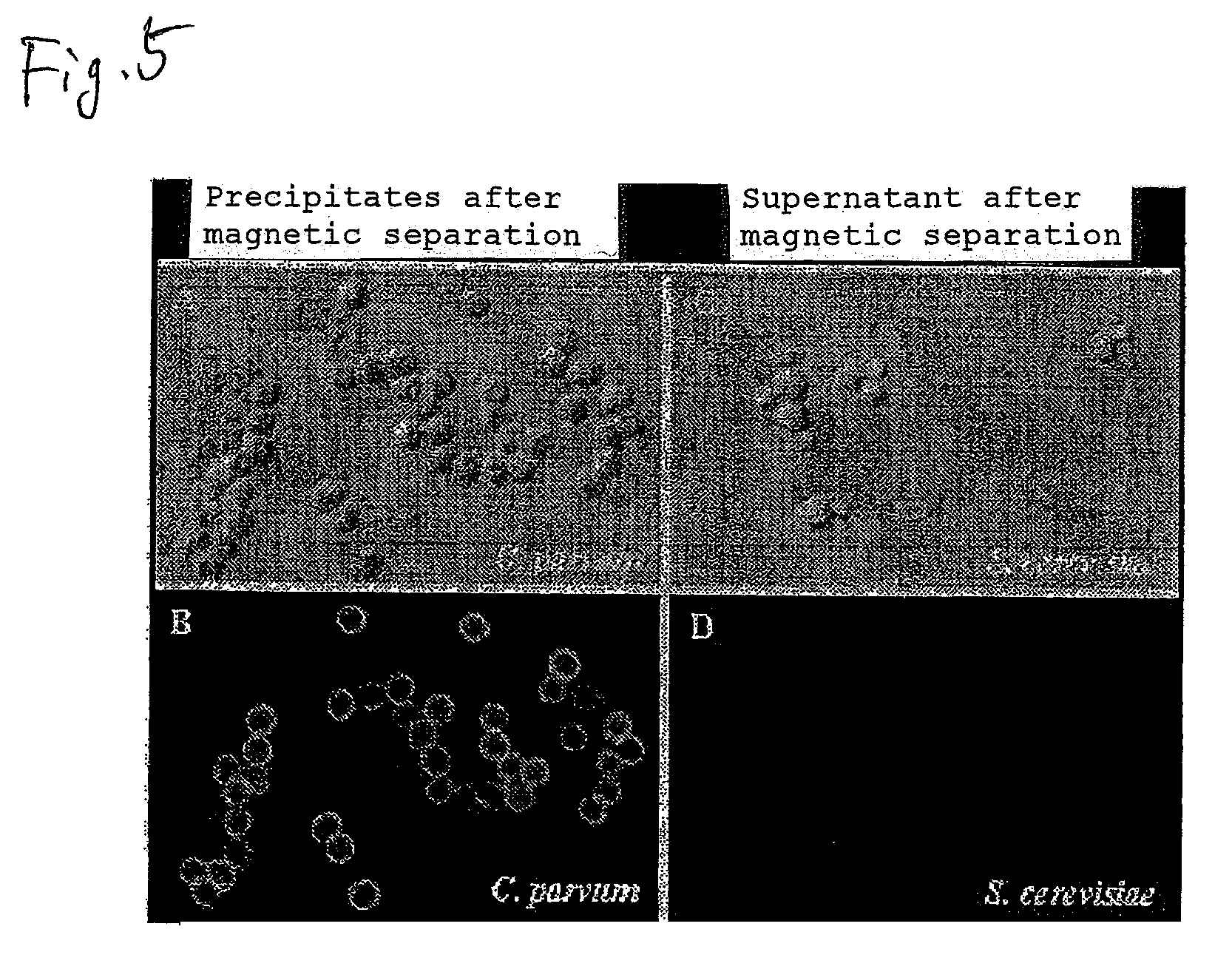
[0093]Fluorescent staining of Cryptosporidium oocysts was attempted in the presence of an excess amount of yeast cells (Saccharomyces cerevisiae). Into 500 μl of TBST buffer were mixed 1×10⁶ yeast cells (see FIG. 4A, which is a bright-field microscope photograph of yeast cells before immunofluorescent labeling) and 3×10⁵ Cryptosporidium oocysts (see FIG. 4B, which is a bright-field microscope photograph of oocysts before immunofluorescent labeling). Then, 50 μl of a commercially available immunofluorescent labeling reagent FITC-Anti-Cryptosporidium IgM (mAb) (Waterborne™, Inc., A400FLR-20X) diluted to 1 / 2000 was added, thereby fluorescent labeling was conducted. Centrifugation at room temperature for 1000 rpm for 5 minutes was conducted and then the precipitate was recovered and the supernatant was discarded. Further, TBST buffer was added and the whole w...


PUM

PropertyMeasurementUnit
particle diameteraaaaaaaaaa
diameteraaaaaaaaaa
specific gravitiesaaaaaaaaaa

Abstract

The present invention provides a method for measuring oocyst of protozoa, such as Cryptosporidium, in an environment sample with high sensitivity at low cost within a short period of time; and a detecting reagent for use therein.Magnetic fine particles of 5 to 500 nm particle diameter having, immobilized thereto, binding factors for specific recognition of oocyst are added to an analyte containing a protozoan oocyst to form oocyst / binding factor / magnetic fine particle complexes by using a binding reaction to the oocyst, the formed complexes are recovered by a magnetic separation, and the protozoan oocysts contained in the complexes are assayed. Further, there is provided, for conducting the above method, a reagent for detecting protozoan oocysts comprising magnetic fine particles of 5 to 500 nm particle diameter having, immobilized thereto, antibodies against oocysts or binding factors for recognizing the antibodies.

Description

TECHNICAL FIELD[0001]The present invention relates to a method for measuring protozoan oocyst and a reagent for detecting the same, capable of inexpensively and conveniently detecting the presence of a protozoan oocyst in various environments such as raw water for tap water, waste water, sewage, natural water and soil.BACKGROUND ART[0002]Tap water, groundwater, stream water, and the like are ingested as drinking water and also there is a possibility that they are unconsciously taken up from mouth, so that it is necessary to pay attention to their treatment in view of good hygiene. In addition to bacteria and suspended particulates, attention has been recently paid to protozoa such as Cryptosporidium. [0003]Cryptosporidium is a digestive tract-parasitic protozoan which is parasitic on mucous membranes of stomach and intestinal tracts to cause diarrhea. Cryptosporidium proliferates with repeating an asexual reproduction term and a sexual reproduction term and oocysts generated from th...

Claims


Application Information

Patent Timeline
Patent Type & Authority Patents(United States)
IPC IPC(8): G01N33/553G01N33/543G01N33/569
CPCG01N33/54326G01N33/56905
Inventor OHNISHI, NORIYUKIFURUKAWA, HIROTAKAHATA, HIDEYUKIMATSUI, KAGEAKINOGAMI, TAKAKONISHIZAWA, HIDEOKONDO, AKIHIKO
Owner KOBE UNIV
Learn More